



fig1
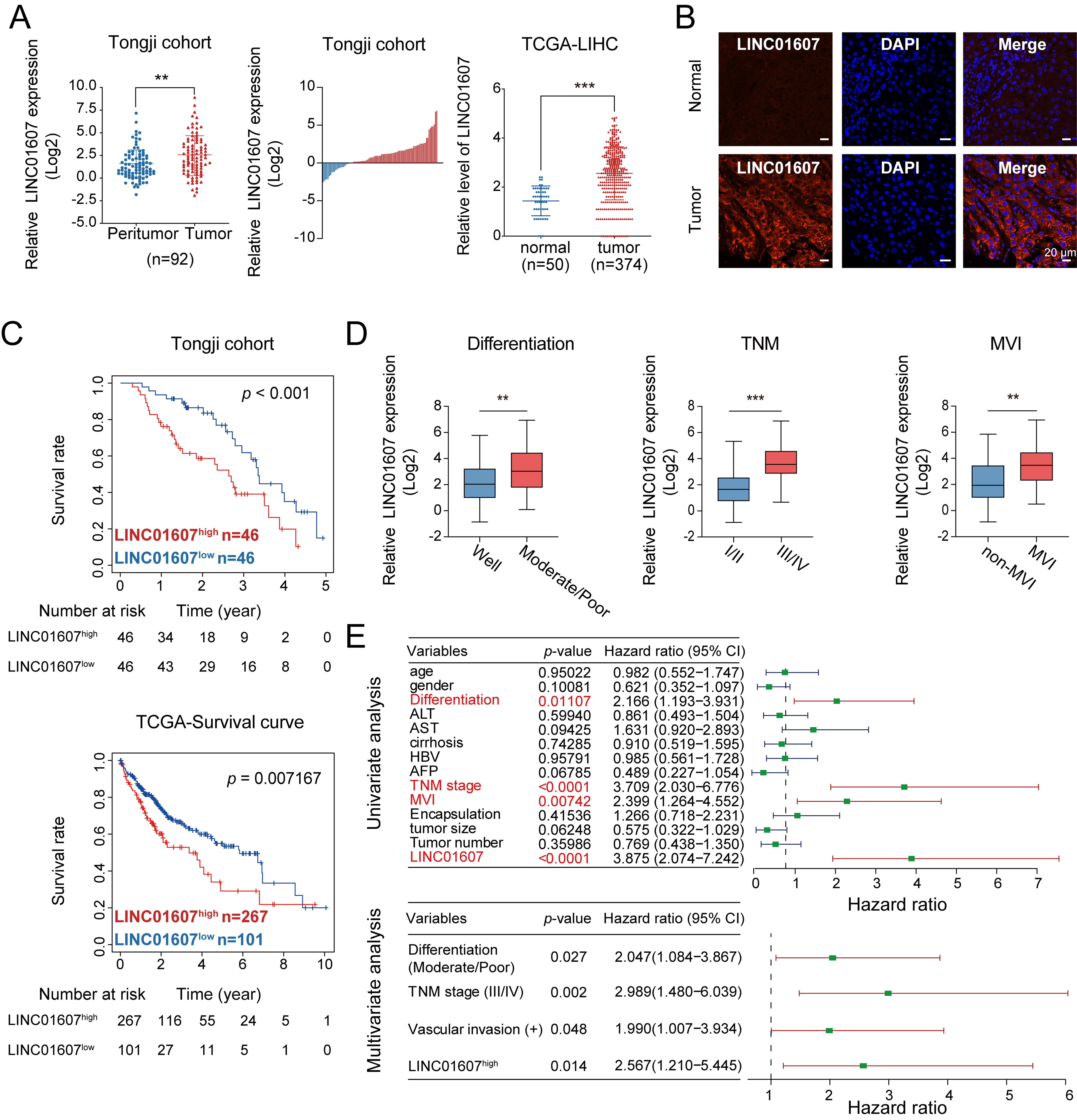
Figure 1. LINC01607 is upregulated in HCC and predicts unfavorable clinical outcomes. (A and B) LINC01607 expression levels in HCC tissues and corresponding peritumor liver tissues were assessed using qRT-PCR (A) and FISH (B). Both the Tongji cohort and TCGA-LIHC dataset showed higher LINC01607 expression in HCC tissues than in corresponding adjacent nontumor tissues; (C) Kaplan–Meier analysis indicated that patients with high LINC01607 expression had shorter overall survival in both the Tongji cohort and TCGA dataset; (D) The correlation between LINC01607 expression and tumor differentiation, TNM staging, and microvascular invasion in HCC patients; (E) Forest plots show univariate and multivariate Cox regression analyses of clinicopathological factors. LINC01607 expression, tumor differentiation, TNM stage, and vascular invasion were independent predictors of reduced overall survival. “n” indicates the number of patient samples. Survival differences were assessed using Kaplan–Meier analysis with the log-rank test. Cox proportional hazards regression was used for univariate and multivariate survival analyses. Other comparisons were performed using Student’s t-test, chi-square test, or Fisher’s exact test, as appropriate. **P < 0.01, ***P < 0.001. HCC: Hepatocellular carcinoma; qRT-PCR: quantitative reverse transcription polymerase chain reaction; FISH: fluorescence in situ hybridization; TCGA-LIHC: The Cancer Genome Atlas Liver Hepatocellular Carcinoma; TNM: Tumor–Node–Metastasis; DAPI: 4′,6-diamidino-2-phenylindole; MVI: microvascular invasion; ALT: alanine aminotransferase; AST: aspartate aminotransferase; HBV: hepatitis B virus; AFP: alpha-fetoprotein.