fig2
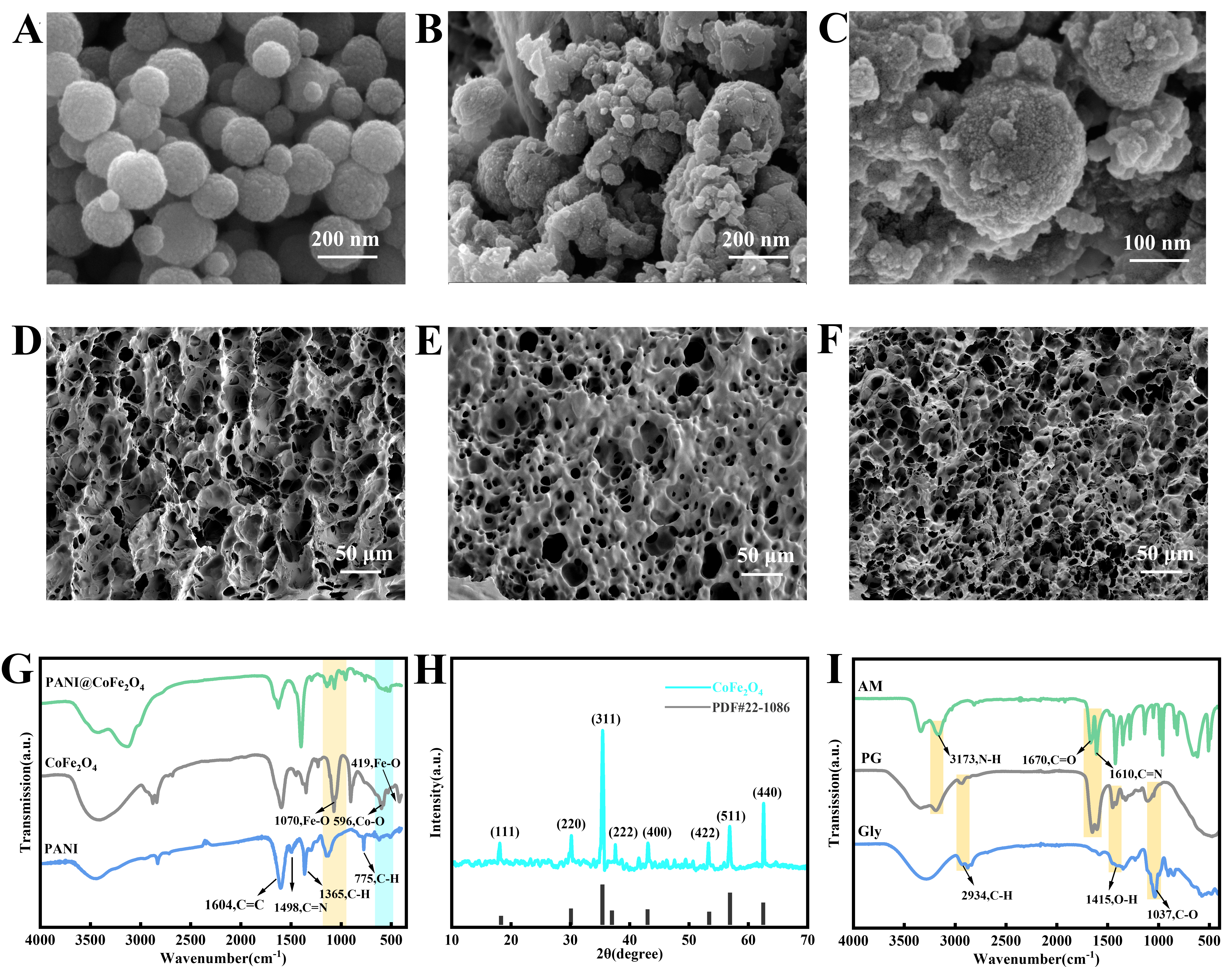
Figure 2. Microstructure and spectroscopic characterization of PANI@CoFe2O4 and PG-based hydrogels; (A) SEM images of CoFe2O4; (B and C) SEM images of PANI@CoFe2O4; (D-F) SEM images of PG, 4-PG/PANI, and 4-PG/P@CoFe; (G) FTIR spectra of PANI, CoFe2O4, and PANI@CoFe2O4; (H) XRD pattern of CoFe2O4; (I) FTIR spectrum of the PG hydrogel. AM: Acrylamide; PG: polyacrylamide/glycerol; SEM: scanning electron microscopy; FTIR: Fourier transform infrared; XRD: X-ray diffraction.