




fig7
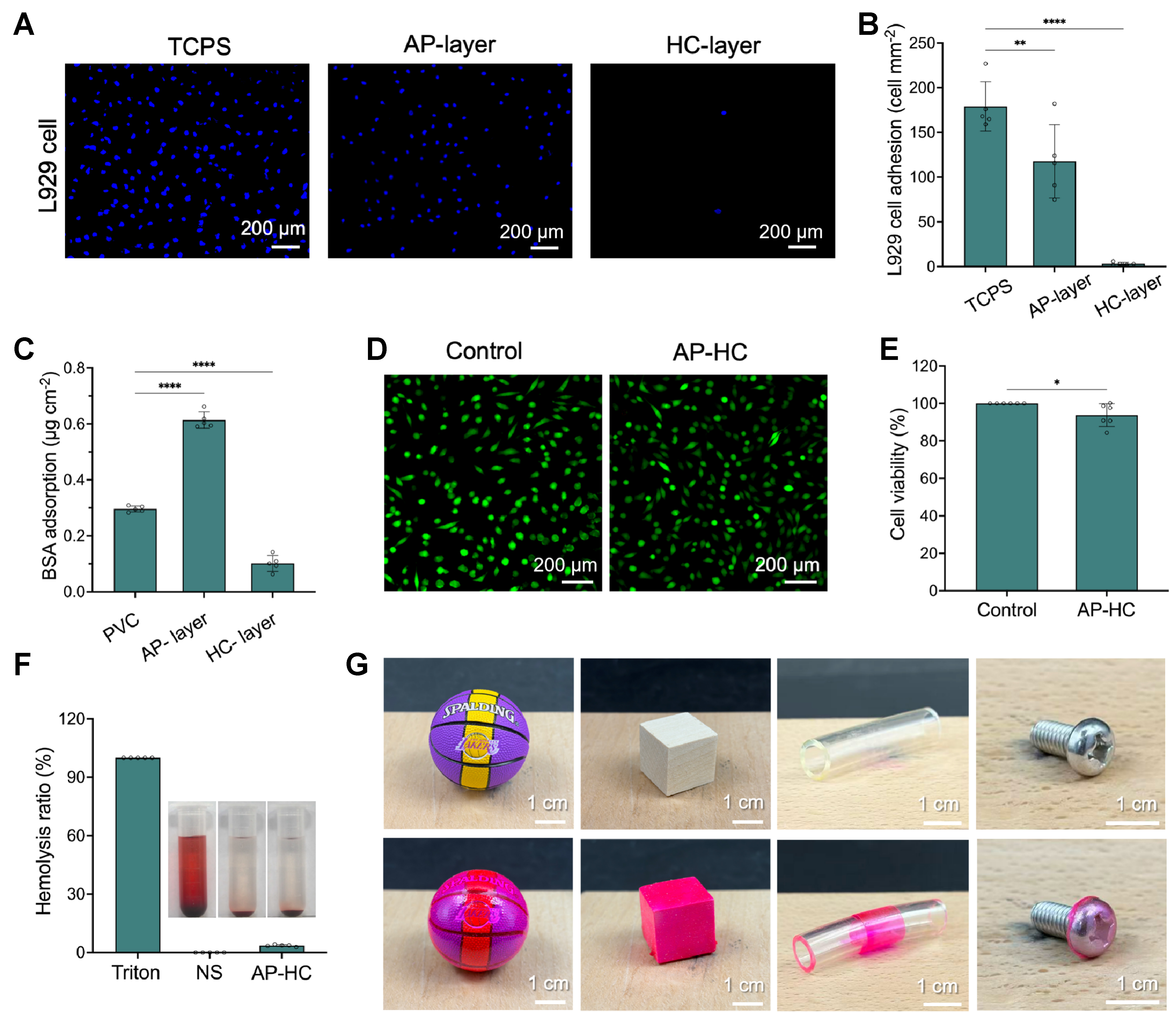
Figure 7. Anti-biofouling performance, biocompatibility, and substrate adaptability of the AP-HC bilayer hydrogel. (A and B) Fluorescence images of Hoechst 33342-stained L929 cells and corresponding quantitative analysis of cell adhesion on the hydrophilic surface of AP-HC-2; (C) Quantified BSA adsorption on PVC, AP-2, and the HC side of AP-HC-2, calculated from the standard curve and expressed as the amount of adsorbed protein per unit exposed surface area; (D and E) Calcein AM/PI fluorescence staining images and cell viability of L929 cells after 24 h of treatment with the AP-HC-2 extract prepared at an extraction ratio of 0.2 g·mL-1; (F) Hemolysis ratio of AP-HC-2; (G) Photographs showing the conformal adhesion of the hydrogel coating on objects with different shapes and materials (The hydrogel was stained with Rhodamine B to improve visual contrast due to its high transparency). Data are presented as mean ± SD; n = 5 independent samples for (B), (C), and (F), and n = 6 independent samples for (E). ****P < 0.0001; **P < 0.01, *P < 0.05. BSA: Bovine serum albumin; PVC: polyvinyl chloride; AM: calcein-AM (calcein acetoxymethyl ester); PI: propidium iodide; SD: standard deviation; TCPS: tissue culture polystyrene; NS: normal saline.