




fig8
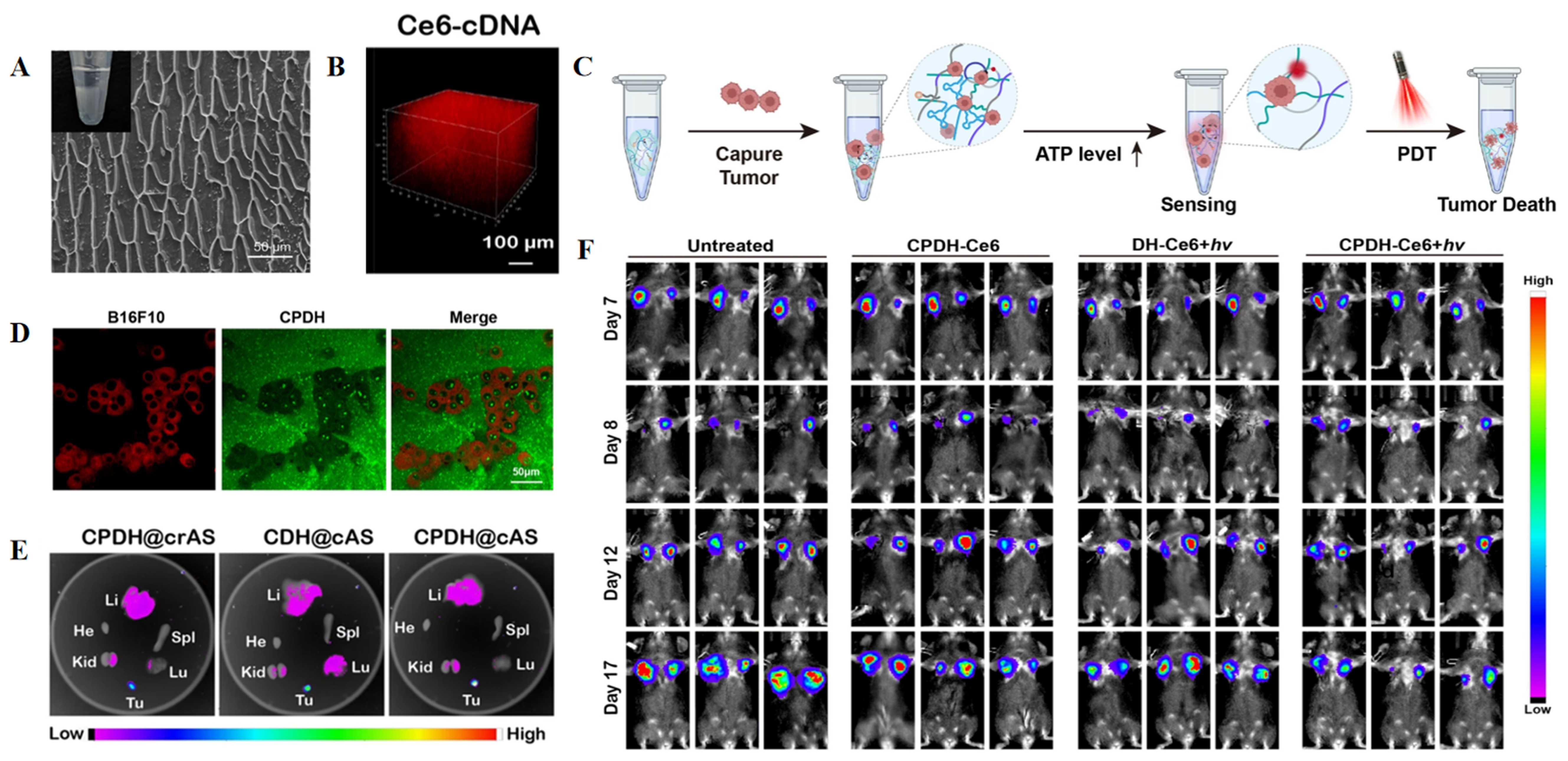
Figure 8. Hydrogel-based theranostic platform for monitoring postoperative tumor recurrence. (A) Cryo-scanning electron microscopy image showing the microstructure of CPDH; (B) 3D stacked fluorescence image showing Ce6-cDNA; (C) Schematic illustration of tumor-cell capture by CPDH-Ce6@cAS, followed by PDT-mediated tumor-cell killing; (D) Confocal fluorescence image of B16F10 cells captured within CPDH-Ce6; (E) Ex vivo fluorescence images of tumors and major organs harvested 48 h after implantation of the indicated hydrogel formulations; (F) Representative bioluminescence images showing the response of B16F10 tumors to local CPDH-Ce6 treatment. (A-F) Adapted with permission from Ref.[249]. In accordance with the Creative Commons Attribution 4.0 International License (CC BY 4.0). © The Author(s) 2023. The original panels were selected, relabeled, and rearranged. CPDH: CpG- and PD-L1 aptamer-containing DNA hydrogel (CpG: unmethylated cytosine-phosphate-guanine motifs; PD-L1: programmed death-ligand 1; DNA: deoxyribonucleic acid); 3D: three-dimensional; Ce6: chlorin e6; cDNA: complementary DNA; cAS: circular ATP sensor; PDT: photodynamic therapy; ATP: adenosine triphosphate.





