

fig3
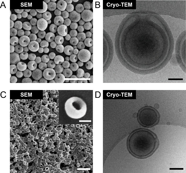
Figure 3. Representative microscopic images of stomatocytes showing their characteristic bowl-shaped morphology and distinctive concave cavity. (A) Morphological characterization of stomatocytes prepared by PEG-b-PS using SEM. Scale bar = 500 nm. Adapted from Shao et al., under the terms of the Creative Commons Attribution 4.0 International License[26]; (B) Cryo-TEM image of PEG-b-PS stomatocytes. Scale bar = 100 nm. Adapted from Wu et al., under the terms of the Creative Commons Attribution 4.0 International License[27]; (C) SEM image of stomatocytes prepared by PEG-PDLLA. Scale bar = 1 μm, insert = 100 nm. Adapted from Shao et al., under the terms of the Creative Commons Attribution 4.0 International License[28]; (D) Bowl-shaped morphology of PEG-PDLLA stomatocytes characterized by cryo-TEM. Scale bar = 200 nm. Adapted from Wang et al., under the terms of the Creative Commons Attribution 4.0 International License[29]. SEM: Scanning electron microscopy; cryo-TEM: cryogenic transmission electron microscopy; PEG-b-PS: poly(ethylene glycol)-block-polystyrene; PEG-PDLLA: poly(ethylene glycol)-block-poly(D,L-lactide).

