

Collagen microgels as ECM-mimetic building blocks: fabrication strategies and regenerative medicine applications
 0
0 Abstract
Collagen microgels are microscale collagen-based materials with tunable particle size, fibrillar structure, crosslinking density, porosity, and loading capacity. By converting collagen from a bulk matrix into discrete microgel units, they provide a useful platform for regulating cell-matrix interactions, mass transport, degradation, and bioactive cargo presentation. This review summarizes recent advances in collagen microgels, beginning with their formation principles, including collagen self-assembly, fibrillogenesis, assisted gelation, and crosslinking-mediated network stabilization. Major preparation methods are then discussed, including mechanical force-driven methods, spray-based manufacturing, microfluidic platforms, and other emerging strategies. The biomedical applications of collagen microgels are further reviewed, with emphasis on cell delivery, growth factor delivery, drug delivery, injectable micro-scaffolds, microgel inks, and organoid culture. Finally, this review outlines key considerations for future collagen microgel design, including collagen source, fabrication control, microgel architecture, and biological response.
Keywords
INTRODUCTION
Collagen is the major structural protein of animal connective tissues and represents approximately 25%-35% of the total protein mass in mammals, making it the most abundant structural component of the extracellular matrix (ECM)[1,2]. Its fundamental molecular architecture consists of three polypeptide α chains wound into a right-handed triple helix, which is stabilized by the repeating Gly-X-Y sequence, where X and Y are frequently occupied by proline and hydroxyproline, respectively[3]. This molecular configuration imparts thermal stability, self-assembly capability, and enzymatic specificity. Based on the composition of α chains and tissue distribution, 28 types of collagen subtypes have been identified. Among them, type I collagen[4], with the molecular composition [α1(I)]2α2(I), is the most commonly used collagen type for biomaterial and microgel fabrication because it is abundant, commercially available, and capable of forming fibrillar networks under mild physiological conditions. Type II collagen is more relevant to cartilage-oriented systems, whereas type III collagen is associated with extensible tissues such as blood vessels, skin, and internal organs[2]. However, compared with type I collagen, these collagen types are less frequently used as primary microgel-forming materials because their sources, purification routes, and tissue-specific applications are less standardized for general biomaterial processing.
Collagen used for microgel fabrication is usually isolated from connective tissues, including rat tail tendon, bovine or porcine skin and tendon, and marine sources such as fish skin, scales, and other fish-processing by-products[4-6]. Native fibrillar collagen is poorly soluble in water because collagen molecules are packed into supramolecular fibrils and stabilized by intermolecular interactions; in mature tissues, pre-existing lysyl oxidase-mediated covalent crosslinks further reduce collagen solubility. Therefore, material-grade collagen is commonly processed into soluble forms by dilute acid extraction or pepsin-assisted extraction. Under cold acidic conditions, protonation of ionizable residues weakens intermolecular interactions, thereby promoting collagen swelling and dispersion. Pepsin digestion further improves solubilization by cleaving non-helical telopeptide regions, while largely preserving the triple-helical domain and reducing antigenicity[5]. The extracted collagen is then commonly purified by salt precipitation, dialysis, and lyophilization before being reconstituted for material fabrication. This distinction is important because soluble collagen is not equivalent to gelatin. Gelatin is generated by partial thermal, acidic, alkaline, or enzymatic denaturation of collagen, during which the triple-helical structure is disrupted into random-coil chains[7]. Compared with gelatin microgels, collagen microgels better retain the native triple-helical structure, fibrillogenic behavior, and collagen-specific cell-recognition motifs. Gelatin microgels, especially methacrylated gelatin systems, are easier to dissolve, process, and photocrosslink, but they do not reproduce the native fibrillar architecture of collagen in the same manner[7,8].
The biomedical value of collagen arises not only from its structural role but also from its intrinsic ECM-like bioactivity. Triple-helical integrin-binding motifs, especially GxOGER-type sequences such as GFOGER[3] and GROGER[9], can be recognized by collagen-binding integrins and initiate receptor-mediated cell adhesion and signaling[10,11]. Although RGD motifs are widely used in biomaterial design, native fibrillar collagen is more accurately characterized by these triple-helical integrin-binding motifs rather than by RGD alone. Collagen-integrin engagement promotes focal adhesion formation and activates downstream signaling pathways associated with cell survival, migration, cytoskeletal tension, and mechanotransduction, including FAK/Src, RhoA/ROCK, MAPK/ERK, and YAP/TAZ-associated responses[12,13]. In parallel, collagen matrices can be enzymatically remodeled by collagenases and matrix metalloproteinases (MMPs), allowing material degradation to be coupled with cell invasion, ECM turnover, and tissue repair[14]. These features distinguish collagen from many polysaccharide-based hydrogels. For example, alginate inherently lacks mammalian cell-adhesive ligands and often requires peptide modification, such as RGD conjugation, to support integrin-mediated adhesion[15]. Hyaluronic acid-based hydrogels are biologically relevant, but they are also frequently chemically modified to tune crosslinking, degradation, mechanics, and cell-interactive properties for tissue engineering applications[16].
Based on these properties, collagen has been developed into a versatile biomaterial platform in the form of hydrogels, porous sponges, films, fibers, and injectable matrices[17]. Among these formats, collagen hydrogels are particularly attractive in regenerative medicine because their high water content and fibrillar architecture partially recapitulate the hydrated ECM microenvironment. Bulk collagen hydrogels are commonly formed through pH neutralization, thermal induction, enzymatic treatment, or chemical crosslinking[18], and have been widely used as tissue engineering scaffolds and three-dimensional cell culture matrices[9]. However, bulk hydrogels still face several limitations. Their relatively large dimensions can restrict oxygen and nutrient transport in thick or cell-dense constructs and may lead to hypoxia or metabolic waste accumulation. Oxygen diffusion coefficients through dense collagen scaffolds have been reported to decrease from approximately 4.5 × 10⁻⁶ cm2/s at 11% collagen density to 1.7 × 10⁻⁶ cm2/s at 34% collagen density, indicating that both matrix density and diffusion distance are critical for cell survival in collagen-based constructs[19]. Bulk collagen hydrogels also commonly show weak mechanics without additional reinforcement[9,20]. These features limit the use of bulk collagen hydrogels in applications that require minimally invasive delivery, shape adaptation to irregular defects, or mechanically robust tissue reconstruction.
Collagen microgels provide a particulate and modular alternative to bulk collagen hydrogels. In this review, collagen microgels are defined as collagen-based hydrogel particles with characteristic dimensions in the micrometer range, typically 1 to 1000 μm[21]. Nanoscale collagen-based gel particles and hybrid microgels are included when collagen serves as the dominant structural or bioactive ECM component. Gelatin-derived or non-native collagen-derived microgel systems are not the main focus of this review, but they are briefly discussed when they provide relevant design principles for collagen microgel engineering, such as interparticle stabilization, printability, and bioink formation. Within the micrometer-size regime, collagen microgel size strongly affects biological performance. Smaller microgels provide shorter diffusion distances and larger surface-area-to-volume ratios, which can reduce mass-transport limitations compared with larger bulk hydrogels[21,22]. For spherical particles, the surface-area-to-volume ratio scales as 6/d; therefore, reducing the particle diameter from 1 mm to 100 μm increases the surface-area-to-volume ratio by approximately 10-fold, while reducing it to 10 μm increases it by approximately 100-fold. This geometric advantage does not necessarily alter the intrinsic diffusivity of the collagen network, which is mainly governed by matrix composition, density, and pore structure. Instead, reducing microgel size shortens the transport path and increases the material-fluid interface for oxygen, nutrients, and metabolic waste exchange[12,21,23]. Larger microgels may provide higher per-particle payload capacity and stronger local retention in some local delivery or implantation settings, although their biodistribution and vascular trapping risk should be considered for systemic administration[22]. When assembled into granular scaffolds, microgels can further generate interconnected interparticle pores that facilitate cell infiltration, matrix deposition, and tissue integration[24].
More importantly, collagen microgels can function as programmable ECM-mimetic building blocks[21]. Their size, stiffness, degradation rate, biochemical composition, and cargo-loading capacity can be adjusted before secondary assembly, allowing different microgel populations to be combined in a modular manner[25]. For example, microgels with distinct crosslinking densities, cell types, or growth factor profiles can be mixed, layered, or spatially organized to generate macroscale constructs with controlled heterogeneity. This modular design principle supports the development of delivery systems, injectable micro-scaffolds, three-dimensional inks, organoid-supporting matrices, and other regenerative medicine platforms[25,26].
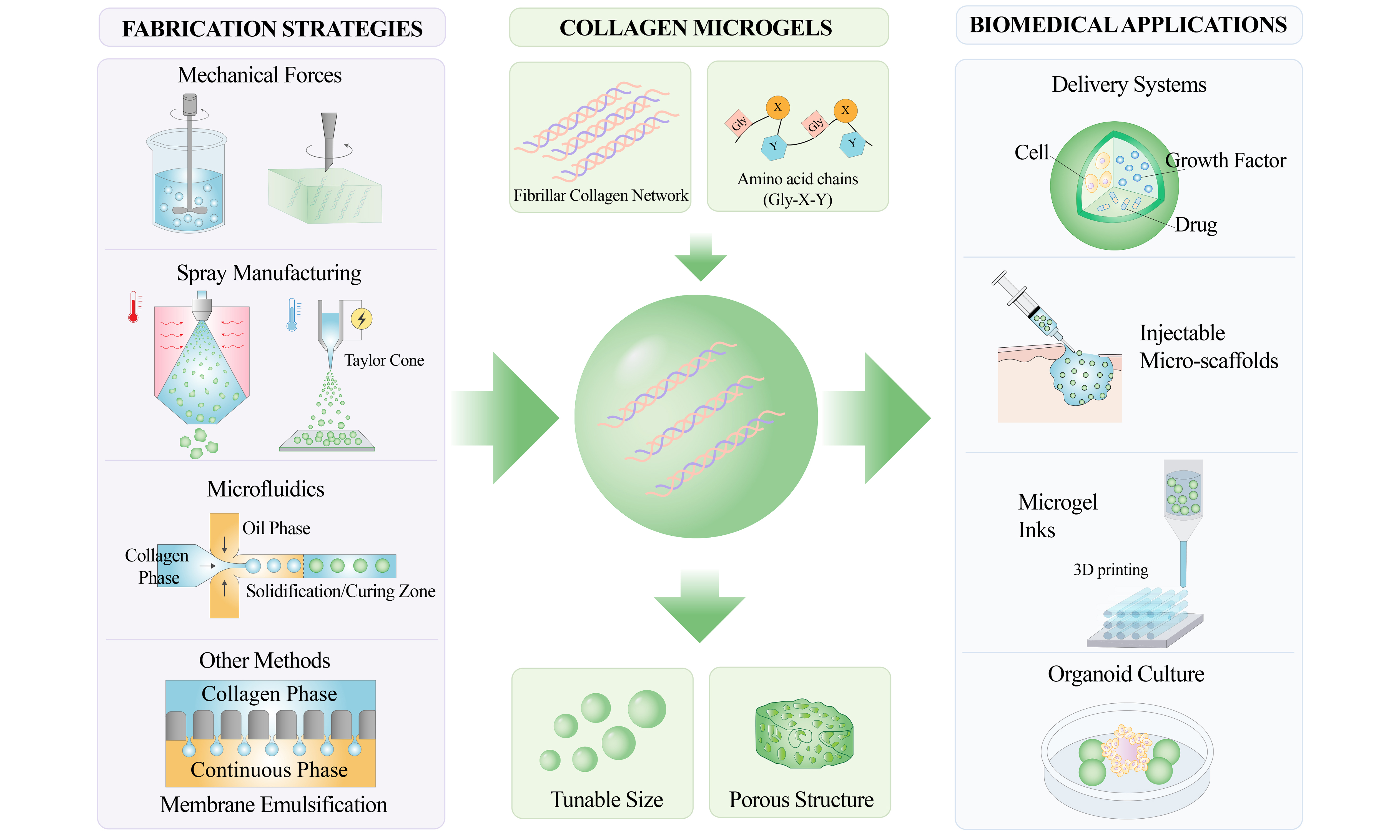
Although collagen microgels have been explored since the 1980s, interest in this field has increased markedly in recent years. Their development has moved from relatively coarse particle production toward more precise and programmable fabrication, and their applications have expanded from simple drug loading and cell culture to more sophisticated strategies for tissue engineering and regenerative medicine. In this review, we summarize recent progress in collagen microgels, focusing on their fabrication strategies, formation mechanisms, and biomedical applications. We also discuss the remaining challenges and future opportunities for developing collagen microgels as ECM-mimetic building blocks for regenerative medicine [Figure 1].
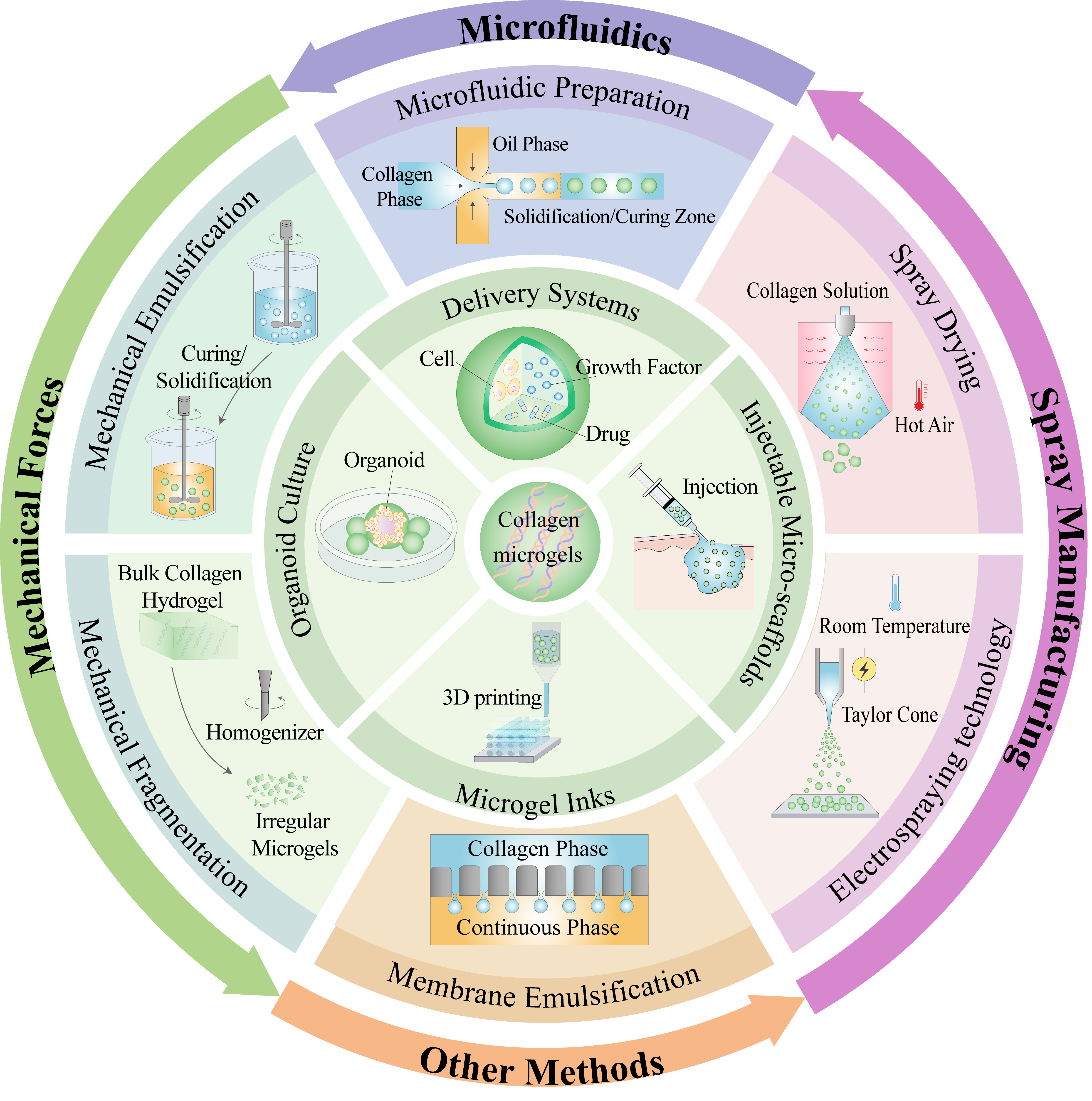
Figure 1. Fabrication strategies and biomedical applications of collagen microgels. Schematic illustration shows the diverse preparation techniques for collagen microgels, including mechanical force-driven methods (emulsification and fragmentation), spray-based manufacturing (spray drying and electrospraying), microfluidic platforms, and alternative approaches (membrane emulsification). The diagram also highlights their applications in regenerative medicine as delivery systems for cells, growth factors, and drugs; injectable micro-scaffolds for tissue repair; microgel inks for 3D printing; and matrices for organoid culture and tissue engineering.
ADVANCES IN THE PREPARATION TECHNOLOGY OF COLLAGEN MICROGELS
Fundamental principles of collagen microgel formation
The fabrication of collagen microgels generally involves two closely coupled processes: the generation of microscale droplets or particles and the stabilization of collagen networks within these confined structures. The former determines the size, morphology, dispersity, and production efficiency of microgels, whereas the latter governs their mechanical integrity, degradation resistance, injectability, diffusion behavior, and ECM-mimetic bioactivity. Therefore, before discussing individual fabrication technologies, it is necessary to clarify the major gelation and stabilization mechanisms underlying collagen microgel formation.
Physical self-assembly and collagen fibrillogenesis
Collagen microgels are distinct from many synthetic microgel systems because collagen itself can undergo spontaneous fibrillogenesis under mild aqueous conditions. In most microgel fabrication protocols, collagen is maintained in a soluble state under acidic and low-temperature conditions and is then induced to assemble by pH neutralization and temperature elevation, often to room or physiological temperature. Under these conditions, collagen molecules gradually self-assemble into fibrillar networks and form physically stabilized hydrogels[27,28]. However, collagen fibrillogenesis should not be viewed as being restricted to a single pH-temperature condition. Dense collagen solutions can form fibrillar gels over broader pH and ionic-strength ranges, and collagen concentration strongly affects the resulting gel architecture and fibril morphology[29]. Therefore, collagen self-assembly in microgel systems should be understood as a physicochemical process governed by pH, temperature, ionic strength, collagen concentration, and confinement[30,31]. This intrinsic behavior provides an important basis for collagen microgel preparation because it enables the construction of ECM-like fibrillar networks while preserving the native triple-helical conformation and biological activity of collagen.
Although collagen fibrillogenesis can occur over a relatively broad physicochemical window, conventional collagen microgel fabrication still requires careful process control[29,32]. In many droplet-based methods, collagen precursors are handled under cold acidic conditions to avoid premature viscosity increase or gelation, followed by neutralization and thermal incubation to induce network formation[33,34]. This approach is mild and bioactive, but its gelation kinetics may be too slow for some microscale manufacturing processes, leading to droplet coalescence, deformation, or loss of structural integrity before sufficient network stabilization occurs. Therefore, temporal coordination between droplet formation and collagen network stabilization remains critical for collagen microgels[35].
Rapid assembly and assisted gelation strategies
Recent advances have focused on developing rapid assembly strategies to overcome the slow kinetics of conventional thermal gelation. Among these strategies, macromolecular crowding has attracted particular attention. Macromolecular crowding agents, such as polyethylene glycol (PEG) and dextran, can reduce the effective free volume of collagen solutions, increase the local effective concentration of collagen molecules, and promote molecular collision, nucleation, and fibril growth. As a result, collagen fibrillogenesis can be significantly accelerated[36].
This strategy has led to significant improvements in collagen microgel preparation. By allowing acidic collagen droplets to come into contact with crowding agents within microfluidic channels[34] or immediately after spray atomization[36], collagen assembly can be rapidly induced within confined droplets. This approach reduces the dependence on conventional pre-neutralization and prolonged thermal incubation, thereby simplifying the preparation process and improving the structural stability of collagen microgels. For example, Samiei et al.[34] used a parallel microfluidic device in which acidic collagen solution was passively broken into droplets within a continuous oil phase. Under the crowding effect of PEG, chaotic convective mixing inside the droplets promoted collagen fibrillogenesis or co-precipitation, leading to the formation of collagen microgels [Figure 2A].
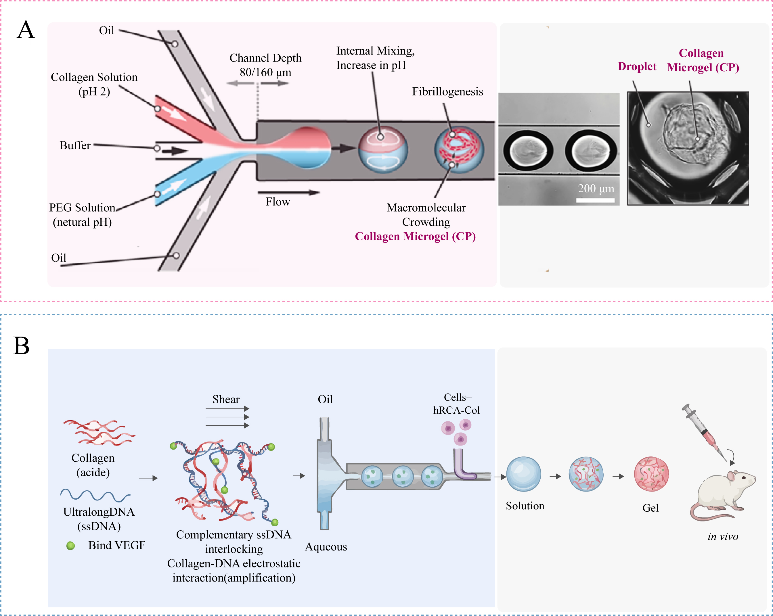
Figure 2. Representative rapid assembly strategies for collagen microgel fabrication. (A) Schematic illustration of collagen microgel formation from acidic collagen solution under PEG-induced macromolecular crowding. The crowding effect accelerates collagen fibril nucleation and assembly, enabling rapid gelation within microfluidic droplets. Figure 2A is adapted in part from Ref.[34]. Reused under the terms of the Creative Commons Attribution License (CC BY); (B) Microfluidic preparation of collagen microgels assisted by ultralong DNA strands. Electrostatic interactions between collagen and DNA, together with the interlocking of complementary ultralong single-stranded DNAs under shear force, markedly accelerate collagen gelation and enable growth factor loading. Figure 2B is newly drawn by the authors based on the fabrication principle reported by Ref.[37]. No original figure elements were reused.
In another representative strategy, Zhao et al.[37] combined the electrostatic interaction between collagen and ultralong DNA strands with the interlocking effect of two complementary ultralong single-stranded DNAs, RCA and c-RCA, under shear force. This design successfully shortened the collagen gelation time from 25 min to 40 s. Moreover, through the multifunctional design of DNA segments, bioactive molecules such as vascular endothelial growth factor (VEGF) could be loaded into the microgel system [Figure 2B]. These studies indicate that rapid assembly strategies can effectively bridge the gap between collagen fibrillogenesis and microscale manufacturing.
Crosslinking chemistry and network stabilization
In addition to physical fibrillogenesis, chemical and supramolecular crosslinking strategies are often introduced to improve the structural stability, mechanical strength, and degradation resistance of collagen microgels. These strategies are especially important when collagen microgels are used as injectable micro-scaffolds, delivery systems, cell carriers, or bioink components, where the microgels must maintain their architecture under mechanical stress and physiological degradation.
Covalent crosslinking is one of the most widely used stabilization strategies. Carbodiimide-mediated[38] or 4-(4,6-dimethoxy-1,3,5-triazin-2-yl)-4-methylmorpholinium chloride (DMTMM)-mediated[39] coupling can activate carboxyl groups on collagen and promote amide bond formation with amino groups, resulting in zero-length crosslinks between collagen molecules. Aldehyde-based crosslinkers, such as glutaraldehyde or oxidized polysaccharides, mainly react with the ε-amino groups of lysine and hydroxylysine residues to form imine or Schiff-base linkages[40]. Genipin[41] can also react with primary amines in collagen and generate bridged networks with relatively lower cytotoxicity than glutaraldehyde. In photocrosslinkable[42] systems, collagen or collagen-derived macromers are first modified with photoactive groups, such as methacrylateps, and then polymerized under light irradiation to achieve spatially and temporally controlled gelation.
Besides conventional chemical crosslinking, enzymatic crosslinking provides a relatively mild and biomimetic route for collagen network stabilization. Transglutaminase-mediated crosslinking has been widely investigated in collagen-based biomaterials. Transglutaminase[43,44] catalyzes acyl-transfer reactions between glutamine and lysine residues, leading to the formation of ε-(γ-glutamyl)lysine isopeptide bonds between collagen molecules. In addition to transglutaminase, lysyl oxidase-related[45] chemistry is also biologically relevant to collagen stabilization, because lysyl oxidase (LOX) initiates native collagen crosslinking in the ECM by oxidatively deaminating lysine or hydroxylysine residues to aldehyde-derived intermediates. These intermediates subsequently participate in intermolecular crosslink formation and contribute to the mechanical maturation of collagen fibrils.
The choice of crosslinking chemistry strongly influences the final performance of collagen microgels. A higher crosslinking density generally improves mechanical stiffness and enzymatic resistance, but excessive crosslinking may reduce swelling capacity, molecular diffusion, and cell-interactive sites. In contrast, mild physical or enzymatic stabilization can better preserve the native bioactivity of collagen, but may provide insufficient long-term stability if the network density is too low. Therefore, the design of collagen microgels requires a balance among network stability, bioactivity, degradation behavior, and process compatibility. Representative gelation and crosslinking mechanisms are summarized in Table 1.
Gelation and network stabilization mechanisms relevant to collagen microgel formation
| Mechanism | Trigger/Reaction | Role in microgel formation | Advantages and limitations | Ref. |
| Physical self-assembly and collagen fibrillogenesis | ||||
| Physical fibrillogenesis of collagen | pH neutralization, temperature elevation, and ionic strength adjustment | Converts soluble collagen molecules into fibrillar networks within droplets or preformed microstructures | Preserves native triple-helical structure and ECM-like bioactivity; however, gelation is relatively slow and requires strict pH/temperature control | [9,27,28,30] |
| Rapid assembly and assisted gelation strategies | ||||
| Macromolecular crowding-assisted collagen assembly | PEG, dextran, or other crowding agents reduce free volume and increase effective collagen concentration | Accelerates collagen fibril nucleation and growth in confined droplets | Improves gelation speed and droplet stabilization; concentration and molecular weight of crowding agents need optimization | [34,36] |
| DNA-assisted rapid collagen gelation | Electrostatic interaction between collagen and ultralong DNA strands, combined with DNA interlocking under shear | Rapidly stabilizes collagen microgels and enables bioactive molecule loading | Greatly shortens gelation time and supports VEGF loading; formulation is more complex than simple collagen gelation | [37] |
| Crosslinking chemistry and network stabilization | ||||
| Amide coupling | EDC/NHS or DMTMM activates collagen -COOH groups and promotes reaction with -NH2 groups | Forms zero-length covalent crosslinks between collagen molecules | Improves mechanical stability and enzymatic resistance; excessive crosslinking may reduce swelling, diffusion, and cell-recognition sites | [9,38,39] |
| Aldehyde-amine crosslinking | Glutaraldehyde or oxidized polysaccharides react with ε-amino groups of lysine/hydroxylysine residues | Forms imine or Schiff-base-linked collagen networks | Efficiently enhances network stability; glutaraldehyde has residual aldehyde and cytotoxicity concerns | [9,40] |
| Genipin-mediated crosslinking | Genipin reacts with primary amines in collagen | Generates genipin-bridged collagen networks | Lower cytotoxicity than glutaraldehyde and improves stability; reaction is slower and may cause color change | [9,41] |
| Photocrosslinking of modified collagen | Methacrylated collagen or collagen-derived macromers undergo light-induced polymerization | Provides rapid and spatially controlled network fixation | Useful for bioinks and patterned constructs; requires chemical modification, photoinitiators, and light exposure | [42] |
| Enzymatic crosslinking | Transglutaminase-mediated reaction between glutamine and lysine residues; LOX-inspired oxidation of lysine residues | Provides biomimetic covalent stabilization of collagen networks | Mild and cytocompatible; reaction efficiency depends on enzyme activity, substrate accessibility, pH, and temperature | [43-45] |
Overall, the preparation methods for collagen microgels share several engineering principles with common microgel systems. However, the intrinsic fibrillogenesis, temperature sensitivity, and crosslinking requirements of collagen make the selection of fabrication strategy particularly important. In the following sections, we discuss the major preparation routes for collagen microgels, including mechanical-force-based, spray-based, and microfluidic methods, as well as phase separation and membrane emulsification, with an emphasis on their advantages and limitations.
Microgel manufacturing under mechanical force
Mechanical emulsification
Mechanical emulsification is a simple and widely accessible method for preparing collagen microgels. In this process, an aqueous collagen solution is dispersed into an oil phase, such as olive oil[46], liquid paraffin[47], or soybean oil, under mechanical stirring. Surfactants such as Span-80, Span-85, or Tween-20 are commonly added to stabilize the resulting water-in-oil (W/O) emulsion. After droplet formation, the collagen phase is stabilized by chemical crosslinking or physical gelation, followed by repeated washing with solvents such as 2-propanol[48], acetone, or Phosphate-Buffered Saline to remove residual oil and surfactants. Chemical crosslinkers[47,48] are frequently used to improve the structural integrity and mechanical stability of collagen microspheres, but residual crosslinkers may raise cytotoxicity concerns and require careful removal. To avoid chemical crosslinking, Snider et al.[46] used the intrinsic room-temperature fibrillogenesis of collagen to prepare collagen microgels through a W/O emulsion process without additional crosslinkers.
However, the gelation state of collagen during emulsification must be carefully controlled. Insufficient fibril formation may lead to droplet rupture or coalescence before the network stabilizes, whereas excessive fibrillogenesis increases precursor viscosity and hinders uniform droplet breakup under stirring. As a result, mechanically emulsified collagen microgels often show broad size distributions and limited sphericity. Incorporating polysaccharides such as chitosan can improve particle morphology by promoting rapid interfacial stabilization through ionic interactions, electrostatic complexation, or complementary network formation[49]. These interactions help maintain droplet integrity during gelation and can produce microgels with improved sphericity. Nevertheless, mechanical emulsification still requires oil phases and surfactants, and incomplete removal of these residues may compromise cell compatibility or interfere with cell membrane integrity.
Mechanical fragmentation
Conventional emulsification methods are often constrained by the relatively low viscosity and collagen concentration of the precursor solution, which makes it difficult to obtain dense collagen microgels. Mechanical fragmentation provides an alternative route. In this method, a high-concentration bulk collagen gel is first prepared to allow collagen fibrillogenesis and network stabilization, and the gel is then physically disrupted into microparticles using a homogenizer, grinder, sieve, or extrusion device[50]. Unlike emulsification, where collagen assembly occurs within individual droplets, mechanical fragmentation divides a preformed collagen network into smaller units. As a result, the fragmented microgels can partially retain the fibrillar architecture, porosity, collagen density, and crosslinking state of the parent hydrogel. This feature is difficult to achieve by direct emulsification of dilute collagen solutions. Hong et al.[51] reported a micro-fragmented collagen/hyaluronic acid hydrogel wound dressing composed of microscale hydrogel units. The enhanced porosity of this fragmented collagen-based system improved hADSC survival and paracrine activity, thereby accelerating wound maturation compared with conventional cell aggregates and bulk collagen hydrogel wound dressings.
However, collagen microparticles produced by mechanical fragmentation often have irregular polygonal or fragmented shapes, with a wide particle size distribution, which is unfavorable for controlled drug release, where uniform size and shape are required. Nonetheless, in tissue filling and cell scaffold applications, the irregular morphology enhances mechanical interlocking among particles, thereby improving the mechanical stability of the hydrogel. Van Der Heide et al.[52] used the mechanical fragmentation method to prepare microgels, which, despite the issue of non-uniform particle size, demonstrated good elastic recovery and shear-thinning properties in terms of biomechanics, and were successfully applied to the delivery of bone morphogenetic protein-7 chemically modified RNA (BMP-7 cmRNA), promoting osteogenic differentiation of human mesenchymal stem cells.
Spray manufacturing method
Spray-based manufacturing methods provide an alternative route for producing dispersed collagen-containing particles. In the context of micrometer-sized collagen microgels, spray-based techniques should be discussed with clear boundaries. Spray drying is a heat-driven continuous powder-production process, whereas electrospraying and electrostatic droplet generation use an electric field to generate charged droplets under relatively mild thermal conditions. Therefore, spray drying is more relevant to collagen-derived powders, gelatin particles, or thermally stabilized collagen systems, while electrospraying and electrostatic droplet generation are more suitable for preparing collagen-containing microparticles, microspheres, or composite microgels in the micrometer-size range.
Spray drying
Spray drying (SD) is a continuous preparation technique that rapidly transforms liquid feed into dry particulate powders through atomization, droplet-to-particle conversion, and particle collection in a hot drying medium[53,54]. The final particle morphology and powder properties are strongly influenced by spray dryer design, feed characteristics, and processing parameters, including inlet temperature, feed flow rate, drying gas flow, atomization mode, feed viscosity, and solid content[53,54]. Because of its high productivity, short processing time, and industrial scalability, SD is widely used for dry particle and powder production. However, its direct application to native collagen microgels remains limited. Native collagen depends on its triple-helical and fibrillar architecture for collagen-specific bioactivity, whereas heat and harsh drying conditions can disrupt the triple helix and promote gelatinization or degradation[55]. Therefore, in the field of micrometer-sized collagen microgels, conventional SD should be regarded mainly as a related powder-forming technology for collagen-derived or thermally stabilized systems rather than as a preferred route for fabricating native ECM-mimetic collagen microgels.
The morphology of spray-dried particles is also influenced by drying kinetics. During rapid solvent evaporation, a partially solidified outer shell may form before the droplet interior is completely dried, resulting in shrinkage, wrinkling, hollow particles, or other non-spherical morphologies[53,54]. These issues are especially relevant for protein-containing feeds because viscosity, solids content, glass-transition behavior, and drying temperature affect shell formation and collapse. Thus, although SD is attractive for continuous manufacturing, its heat-driven nature and drying-induced morphological changes may limit its suitability for preparing native collagen microgels that require preservation of fibrillar microarchitecture and collagen-specific bioactivity. Low-temperature spray-based techniques provide a promising alternative for thermally sensitive collagen-containing microgels.
Electrospraying technology
In contrast, electrospraying and electrostatic droplet generation are more compatible with micrometer-scale collagen microgel manufacturing because they can generate droplets under mild or ambient thermal conditions. In electrospraying, a high-voltage electric field deforms the liquid meniscus into a Taylor cone. When electrostatic forces overcome surface tension, charged droplets are ejected and subsequently solidify during flight or upon collection[56]. The final particle size and morphology are affected by solution viscosity, conductivity, surface tension, flow rate, applied voltage, needle-collector distance, and the collection medium[56]. For collagen-containing systems, this non-thermal feature reduces the risk of heat-induced denaturation compared with SD, although collagen structure and fibrillogenesis still depend on solvent composition, pH, ionic strength, collagen concentration, and post-gelation conditions.
Electrospraying has been used to prepare micrometer-sized collagen-containing particles. Jayakumar et al.[57] prepared antibacterial collagen-pectin microparticles by electrospraying collagen dissolved in acetic acid. They showed that particle formation depended strongly on acetic acid concentration, viscosity, conductivity, and NaCl content, and the optimized collagen-pectin microparticles were smaller than 150 μm when sprayed using a 0.65 mm nozzle. This example demonstrates that electrospraying can process collagen-containing feeds into micron-scale particles, but it also highlights the narrow process window caused by the rheological and electrical properties of collagen solutions. Electrostatic droplet generation has also been used to prepare collagen-containing composite microgels, especially alginate-collagen microspheres. In these systems, alginate provides rapid Ca2+-mediated ionic fixation, while collagen undergoes fibrillogenesis during subsequent incubation. The key challenge is to balance fast alginate gelation with slower collagen fiber assembly within the same microsphere. Lehnert et al.[28] prepared alginate-collagen microspheres using an electrostatic droplet generator and showed that collagen fibrillogenesis inside the gelled alginate matrix was strongly influenced by the gelling buffer. In particular, the ratio and amount of calcium, sodium, and chloride ions, as well as the presence of small polar molecules such as glycine, affected the development of the collagen fiber network.
Overall, spray-based methods occupy different positions in collagen microgel manufacturing. Spray drying enables high-throughput and industrially mature powder production, but it is more suitable for collagen-derived powders, gelatin particles, or thermally stabilized systems than for native collagen microgels. Electrospraying and electrostatic droplet generation are more relevant to micrometer-scale collagen-containing particles and composite microgels, as they enable droplet formation under milder thermal conditions. Their main limitations are lower throughput, high sensitivity to feed viscosity and conductivity, and the need to coordinate droplet formation with collagen fibrillogenesis or hybrid network gelation. Future development should therefore focus on low-temperature spray processing, parallelized electrohydrodynamic devices, and optimized gelling buffers that preserve collagen fibrillar assembly while enabling scalable microgel production.
Microfluidic manufacturing method
Microfluidic manufacturing is an important bottom-up strategy for preparing collagen microgels with controlled size, morphology, and biological function. In principle, droplet-based microfluidic fabrication is also a W/O emulsion method, in which an aqueous collagen precursor phase is dispersed into a continuous oil phase inside microchannels[33]. Compared with conventional bulk emulsification, however, microfluidic emulsification generates droplets under well-defined flow conditions, such as those in T-junction, flow-focusing, or co-flow devices. Droplet breakup is governed by the interplay among interfacial tension, viscous shear, flow-rate ratio, capillary number, and channel geometry, allowing relatively monodisperse collagen-containing droplets to be produced and subsequently converted into microgels by physical gelation or chemical stabilization[58,59]. This controlled W/O microemulsion process reduces batch-to-batch variation and enables the integration of droplet generation, gelation, extraction, and cell encapsulation within a single platform[33,59]. Microfluidic microgels have also been widely recognized as useful tissue-engineering building blocks because their high surface-to-volume ratio improves mass transport, while their tunable size and composition allow spatially controlled assembly into larger constructs[58].
Several gelation strategies have been combined with microfluidic W/O droplet formation. Thermal gelation is the most direct route for native collagen because collagen molecules can self-assemble into fibrillar networks upon neutralization and incubation at physiological temperature. Matsunaga et al.[60] used a microfluidic system to prepare size-controlled collagen gel beads as cell-bearing tissue-building units, which could be assembled into macroscopic three-dimensional tissue architectures. Hong et al.[33] developed an integrated microfluidic material-processing chip in which monodisperse collagen microdroplets were generated at a T-junction between aqueous collagen and mineral oil flows, then immediately heated to 37 °C to induce collagen fiber assembly in a gelation channel, and finally extracted into cell culture medium. This on-chip process reduced droplet coalescence, improved microsphere uniformity, and maintained high cell viability.
Nevertheless, the relatively slow gelation of collagen remains a major challenge for droplet microfluidics, because W/O droplets may coalesce or deform before sufficient network stabilization occurs. To address this limitation, Zhao et al.[37] developed a surfactant-free microfluidic strategy in which ultralong single-stranded DNA interlocked collagen under microfluidic shear [Figure 2B]. This design shortened collagen gelation by more than 30-fold to approximately 40 s, while maintaining the viscoelasticity, biodegradability, and biochemical properties of native collagen. Incorporating VEGF aptamer sequences into the DNA component, the resulting angiogenic collagen microgels further supported vascularization, wound healing, and liver regeneration in vivo.
Microfluidics also allows precise regulation of microgel geometry and biological function. Yamada et al.[61] produced unique disc-shaped condensed type I collagen microparticles using droplet microfluidics or membrane emulsification followed by solvent-induced water extraction. The resulting particles reached a final collagen concentration exceeding 10% and supported primary rat hepatocyte-based composite spheroid formation. In another recent example, Samiei et al.[34] prepared irregularly shaped fibrillar collagen and collagen-glycosaminoglycan microgels in a continuous oil phase using a multilayer droplet microfluidic device. This system achieved production rates of up to 5500 microgels s⁻¹, allowed average microgel size tuning from 40 to 170 μm, and generated microgels that promoted the attachment and proliferation of human fibroblasts and mesenchymal stromal/stem cells.
Recent studies have further expanded microfluidic collagen microgel fabrication beyond the fabrication of compact spherical particles. Singh et al.[62] reported a scalable strategy for preparing macroporous collagen microgels from high-concentration collagen solutions. In this approach, viscous collagen solutions up to 10 mg mL-1 were converted into droplets using a parallelized microfluidic device via air-bubble-induced breakup in a continuous oil phase. After gelation, oil removal, washing, and ice templating, macroporous collagen microgels with independently tunable diameter and porosity were obtained. The resulting microgels had diameters of approximately 175-250 μm and porosities of 58%-76%, providing collagen building blocks suitable for granular bioinks and tissue-engineering applications.
Despite these advantages, microfluidic fabrication still faces barriers to large-scale production. Conventional droplet microfluidic platforms often require specialized chip fabrication, precise sealing, stable flow control, and careful matching between droplet formation and gelation kinetics. Compared with spray drying or bulk emulsification, many chip-based microfluidic systems still operate through a droplet-by-droplet mode; therefore, high microgel quality is often achieved at the expense of limited throughput. Kamperman et al.[22] noted that conventional chip-based droplet microfluidics is well suited for monodisperse cell-laden microgels but remains constrained by limited per-nozzle throughput, whereas spray-based technologies can reach higher production ranges but usually produce more polydisperse particles. Parallelization and multilayer device designs can substantially improve productivity, as shown by the high-throughput collagen-based microgel production reported by Samiei et al.[34]. For collagen specifically, slow fibrillogenesis and temperature-sensitive gelation impose additional constraints. Premature gelation may clog microchannels, whereas delayed gelation may lead to droplet coalescence or deformation. Therefore, future microfluidic platforms for collagen microgels should integrate high-throughput W/O droplet generation with rapid, mild, and well-controlled collagen network stabilization.
Other manufacturing methods
In addition to the aforementioned methods, collagen microgels or collagen-based microscale gel particles can also be prepared using phase separation and membrane emulsification (a summary of collagen microgel preparation techniques is provided in Table 2). Phase separation usually involves inducing polymer-rich and polymer-poor domains from a homogeneous precursor solution by changing temperature, solvent composition, or nonsolvent content, followed by solidification or crosslinking to preserve the resulting porous structure. This strategy is particularly useful for generating porous protein-based microscale materials. For example, water-transport-induced liquid-liquid phase separation (LLPS) has been used to prepare recombinant protein microgels with controllable formation behavior[63], although its application to native collagen microgels remains limited. For collagen systems, Keshaw et al.[64] prepared microporous collagen spheres by thermally induced phase separation (TIPS), followed by freeze-drying and vapor-phase crosslinking. These collagen spheres promoted VEGF secretion by human colonic myofibroblasts and enhanced angiogenesis in the chick chorioallantoic membrane model.
Summary of collagen microgel preparation techniques
| Method | Brief description | Advantages | Disadvantages | Ref. |
| Mechanical force control | ||||
| Mechanical Emulsification | Uses mechanical force to form a stable water/oil interface, allowing collagen to form microgels post phase separation | Mature technology; scalable for large-scale production | Complicated post-processing; poor size uniformity; inherent protein denaturation and aggregation issues | [46-48,50,65] |
| Mechanical Fragmentation | Utilizes mechanical forces such as high pressure or ultrasound to break bulk collagen into microgels | Simple operation; conventional equipment; suitable for large-scale production | Difficult to precisely control particle size distribution; potential collagen structure damage. | [50-52] |
| Spray manufacturing | ||||
| Spray Drying | Atomizes collagen-containing feed into droplets and rapidly dries them with hot air to form dry microparticles or powders | Continuous, mature, scalable, and efficient | High temperatures limit its application; low-temperature spray drying can mitigate it | [53,66] |
| Electrospraying technology | Atomizes collagen solution under an electric field to form microgels | Uniform particle size; strong controllability | Relatively low production efficiency; requires strict control of solution properties | [28,67,68] |
| Microfluidics/Fluid dynamics methods | ||||
| Microfluidic Preparation | Forms collagen microgels by precisely controlling fluid flow within a microfluidic chip | High precision; uniform particle size; precise control of particle morphology and size | High equipment costs; expensive chips; requires fine control to prevent clogging | [34,37,55,61,62] |
| Other Methods | ||||
| Phase Separation | Induces collagen to separate from the solution and form microgels by altering solvent conditions | No external mechanical stimulation required; suitable for precise control of microgel morphology | Complex process; solvent selection is sensitive and can affect collagen properties | [63,64] |
| Membrane Emulsification | Uses a microporous membrane as a “mold” to form droplets under pressure as the dispersion phase passes through the membrane | Uniform size; simple operation; easy to scale up production | Requires specialized membrane components and high-pressure pumps; high maintenance costs | [61] |
Membrane emulsification uses pressure to drive the collagen dispersion phase through a microporous membrane with uniform pore sizes, forming droplets on the membrane surface, which then enter the continuous phase. This method differs significantly from traditional mechanical shear emulsification, with its core advantage being high monodispersity and low shear stress. Due to the mild preparation process, this technique maximally avoids collagen fiber denaturation caused by physical shear, thus preserving the native triple-helix structure. Yamada et al.[61] used membrane emulsification to inject diluted type I collagen solution into a methyl acetate continuous phase, successfully achieving large-scale preparation of cell-sized collagen microgels, which were applied in the construction of primary liver cell spheroids. However, the high viscosity of the collagen solution and the protein adsorption properties of collagen can easily lead to membrane pore clogging, which imposes high requirements on the surface treatment and cleaning processes of the membrane components.
BIOMEDICAL ENGINEERING AND REGENERATIVE MEDICINE
Regenerative medicine aims to restore tissue structure and function by combining biomaterials, cells, bioactive molecules, and tissue-engineering strategies. Collagen is particularly attractive in this context because it is a major ECM protein and provides both structural and biochemical cues for cells. Its fibrillar architecture supports cell attachment and matrix organization, while collagen-specific integrin-binding motifs, growth-factor-binding capacity, and enzymatically degradable sites allow collagen matrices to participate in cell adhesion, migration, remodeling, and tissue repair[10, 69, 70]. However, conventional bulk collagen hydrogels and scaffolds still face practical limitations. Their continuous matrix structure can restrict mass transport in thick or cell-dense constructs, and their macroscopic geometry may limit injection, conformal filling of irregular defects, and fine spatial control of the cell microenvironment.
Collagen microgels provide a particulate and modular format that helps address these limitations. They retain the intrinsic bioactivity and biodegradability of collagen while introducing microscale control over size, stiffness, porosity, and cargo loading. As a result, collagen microgels can act as local reservoirs for cells, growth factors, or drugs; assemble into injectable porous scaffolds after delivery; and serve as bioactive granular components in printable inks[25]. Their modularity also makes it possible to combine microgel populations with different compositions or payloads, enabling more spatially organized microenvironments for tissue engineering and organoid-related culture systems[21,23,25].
Delivery systems
Delivering therapeutic molecules or cells to target tissues and controlling their spatiotemporal release behavior are important strategies to improve efficacy and reduce side effects. Conventional drug delivery systems often suffer from limited site-specific accumulation, rapid clearance, uncontrolled release, or insufficient local therapeutic concentrations, which can reduce efficacy and increase off-target effects[71,72]. Free growth factors are also limited by short half-life, instability, and poor retention in vivo, while directly injected cells often show low viability and transient retention because of enzymatic degradation, mechanical stress, and the lack of a supportive local microenvironment[73,74]. Collagen microgels, with their intrinsic bioactivity, porous structure, and controllable biodegradability, have therefore emerged as promising carriers for cells, growth factors, and drugs[75,76].
Cell delivery
Collagen microgels can provide a three-dimensional ECM-like microenvironment for local cell delivery and tissue repair. Compared with direct cell injection, microgel-based carriers can improve cell handling, provide temporary matrix support, and help maintain cell viability by offering cell-adhesive sites, hydrated networks, and favorable nutrient and waste exchange[21,23]. Cells can be encapsulated within collagen microgels, attached to the microgel surface, or distributed within the interparticle pores after microgel assembly, depending on the fabrication strategy and target tissue.
In bone tissue regeneration, collagen microgels are used for the delivery of mesenchymal stem cells (MSCs). Studies have shown[77] that encapsulating MSCs in collagen microgels allows the integrin binding sites in the collagen matrix to interact with receptors on the cell surface, activating osteogenesis-related signaling pathways. This promotes the formation of new bone tissue while supporting cell survival and osteogenic differentiation. Meanwhile, in the field of skin wound repair[37,77,78], collagen microgels can support the adhesion, proliferation, and migration of fibroblasts and keratinocytes. For example, in a diabetic wound healing model, gelatin microgels encapsulating Rat Adipose-Derived Stem Cells (rADSCs) have been shown to significantly accelerate angiogenesis and epithelialization, promoting wound closure [Figure 3A][79]. In cardiovascular tissue repair[80], collagen microgel materials in stem cell therapies have the potential to mitigate arrhythmic side effects. Recombinant human collagen microgels themselves do not affect cardiac conduction, and they effectively alleviate conduction slowing caused by pig adipose-derived mesenchymal stem cells, which is significant for the development of safer stem cell-based cardiac therapies. In the field of neuroregeneration, a widely recognized challenge, collagen microgels also exhibit unique advantages. Research shows[81] that in in vitro co-culture systems, collagen microspheres adhering to oligodendrocyte precursor cells (OPCs) not only support the differentiation of OPCs into oligodendrocytes (OLs), but these OLs are also capable of extending processes that effectively wrap around the axons of dorsal root ganglion (DRG) neurons, promoting myelination [Figure 3B]. Whether cells are encapsulated within collagen microgels or adhere and grow on the surface and interior of the collagen microgels, the microgels maintain good cell viability [Figure 3].
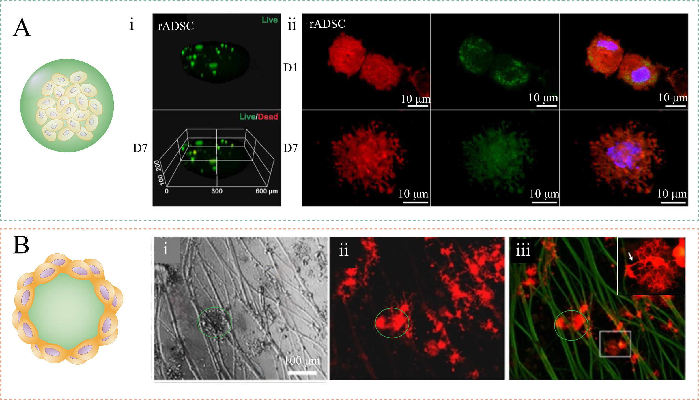
Figure 3. Collagen and collagen-derived microscale carriers for cell delivery. (A) Cell viability detection and confocal imaging of rADSCs encapsulated in crosslinked gelatin microspheres: (i) Live/dead assay demonstrated that most cells were still viable. (ii) After 7 days of culture, rADSCs spread out, and a large number of filopodia structures formed. Figure 3A is adapted in part from Ref.[79]. Reused under the terms of the Creative Commons Attribution License (CC BY); (B) Collagen microspheres carrying DRGs and OPCs co-culture. (i) The green-circled area indicates the collagen microspheres. (ii) and (iii) On the collagen microspheres, OPCs differentiate into OLs, and the OLs surround the DRGs. Figure 3B is adapted in part from Ref.[81]. Reused under the terms of the Creative Commons Attribution License (CC BY). rADSC: Rat Adipose-Derived Stem Cell; DRG: dorsal root ganglion; OPC: oligodendrocyte precursor cell; OL: oligodendrocyte.
Growth factor delivery
Tissue regeneration is regulated by coordinated growth factor signaling over time and space. In bone repair, for example, angiogenic factors such as VEGF contribute to early vascularization, whereas osteogenic factors such as BMP-2 promote osteogenic differentiation and bone matrix formation. Sequential or staged delivery of VEGF and BMP-2 has therefore been explored to better coordinate angiogenesis and osteogenesis during bone regeneration[82]. However, direct administration of soluble growth factors is inefficient in many regenerative settings because these proteins often have short in vivo half-lives, are susceptible to proteolytic degradation or inactivation, and may diffuse away from the target site. For BMP-2 in particular, uncontrolled burst release and poor retention can necessitate supraphysiological doses, thereby increasing costs and the risk of adverse effects[83,84].
Collagen microgels can serve as growth factor delivery carriers, but their role should be described carefully. Native collagen provides a hydrated fibrillar network and an enzymatically degradable matrix that can physically entrap growth factors and release them as the matrix swells, degrades, or remodels. However, unmodified collagen does not provide strong growth factor-specific binding in the same way as ECM components such as heparan sulfate, fibronectin, or other affinity-bearing matrix molecules[9]. Therefore, growth factor retention in collagen microgels is usually improved by regulating microgel size, porosity, and crosslinking density, or by introducing affinity components such as glycosaminoglycans, heparin-like groups, alginate, aptamers, or engineered binding motifs[9,85]. These modifications can reduce initial burst release, prolong local retention, and better match growth factor presentation with tissue repair kinetics.
A representative example was reported by Mumcuoglu et al.[86], who developed an injectable BMP-2 delivery system based on recombinant collagen-derived microspheres embedded in alginate. The system was designed to reduce the supraphysiological BMP-2 dose used in clinical settings and achieved time- and dose-dependent bone formation in both ectopic and calvarial defect models. In related collagen-based microsphere systems, site-directed immobilization of engineered BMP-2 variants further improved retention on collagen-based scaffolds and altered the morphology of newly formed bone, showing that release mode and ligand presentation strongly influence the biological outcome[84]. These studies indicate that collagen or collagen-derived microgels are most effective for growth factor delivery when their physical network is combined with controlled crosslinking, affinity binding, or engineered immobilization strategies, rather than relying only on nonspecific adsorption to collagen.
Drug delivery
Collagen microgels have attracted increasing attention as drug delivery platforms because they combine intrinsic bioactivity, biodegradability, and structural tunability[87]. As collagen is a major component of the mammalian ECM, collagen-based carriers can provide favorable biological recognition and enzymatic degradability in vivo, which may reduce foreign-body responses and improve compatibility in complex physiological environments[88]. Compared with many synthetic polymers or bioinert polysaccharide carriers, collagen microgels offer matrix-like interactions with cells and tissues, making them particularly suitable for localized and sustained delivery[55]. Moreover, their physicochemical properties can be precisely regulated during fabrication. Parameters such as particle size, pore architecture, crosslinking density, degradation rate, and mechanical stiffness can be adjusted according to the physicochemical properties of therapeutic cargos, the biological requirements of the target tissue, and the desired release kinetics[67].
One clinically relevant advantage of collagen-derived micro/nanogels is their ability to shift from simple diffusion-controlled release to microenvironment-responsive release. Pathological tissues often exhibit abnormal biochemical cues, including elevated protease activity, acidic pH, oxidative stress, or increased intracellular reducing potential[89,90]. By incorporating enzyme-cleavable segments, redox-sensitive linkages, or other stimuli-responsive motifs, collagen-derived carriers can achieve site-specific degradation and triggered cargo release. For example, Zhao et al.[89] developed redox-sensitive gelatin/silica-aptamer nanogels for targeted siRNA delivery. In this system, siRNA was conjugated to gelatin/silica hybrid nanogels through disulfide linkages, while the AS1411 aptamer provided tumor-cell-targeting capability. After cellular internalization, the high intracellular glutathione level promoted disulfide cleavage and triggered cytosolic siRNA release. This design illustrates how collagen-derived nanogels can integrate biological affinity, active targeting, and intracellular stimulus-responsive release into a single delivery system.
In addition to their degradable network structure, collagen microgels contain chemically addressable groups, particularly amine and carboxyl groups, together with hydroxyl-bearing residues, which provide reactive sites for crosslinking, ligand conjugation, and drug immobilization[91,92]. These groups can be used to conjugate targeting ligands, peptides, aptamers, or antibodies onto the microgel surface. Such modifications enable receptor-mediated recognition of specific cells or diseased tissues, thereby increasing effective drug exposure at the target site while reducing off-target distribution and systemic toxicity[93,94]. This strategy may improve the therapeutic index by increasing drug exposure at the target site while limiting off-target accumulation and systemic toxicity, which is particularly important for therapeutic cargoes with narrow safety margins or strong dose-dependent toxicity[93,95]. Therefore, the integration of endogenous bioactivity, structural tunability, environmental responsiveness, and ligand-mediated targeting makes collagen microgels a versatile platform for precision drug delivery, regenerative medicine, and tissue engineering.
Injectable micro-scaffolds
Traditional preformed scaffolds often require surgical implantation, which can increase tissue injury and is not always suitable for defects with irregular geometry. Collagen microgels provide an injectable scaffold format as they can be delivered as concentrated particulate suspensions while retaining the cell-adhesive and enzymatically degradable features of collagen. At sufficient packing density, microgel suspensions can flow under shear during injection and recover a solid-like state after placement through particle jamming, interparticle interactions, or secondary crosslinking[24,96]. This behavior allows collagen microgels to fill tissue defects of complex shapes and to form a localized matrix upon delivery.
After injection or placement, densely packed collagen microgels form a scaffold in which pores are generated mainly by the interstitial spaces between adjacent particles[96]. This microporous structure provides pathways for cell infiltration, cell spreading, nutrient transport, waste removal, and vascular ingrowth[24,96]. This is an important distinction from bulk collagen hydrogels, where cell invasion often depends on gradual matrix degradation and remodeling. In microgel-based scaffolds, the pore structure can be adjusted by changing microgel size, particle morphology, packing density, and the method used to stabilize the assembled particles[96,97]. Scaffold porosity and geometry may also affect the early host response, including macrophage recruitment and polarization, although this response is jointly regulated by material composition, degradation products, and local tissue context[98,99].
Several collagen-based microgel systems have been used in tissue repair models. Zhao et al.[37] prepared injectable angiogenic collagen microgels using ultra-long DNA interlocking and VEGF aptamer functionalization. The approach shortened collagen gelation to about 40 s while preserving the viscoelasticity, biodegradability, and biochemical properties of native collagen. Cell-laden angiogenic microgels improved skin wound healing and liver regeneration compared with non-angiogenic or acellular microgels. Chung et al.[100] prepared collagen microgels (CMGs) by micro-fragmenting a collagen-hyaluronic acid (HA) polyionic complex. When cultured with human adipose-derived stem cells (hASCs), the CMGs promoted integrin-mediated cell-matrix interactions and formed injectable CMG-hASC constructs with a microporous microarchitecture and improved mass transfer. Compared with cell aggregates, CMG-hASCs showed higher cell survival and angiogenic activity in vitro, and improved blood perfusion and limb salvage in a mouse critical limb ischemia model. Li et al.[101] reported an injectable collagen-microgel/tannic acid/nano-hydroxyapatite granular hydrogel for wound repair, in which collagen microgel size affected scaffold porosity and self-healing behavior.
Overall, collagen microgels represent a useful format for injectable micro-scaffolds when the repair site requires minimally invasive delivery, defect-shape adaptation, and early cell infiltration. Their assembled structure provides two complementary levels of support: the collagen microgel phase offers cell-adhesive and degradable matrix cues, whereas the interparticle spaces form a microporous network for mass transport and tissue ingrowth. For this reason, collagen micro-scaffold design should be guided by the target tissue rather than by a single material parameter. Microgel size, packing density, interparticle bonding, and degradation behavior need to be adjusted according to the required degree of cell retention, vascularization, and matrix remodeling.
Microgel inks
Microgel inks are formed by densely packed hydrogel microparticles and therefore show rheological behavior different from that of precursor-solution-based bioinks[96]. At rest, particle jamming and interparticle interactions allow the ink to behave as a soft solid with a finite yield stress, which helps the printed filament maintain its shape after deposition[97]. Under shear, the particles rearrange and slide past each other, leading to a decrease in apparent viscosity during extrusion. This shear-thinning behavior is useful for cell-laden printing, but cell viability is still affected by nozzle diameter, extrusion pressure, printing speed, residence time, and post-printing crosslinking conditions[102,103].
For collagen microgel inks, the relevant structural feature is the coexistence of intraparticle and interparticle networks. Each collagen microgel retains an internal fibrillar matrix, whereas packed microgels generate interstitial pores between adjacent particles[23,34]. The size and connectivity of these pores are affected by microgel diameter, particle-size distribution, deformability, packing density, and post-printing stabilization[97]. These interparticle pores provide additional pathways for nutrient transport, waste removal, and cell migration, while diffusion within each microgel remains governed by its internal collagen networ[104]. Therefore, the transport behavior of printed collagen microgel constructs depends on both the internal matrix structure of individual particles and the pore architecture generated by particle packing[105].
Collagen-containing and collagen-derived microgel systems have been investigated as bioinks or bioink-relevant granular materials. Van der Heide et al.[52] reported three-dimensional (3D)-printable tyramine-modified hyaluronic acid-collagen (THA-Col) granular hydrogels for delivery of BMP-7 cmRNA [Figure 4A]. Samiei et al.[34] prepared fibrillar collagen and collagen-glycosaminoglycan (GAG) microgels using droplet microfluidics; the resulting densely packed microgel suspensions showed shear-thinning behavior and were evaluated as injectable biomaterials and potential collagen-based bioinks [Figure 4B].
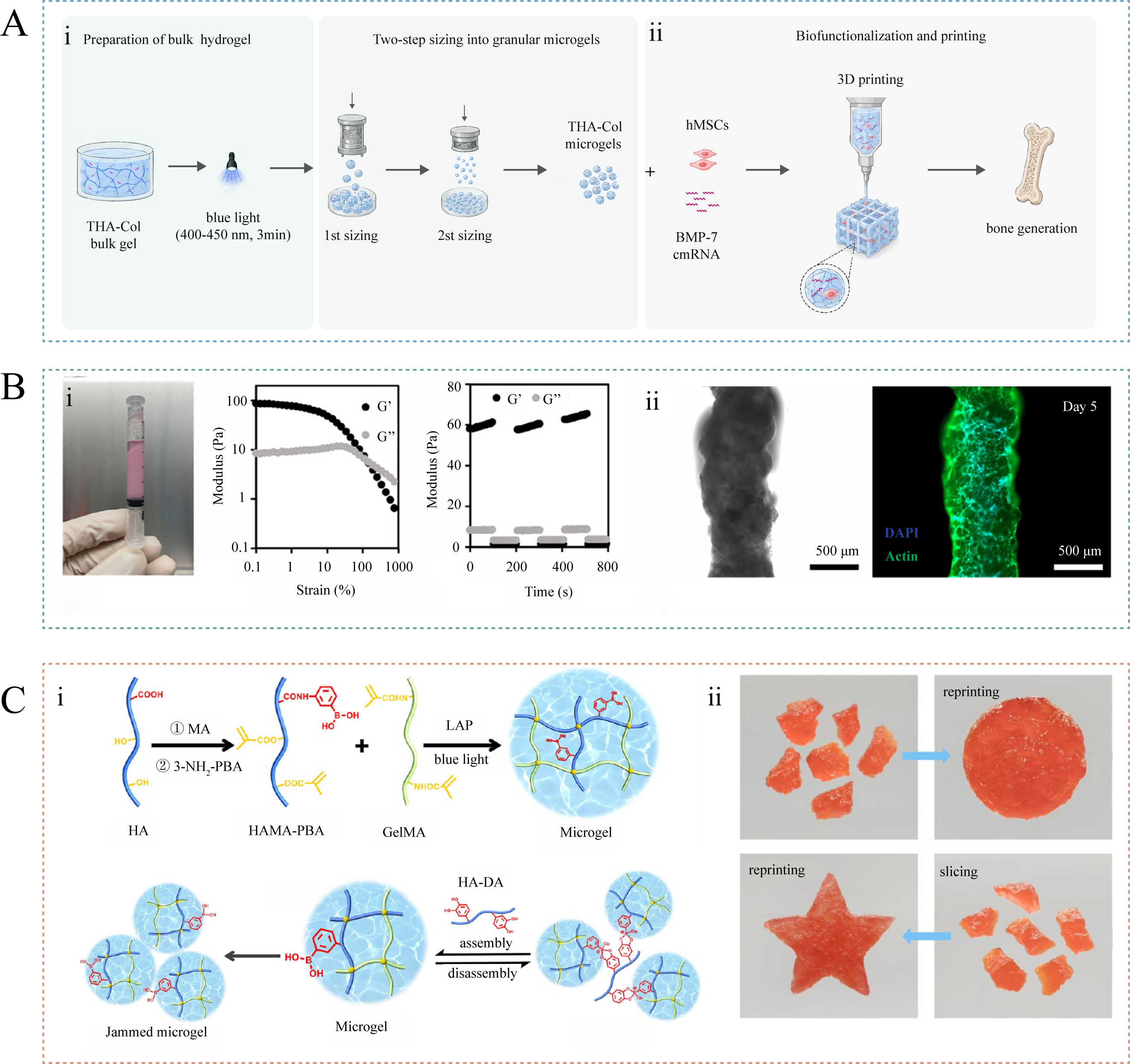
Figure 4. Collagen-containing and collagen-relevant microgel-based inks for three-dimensional printing and tissue assembly. (A) Preparation process (i) and printed product (ii) of a 3D printable particulate hydrogel based on THA-Col. Figure 4A is newly drawn by the authors based on the fabrication principle reported by Ref.[52]. No original figure elements were reused; (B) Printability (i) and cell-laden printing capability (ii) of collagen-based Collagen-GAG Microgel. Figure 4B is adapted in part from Ref.[34]. Reused under the terms of the Creative Commons Attribution License (CC BY); (C) Synthesis (i) and reprintability (ii) of DC-MA bioink. Dynamic covalent bonding between dopamine-modified HA and phenylboronic acid ester enables high printability, self-healing, and reprintability without secondary crosslinking. Figure 4C is adapted in part with permission from Ref.[106]. Copyright 2022, American Chemical Society. THA-Col: Tyramine-modified hyaluronic acid-collagen; 3D: three-dimensional; DAPI: 4',6-diamidino-2-phenylindole; MA: methacrylic anhydride; HA-DA: dopamine-modified hyaluronic acid; GelMA: methacrylated gelatin; LAP: lithium phenyl-2,4,6-trimethylbenzoylphosphinate; HAMA-PBA: phenylboronic acid-modified methacrylated hyaluronic acid: DC-MA: dynamic cross-linked microgel assembly;
A practical challenge for microgel inks is balancing extrusion flow with post-printing shape retention. Weak interparticle cohesion can facilitate flow but may reduce filament stability after deposition. Interparticle bonding can therefore be used to improve print fidelity while preserving microporosity. Although not based on native collagen microgels, Feng et al.[106] provided a relevant collagen-derived example by assembling methacrylated gelatin (GelMA)-containing microgels through reversible boronate ester bonding between phenylboronic acid-modified methacrylated hyaluronic acid (HAMA-PBA) and dopamine-modified hyaluronic acid (HA-DA). The resulting dynamic cross-linked microgel assembly (DC-MA) bioink showed improved printability, microporosity, tissue adhesion, self-healing, and reprintability [Figure 4C].
Organoid culture
Traditional organoid culture still relies heavily on Matrigel, a basement membrane extract derived from Engelbreth-Holm-Swarm mouse sarcoma. Matrigel contains major basement-membrane components, including laminin, collagen IV, entactin/nidogen, and heparan sulfate proteoglycans, as well as variable amounts of growth factors. These biochemical cues partly explain its broad use in initiating and maintaining epithelial organoids. However, its tumor-derived and xenogeneic origin, complex and incompletely defined composition, batch-to-batch variability, and potential biosafety concerns limit its use in standardized disease modeling, drug screening, and translational applications[107]. Synthetic ECM substitutes, such as polyethylene glycol-based hydrogels, provide better control over stiffness, degradability, adhesive ligands, and biochemical presentation, but they usually require additional functionalization to reproduce cell-adhesive and cell-remodelable matrix cues[108,109].
Collagen-based matrices occupy an intermediate position between Matrigel and fully synthetic systems. Type I collagen does not recapitulate the full basement membrane composition of Matrigel, but it forms a fibrillar, cell-remodelable matrix that can transmit cell-generated traction forces. This feature is relevant for organoid and assembloid models in which tissue morphology is regulated not only by soluble factors, but also by matrix remodeling and mechanical tension. For example, intestinal epithelial organoids cultured in floating type I collagen gels can align and fuse into macroscopic tube-like structures, whereas organoids in Matrigel mainly form cystic budding structures[110] [Figure 5A]. This difference suggests that type I collagen is useful for modeling traction-mediated morphogenesis, especially when organoid organization depends on matrix remodeling rather than only basement-membrane biochemical cues.
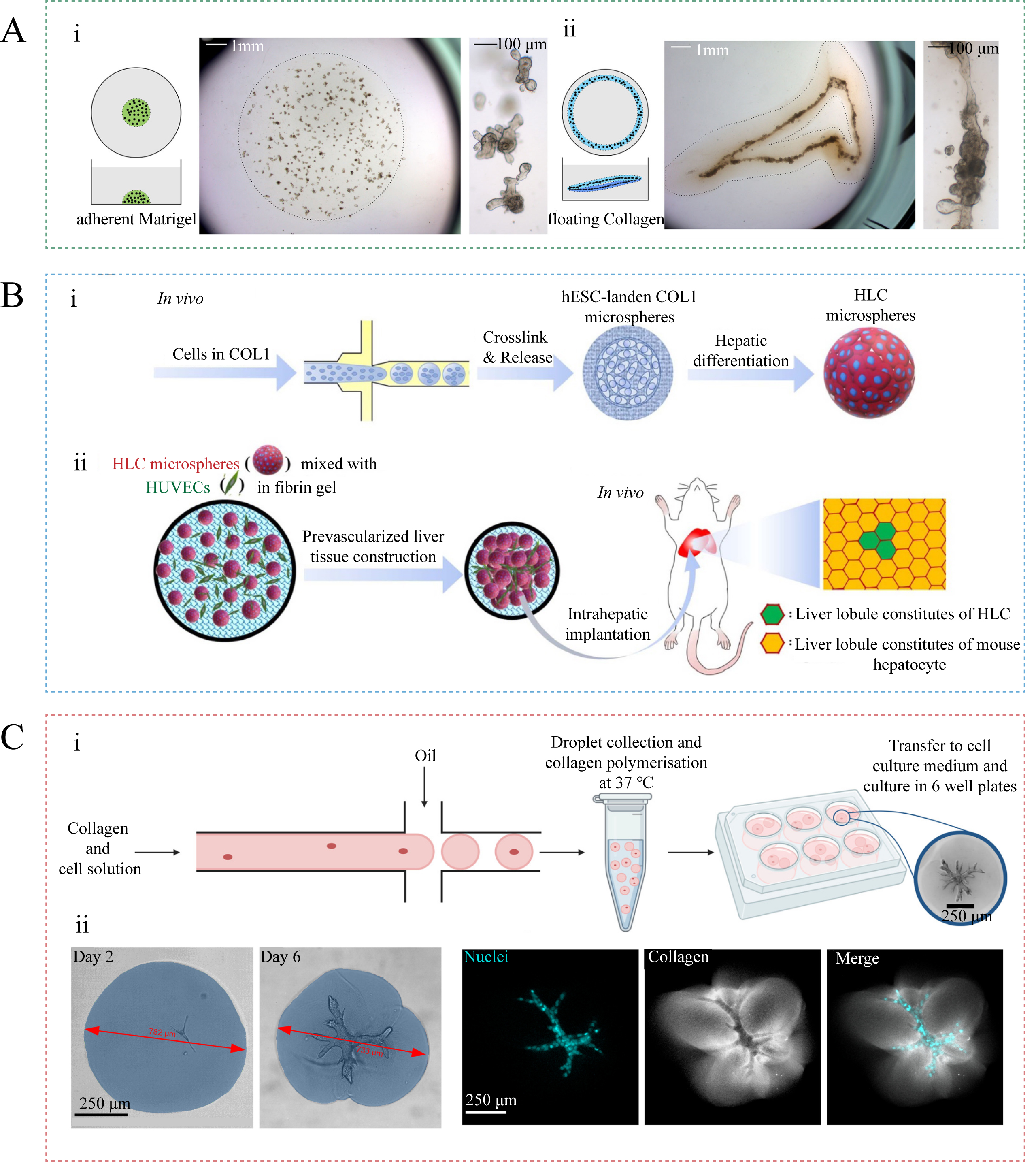
Figure 5. Collagen-based matrices and microgels for organoid and assembloid-oriented culture. (A) Comparison of small intestinal organoids cultured in traditional Matrigel and in floating type I collagen gel rings: (i) Organoids in traditional Matrigel mainly grow as single budding cysts. (ii) In floating collagen gel rings, the organoids self-organize into continuous macroscopic tubular structures that better mimic native intestinal architecture through mechanical tension-mediated morphogenesis. Figure 5A is adapted in part with permission from Ref.[110]. Copyright 2017, The Company of Biologists Ltd; (B) Schematic diagram of the preparation of HLC microspheres and the culture of liver organoids: (i) Microfluidic fabrication of collagen microspheres encapsulating hESCs and a schematic diagram of hESC differentiation into HLCs. (ii) The HLC microspheres were further assembled with human umbilical vein endothelial cells (HUVECs) to form prevascularized liver tissue (PLT), which was implanted into a mouse liver. Figure 5B is adapted in part from Ref.[111]. Reused under the terms of the Creative Commons Attribution License (CC BY); (C) Cell-laden collagen microgels for the culture of complex branched organoids. (i) Microfluidic fabrication of collagen microgel encapsulating pancreatic ductal adenocarcinoma cells and culture of complex branched organoids. (ii) Deformation of collagen microgels and formation of branched structures under the traction of organoids demonstrating the capacity of microgel confinement to guide organoid morphogenesis. Figure 5C is adapted in part from Ref.[112]. Reused under the terms of the Creative Commons Attribution License (CC BY). hESC: Human embryonic stem cell; HLC: hepatocyte-like cell; COL1: Collagen type I.
Collagen microgels translate the fibrillar collagen matrix into discrete culture compartments. Compared with bulk collagen gels, this format reduces the matrix volume surrounding each cell aggregate or organoid and shortens the diffusion distance for soluble cues and metabolic exchange[111]. It also allows individual organoid units to be generated, cultured, transferred, or assembled with improved size control[112]. These features are useful for organoid systems in which matrix confinement, local remodeling, and subsequent assembly of cell-laden units are required. The performance of such systems is therefore governed by the dimensions and composition of the microgels, including collagen concentration, fibril architecture, stiffness, degradability, and the manner in which individual microgels are packed or assembled.
This concept has been applied in organoid-relevant culture systems[37,113,114]. Deng et al.[111] generated type I collagen microspheres by microfluidics and encapsulated human embryonic stem cells (hESCs) for hepatic differentiation. Compared with bulk collagen hydrogels and two-dimensional culture, the microsphere format promoted the formation of more mature hepatocyte-like cells (HLCs). These HLC microspheres were then combined with endothelial cells to form prevascularized liver tissue (PLT), which engrafted in mouse liver and improved hepatic function in vivo [Figure 5B]. Ruider et al.[112] used collagen microgels to confine pancreatic ductal adenocarcinoma cells and showed that microgel size influenced the emergence of branched organoid structures [Figure 5C]. These studies show that collagen microgels can regulate organoid development through matrix confinement, local remodeling, and the assembly of cell-laden units.
The development of organoid matrices is moving from permissive culture substrates toward instructive and modular microenvironments[115]. In this context, collagen microgels do not simply offer another substitute for Matrigel; they provide a matrix format in which fibrillar collagen cues, microscale confinement, and unit-by-unit assembly can be combined[111]. This is particularly relevant for organoid and assembloid systems in which morphogenesis depends on cell-matrix traction, local matrix remodeling, spatial confinement, or the integration of multiple cell-laden modules. Compared with Matrigel, collagen microgels provide less basement-membrane biochemical complexity but greater control over collagen-rich matrix architecture. Compared with fully synthetic ECM hydrogels, they offer less molecular precision but retain native fibrillar and degradable matrix features. A reasonable direction is to integrate collagen microgels with defined adhesive or basement-membrane-mimetic cues[116,117], tunable or dynamic crosslinking chemistries[118], and microfluidic perfusion or organoid-on-chip platforms[114]. Such hybrid systems may improve the reproducibility and physiological relevance of organoid models while preserving the fibrillar and remodelable features of collagen.
CONCLUSION AND OUTLOOK
Collagen microgels provide a useful way to reorganize collagen from a continuous bulk matrix into discrete microscale building blocks. This change in material format is important because collagen assembly, crosslinking, particle size, porosity, degradation, and bioactive loading can be adjusted at the level of individual microgels. As a result, collagen microgels can be adapted for drug and cell delivery, injectable micro-scaffolds, granular printing inks, and organoid culture matrices. Across these applications, performance is determined not by collagen composition alone, but by how microscale structure controls local mechanics, mass transport, cell-matrix interaction, and tissue remodeling.
The next stage of this field should focus on improving the reproducibility, interpretability, and clinical relevance of collagen microgels. Recombinant human collagen may help reduce source variability, but its value will depend on whether it can reproduce the post-translational modification, triple-helix stability, fibril formation, and processing behavior required for functional microgels. For translational use, acellular, drug-loaded, and cell-laden microgels will also need to be evaluated according to their specific composition and biological function rather than treated as a single material class. More attention should be given to host responses, including macrophage polarization, chronic inflammation, fibrosis, and constructive remodeling, because these processes often determine long-term outcomes after implantation. In the longer term, data-driven approaches, including artificial intelligence (AI) and machine learning (ML), may help connect collagen source, fabrication parameters, microgel structure, and biological performance, but only if supported by standardized experimental datasets and mechanistic validation.
DECLARATIONS
Authors’ contributions
Conception and manuscript revision: Qu, X; Liu, X; Qiu, J.
Manuscript writing: Peng, Q; Liu, X.
Availability of data and materials
Not applicable.
AI and AI-assisted tools statement
During the preparation of this manuscript, the AI tool ChatGPT (version 5.2, released 2025-12-11) was used solely for language editing. The tool did not influence the study design, data collection, analysis, interpretation, or the scientific content of the work. All authors take full responsibility for the accuracy, integrity, and final content of the manuscript.
Financial support and sponsorship
Support from the National Natural Science Foundation of China (32425031), the Science and Technology Innovation Project of Shanghai Science and Technology Committee (24CL2900800, 25CL2900700), the Zhejiang Natural Science Foundation (Z25E030005).
Conflicts of interest
Qu, X. is an Associate Editor of the journal Micro Nano Science. Qu, X. was not involved in any steps of the editorial process, including reviewers’ selection, manuscript handling, or decision-making. The other authors declare no conflicts of interest.
Ethical approval and consent to participate
Not applicable.
Consent for publication
Not applicable.
Copyright
© The Author(s) 2026.
REFERENCES
1. Zheng, M.; Wang, X.; Chen, Y.; et al. A review of recent progress on collagen-based biomaterials. Adv. Healthc. Mater. 2023, 12, e2202042.
3. Shoulders, M. D.; Raines, R. T. Collagen structure and stability. Annu. Rev. Biochem. 2009, 78, 929-58.
4. Amirrah, I. N.; Lokanathan, Y.; Zulkiflee, I.; Wee, M. F. M. R.; Motta, A.; Fauzi, M. B. A Comprehensive review on collagen type I development of biomaterials for tissue engineering: from biosynthesis to bioscaffold. Biomedicines 2022, 10, 2307.
5. Oslan, S. N. H.; Shapawi, R.; Mokhtar, R. A. M.; Noordin, W. N. M.; Huda, N. Characterization of acid- and pepsin-soluble collagen extracted from the skin of purple-spotted bigeye snapper. Gels 2022, 8, 665.
6. Jafari, H.; Lista, A.; Siekapen, M. M.; et al. Fish collagen: extraction, characterization, and applications for biomaterials engineering. Polymers 2020, 12, 2230.
7. Bigi, A.; Panzavolta, S.; Rubini, K. Relationship between triple-helix content and mechanical properties of gelatin films. Biomaterials 2004, 25, 5675-80.
8. Pawelec, K. M.; Best, S. M.; Cameron, R. E. Collagen: a network for regenerative medicine. J. Mater. Chem. B. 2016, 4, 6484-96.
9. Sarrigiannidis, S.; Rey, J.; Dobre, O.; González-García, C.; Dalby, M.; Salmeron-Sanchez, M. A tough act to follow: collagen hydrogel modifications to improve mechanical and growth factor loading capabilities. Mater. Today. Bio. 2021, 10, 100098.
10. Knight, C. G.; Morton, L. F.; Peachey, A. R.; Tuckwell, D. S.; Farndale, R. W.; Barnes, M. J. The collagen-binding A-domains of Integrins α1β1 and α2β1recognize the same specific amino acid sequence, GFOGER, in native (triple-helical) collagens. J. Biol. Chem. 2000, 275, 35-40.
11. Raynal, N.; Hamaia, S. W.; Siljander, P. R.; et al. Use of synthetic peptides to locate novel integrin α2β1-binding motifs in human collagen III. J. Biol. Chem. 2006, 281, 3821-31.
12. Sun, Z.; Guo, S. S.; Fässler, R. Integrin-mediated mechanotransduction. J. Cell. Biol. 2016, 215, 445-56.
13. Panciera, T.; Azzolin, L.; Cordenonsi, M.; Piccolo, S. Mechanobiology of YAP and TAZ in physiology and disease. Nat. Rev. Mol. Cell. Biol. 2017, 18, 758-70.
14. Page-Mccaw, A.; Ewald, A. J.; Werb, Z. Matrix metalloproteinases and the regulation of tissue remodelling. Nat. Rev. Mol. Cell. Biol. 2007, 8, 221-33.
15. Rowley, J. A.; Mooney, D. J. Alginate type and RGD density control myoblast phenotype. J. Biomed. Mater. Res. 2002, 60, 217-23.
16. Burdick, J. A.; Prestwich, G. D. Hyaluronic acid hydrogels for biomedical applications. Adv. Mater. 2011, 23, H41-56.
17. Sorushanova, A.; Delgado, L. M.; Wu, Z.; et al. The collagen suprafamily: from biosynthesis to advanced biomaterial development. Adv. Mater. 2018, 31, 1801651.
18. Gu, L.; Shan, T.; Ma, Y.; Tay, F. R.; Niu, L. Novel biomedical applications of crosslinked collagen. Trends. Biotechnol. 2019, 37, 464-91.
19. Cheema, U.; Rong, Z.; Kirresh, O.; Macrobert, A. J.; Vadgama, P.; Brown, R. A. Oxygen diffusion through collagen scaffolds at defined densities: implications for cell survival in tissue models. J. Tissue. Eng. Regen. Med. 2012, 6, 77-84.
20. Raub, C.; Putnam, A.; Tromberg, B.; George, S. Predicting bulk mechanical properties of cellularized collagen gels using multiphoton microscopy. Acta. Biomater. 2010, 6, 4657-65.
21. Daly, A. C.; Riley, L.; Segura, T.; Burdick, J. A. Hydrogel microparticles for biomedical applications. Nat. Rev. Mater. 2020, 5, 20-43.
22. Kamperman, T.; Karperien, M.; Le Gac, S.; Leijten, J. Single-cell microgels: technology, challenges, and applications. Trends. Biotechnol. 2018, 36, 850-65.
23. Xuan, L.; Hou, Y.; Liang, L.; et al. Microgels for cell delivery in tissue engineering and regenerative medicine. Nano-Micro. Lett. 2024, 16, 218.
24. Griffin, D. R.; Weaver, W. M.; Scumpia, P. O.; Di Carlo, D.; Segura, T. Accelerated wound healing by injectable microporous gel scaffolds assembled from annealed building blocks. Nat. Mater. 2015, 14, 737-44.
25. Feng, Q.; Li, D.; Li, Q.; Cao, X.; Dong, H. Microgel assembly: fabrication, characteristics and application in tissue engineering and regenerative medicine. Bioact. Mater. 2022, 9, 105-19.
26. Muir, V. G.; Qazi, T. H.; Shan, J.; Groll, J.; Burdick, J. A. Influence of microgel fabrication technique on granular hydrogel properties. ACS. Biomater. Sci. Eng. 2021, 7, 4269-81.
27. Harris, J. R.; Soliakov, A.; Lewis, R. J. In vitro fibrillogenesis of collagen type I in varying ionic and pH conditions. Micron 2013, 49, 60-8.
28. Lehnert, S.; Sikorski, P. Tailoring the assembly of collagen fibers in alginate microspheres. Mat. Sci. Eng. C. 2021, 121, 111840.
29. Gobeaux, F.; Mosser, G.; Anglo, A.; et al. Fibrillogenesis in dense collagen solutions: a physicochemical study. J. Mol. Biol. 2008, 376, 1509-22.
30. Achilli, M.; Mantovani, D. Tailoring mechanical properties of collagen-based scaffolds for vascular tissue engineering: the effects of pH, temperature and ionic strength on gelation. Polymers 2010, 2, 664-80.
31. Cheng, X.; Gurkan, U. A.; Dehen, C. J.; et al. An electrochemical fabrication process for the assembly of anisotropically oriented collagen bundles. Biomaterials 2008, 29, 3278-88.
32. Holder, A. J.; Badiei, N.; Hawkins, K.; Wright, C.; Williams, P. R.; Curtis, D. J. Control of collagen gel mechanical properties through manipulation of gelation conditions near the sol-gel transition. Soft. Matter. 2018, 14, 574-80.
33. Hong, S.; Hsu, H.; Kaunas, R.; Kameoka, J. Collagen microsphere production on a chip. Lab. Chip. 2012, 12, 3277.
34. Samiei, E.; Veres, T.; Günther, A. Microfluidic synthesis of collagen-based microgels for tissue engineering applications. Nano. Select. 2025, 6, e202400121.
35. Antoine, E. E.; Vlachos, P. P.; Rylander, M. N. Review of collagen I hydrogels for bioengineered tissue microenvironments: characterization of mechanics, structure, and transport. Tissue. Eng. Part. B-Re. 2014, 20, 683-96.
36. Gong, X.; Wen, Z.; Liang, Z.; et al. Instant assembly of collagen for tissue engineering and bioprinting. Nat. Mater. 2025, 24, 1307-18.
37. Zhao, H.; Wang, Z.; Jiang, S.; et al. Microfluidic synthesis of injectable angiogenic microgels. Cell. Rep. Phys. Sci. 2020, 1, 100047.
38. Davidenko, N.; Schuster, C.; Bax, D.; et al. Control of crosslinking for tailoring collagen-based scaffolds stability and mechanics. Acta. Biomater. 2015, 25, 131-42.
39. Beghetto, V.; Gatto, V.; Conca, S.; Bardella, N.; Scrivanti, A. Polyamidoamide dendrimers and cross-linking agents for stabilized bioenzymatic resistant metal-free bovine collagen. Molecules 2019, 24, 3611.
40. Olde Damink, L. H. H.; Dijkstra, P. J.; Van Luyn, M. J. A.; Van Wachem, P. B.; Nieuwenhuis, P.; Feijen, J. Glutaraldehyde as a crosslinking agent for collagen-based biomaterials. J. Mater. Sci:. Mater. Med. 1995, 6, 460-72.
41. Macaya, D.; Ng, K. K.; Spector, M. Injectable collagen-genipin gel for the treatment of spinal cord injury: in vitro studies. Adv. Funct. Mater. 2011, 21, 4788-97.
42. Drzewiecki, K. E.; Malavade, J. N.; Ahmed, I.; Lowe, C. J.; Shreiber, D. I. A thermoreversible, photocrosslinkable collagen bio-ink for free-form fabrication of scaffolds for regenerative medicine. Technology 2017, 05, 185-95.
43. Orban, J. M.; Wilson, L. B.; Kofroth, J. A.; El-Kurdi, M. S.; Maul, T. M.; Vorp, D. A. Crosslinking of collagen gels by transglutaminase. J. Biomed. Mater. Res. 2004, 68A, 756-62.
44. Chen, R.; Ho, H.; Sheu, M. Characterization of collagen matrices crosslinked using microbial transglutaminase. Biomaterials 2005, 26, 4229-35.
45. Eyre, D. R.; Weis, M.; Rai, J. Analyses of lysine aldehyde cross-linking in collagen reveal that the mature cross-link histidinohydroxylysinonorleucine is an artifact. J. Biol. Chem. 2019, 294, 6578-90.
46. Snider, C.; Bellrichard, M.; Meyer, A.; Kannan, R.; Grant, D.; Grant, S. A novel crosslinker-free technique toward the fabrication of collagen microspheres. J. Biomed. Mater. Res. 2020, 108, 2789-98.
47. Berthold, A.; Cremer, K.; Kreuter, J. Collagen microparticles: carriers for glucocorticosteroids. Eur. J. Pharm. Biopharm. 1998, 45, 23-9.
48. Rössler, B.; Kreuter, J.; Scherer, D. Collagen microparticles: preparation and properties. J. Microencapsulation. 1995, 12, 49-57.
49. Teng, S.; Liang, M.; Wang, P.; Luo, Y. Biomimetic composite microspheres of collagen/chitosan/nano-hydroxyapatite: In-situ synthesis and characterization. Materials. Mater. Sci. Eng. C. 2016, 58, 610-3.
50. Devernois, E.; Coradin, T. A top-down approach to dense collagen I-chitosan microgels and granular hydrogels. ChemRxiv 2025. Available online: https://doi.org/10.26434/chemrxiv-2025-rrcq8. [accessed 6 July 2026].
51. Hong, C.; Lee, Y.; Chung, H.; et al. Micro-fragmented collagen hydrogel wound dressing: Enhanced porosity facilitates elevated stem cell survival and paracrine effects for accelerated wound maturation. Mater. Today. Bio. 2025, 32, 101678.
52. Van Der Heide, D.; Del Toro Runzer, C.; Della Bella, E.; et al. Implementing BMP-7 chemically modified RNA for bone regeneration with 3D printable hyaluronic acid-collagen granular gels. Adv. Healthc. Mater. 2025, 14, 2405047.
53. Barberi, G.; Biscari, G.; Palumbo, F. S.; et al. Exploring marine collagen application in the formulation of microparticles as scaffold doping agents in tissue engineering. J. Drug. Delivery. Sci. Technol. 2025, 114, 107433.
54. Santos, D.; Maurício, A. C.; Sencadas, V.; Santos, J. D.; Fernades, M. H.; Gomes, R. S. In: Biomaterials - physics and chemistry - new edition. Spray drying: an overview. IntechOpen, 2018; pp 9-36.
55. Du, B.; Feng, S.; Wang, J.; et al. Collagen-based micro/nanogel delivery systems: manufacturing, release mechanisms, and biomedical applications. Chin. Med. J. (Engl). 2025, 138, 1135-52.
56. Wang, J.; Jansen, J. A.; Yang, F. Electrospraying: possibilities and challenges of engineering carriers for biomedical applications - a mini review. Front. Chem. 2019, 7, 258.
57. Jayakumar, G. C.; Usharani, N.; Kawakami, K.; Rao, J. R.; Nair, B. U. Preparation of antibacterial collagen-pectin particles for biotherapeutics. RSC. Adv. 2014, 4, 42846-54.
58. Jiang, W.; Li, M.; Chen, Z.; Leong, K. W. Cell-laden microfluidic microgels for tissue regeneration. Lab. Chip. 2016, 16, 4482-506.
59. Wei, Z.; Wang, S.; Hirvonen, J.; Santos, H. A.; Li, W. Microfluidics fabrication of micrometer-sized hydrogels with precisely controlled geometries for biomedical applications. Adv. Healthc. Mater. 2022, 11, 2200846.
60. Matsunaga, Y. T.; Morimoto, Y.; Takeuchi, S. Molding cell beads for rapid construction of macroscopic 3D tissue architecture. Adv. Mater. 2011, 23, H90-4.
61. Yamada, M.; Hori, A.; Sugaya, S.; et al. Cell-sized condensed collagen microparticles for preparing microengineered composite spheroids of primary hepatocytes. Lab. Chip. 2015, 15, 3941-51.
62. Singh, S.; Chu, W. Y.; Ostadsharif Memar, R.; De Carlo, A.; Veres, T.; Günther, A. Scalable preparation of macroporous collagen microgels by air bubble-induced breakup and ice templating. React. Chem. Eng. 2024, 9, 2584-98.
63. Chen, M. W.; Fan, D.; Liu, X.; et al. Water Transport-induced liquid-liquid phase separation facilitates gelation for controllable and facile fabrication of physically crosslinked microgels. Adv. Mater. 2024, 36, 2405109.
64. Keshaw, H.; Thapar, N.; Burns, A. J.; et al. Microporous collagen spheres produced via thermally induced phase separation for tissue regeneration. Acta. Biomater. 2010, 6, 1158-66.
65. Miyata, T.; Namiki, S. Substrate consisting of regenerated collagen fibrils and method of manufacturing same. US4565580A, 1986.
66. Zeng, Q.; Zhang, M.; Adhikari, B. P.; Mujumdar, A. S. Effect of drying processes on the functional properties of collagen peptides produced from chicken skin. Drying. Technol. 2013, 31, 1653-60.
67. Nagarajan, U.; Kawakami, K.; Zhang, S.; Chandrasekaran, B.; Unni Nair, B. Fabrication of solid collagen nanoparticles using electrospray deposition. Chem. Pharm. Bull. 2014, 62, 422-8.
68. Yao, R.; Zhang, R.; Lin, F.; Luan, J. Injectable cell/hydrogel microspheres induce the formation of fat lobule-like microtissues and vascularized adipose tissue regeneration. Biofabrication 2012, 4, 045003.
69. Bielajew, B. J.; Hu, J. C.; Athanasiou, K. A. Collagen: quantification, biomechanics and role of minor subtypes in cartilage. Nat. Rev. Mater. 2020, 5, 730-47.
70. Parenteau-Bareil, R.; Gauvin, R.; Berthod, F. Collagen-based biomaterials for tissue engineering applications. Materials 2010, 3, 1863-87.
71. Adepu, S.; Ramakrishna, S. Controlled drug delivery systems: current status and future directions. Molecules 2021, 26, 5905.
72. Ren, X.; Zhao, M.; Lash, B.; Martino, M. M.; Julier, Z. Growth factor engineering strategies for regenerative medicine applications. Front. Bioeng. Biotechnol. 2020, 7, 469.
73. Mitchell, A. C.; Briquez, P. S.; Hubbell, J. A.; Cochran, J. R. Engineering growth factors for regenerative medicine applications. Acta. Biomater. 2016, 30, 1-12.
74. Sivaraj, D.; Chen, K.; Chattopadhyay, A.; et al. Hydrogel scaffolds to deliver cell therapies for wound healing. Front. Bioeng. Biotechnol. 2021, 9, 660145.
75. Sulaiman, S. B.; Idrus, R. B. H.; Hwei, N. M. Gelatin microsphere for cartilage tissue engineering: current and future strategies. Polymers 2020, 12, 2404.
76. Sharma, K.; Porat, Z.; Gedanken, A. Designing natural polymer-based capsules and spheres for biomedical applications - a review. Polymers 2021, 13, 4307.
77. Chan, B. P.; Hui, T. Y.; Wong, M. Y.; Yip, K. H. K.; Chan, G. C. F. Mesenchymal stem cell-encapsulated collagen microspheres for bone tissue engineering. Tissue. Eng. Part. C. Methods. 2010, 16, 225-35.
78. Nii, T. Strategies using gelatin microparticles for regenerative therapy and drug screening applications. Molecules 2021, 26, 6795.
79. Shi, M.; Gao, Y.; Lee, L.; et al. Adaptive gelatin microspheres enhanced stem cell delivery and integration with diabetic wounds to activate skin tissue regeneration. Front. Bioeng. Biotechnol. 2022, 10, 813805.
80. Smit, N. W.; Ten Sande, J. N.; Parvizi, M.; et al. Recombinant human collagen-based microspheres mitigate cardiac conduction slowing induced by adipose tissue-derived stromal cells. PLoS. ONE. 2017, 12, e0183481.
81. Yao, L.; Phan, F.; Li, Y. Collagen microsphere serving as a cell carrier supports oligodendrocyte progenitor cell growth and differentiation for neurite myelination in vitro. Stem. Cell. Res. Ther. 2013, 4, 109.
82. Liu, K.; Meng, C.; Lv, Z.; et al. Enhancement of BMP-2 and VEGF carried by mineralized collagen for mandibular bone regeneration. Regener. Biomater. 2020, 7, 435-40.
83. Mumcuoglu, D.; Fahmy-Garcia, S.; Ridwan, Y.; et al. ; FUJIFILM Manufacturing Europe B. V., Oudenstaart 1, 5047 TK Tilburg, the Netherlands. Injectable BMP-2 delivery system based on collagen-derived microspheres and alginate induced bone formation in a time- and dose-dependent manner. Eur. Cell. Mater. 2018, 35, 242-54.
84. Siverino, C.; Fahmy-garcia, S.; Mumcuoglu, D.; et al. Site-directed immobilization of an engineered bone morphogenetic protein 2 (BMP2) variant to collagen-based microspheres induces bone formation in vivo. Int. J. Mol. Sci. 2022, 23, 3928.
85. Hachim, D.; Whittaker, T. E.; Kim, H.; Stevens, M. M. Glycosaminoglycan-based biomaterials for growth factor and cytokine delivery: making the right choices. J. Controlled. Release. 2019, 313, 131-47.
86. Mumcuoglu, D.; De Miguel, L.; Jekhmane, S.; et al. Collagen I derived recombinant protein microspheres as novel delivery vehicles for bone morphogenetic protein-2. Mat. Sci. Eng. C. 2018, 84, 271-80.
87. Li, Q.; Chang, B.; Dong, H.; Liu, X. Functional microspheres for tissue regeneration. Bioact. Mater. 2023, 25, 485-99.
88. Jeevithan, E.; Qingbo, Z.; Bao, B.; Wu, W. Biomedical and pharmaceutical application of fish collagen and gelatin: a review. J. Nutr. Ther. 2013, 2, 218-27.
89. Zhao, X.; Xi, Y.; Zhang, Y.; et al. Redox-sensitive gelatin/silica-aptamer nanogels for targeted siRNA delivery. Nanoscale. Res. Lett. 2019, 14, 273.
90. Liu, Y.; Chen, L.; Shi, Q.; Zhao, Q.; Ma, H. Tumor microenvironment-responsive polypeptide nanogels for controlled antitumor drug delivery. Front. Pharmacol. 2021, 12, 748102.
91. Pires Figueiredo, M.; Rodríguez-Fernández, S.; Copes, F.; Mantovani, D. Review of collagen type I-based hydrogels: focus on composition-structure-properties relationships. npj. Biomed. Innov. 2025, 2, 16.
92. Guzdek, B.; Fołta, K.; Staniek, N.; Stolarczyk, M.; Krukiewicz, K. Collagen-based drug delivery agents for glioblastoma multiforme treatment. Int. J. Mol. Sci. 2025, 26, 6513.
93. Rosenblum, D.; Joshi, N.; Tao, W.; Karp, J. M.; Peer, D. Progress and challenges towards targeted delivery of cancer therapeutics. Nat. Commun. 2018, 9, 1410.
94. Gao, F.; Yin, J.; Chen, Y.; Guo, C.; Hu, H.; Su, J. Recent advances in aptamer-based targeted drug delivery systems for cancer therapy. Front. Bioeng. Biotechnol. 2022, 10, 972933.
95. Mitchell, M. J.; Billingsley, M. M.; Haley, R. M.; Wechsler, M. E.; Peppas, N. A.; Langer, R. Engineering precision nanoparticles for drug delivery. Nat. Rev. Drug. Discovery. 2021, 20, 101-24.
96. Riley, L.; Schirmer, L.; Segura, T. Granular hydrogels: emergent properties of jammed hydrogel microparticles and their applications in tissue repair and regeneration. Curr. Opin. Biotechnol. 2019, 60, 1-8.
97. Truong, N. F.; Kurt, E.; Tahmizyan, N.; et al. Microporous annealed particle hydrogel stiffness, void space size, and adhesion properties impact cell proliferation, cell spreading, and gene transfer. Acta. Biomater. 2019, 94, 160-72.
98. Widener, A. E.; Roberts, A.; Phelps, E. A. Granular hydrogels for harnessing the immune response. Adv. Healthc. Mater. 2024, 13, 2303005.
99. Yin, Y.; He, X.; Wang, J.; et al. Pore size-mediated macrophage M1-to-M2 transition influences new vessel formation within the compartment of a scaffold. Applied. Materials. Today. 2020, 18, 100466.
100. Chung, H.; Choi, J.; Hong, C.; et al. A micro-fragmented collagen gel as a cell-assembling platform for critical limb ischemia repair. Bioact. Mater. 2024, 34, 80-97.
101. Li, Y.; Bai, X.; Ren, C.; Ma, Y.; Liu, Y. Construction of injectable collagen-microgel/tannic acid/nano-hydroxyapatite granular hydrogel and evaluation of its potential in wound healing. J. Bioact. Compat. Polym. 2023, 38, 325-39.
102. Tuftee, C.; Alsberg, E.; Ozbolat, I. T.; Rizwan, M. Emerging granular hydrogel bioinks to improve biological function in bioprinted constructs. Trends. Biotechnol. 2024, 42, 339-52.
103. Yu, S.; Luo, Y.; Chen, S.; Fan, J.; Zhang, H. Quantitative assessment of hydrogel printability in extrusion bioprinting. Gels 2026, 12, 189.
104. Ma, S.; Mukherjee, N.; Mikhailova, E.; Bayley, H. Gel microrods for 3D tissue printing. Adv. Biosys. 2017, 1, 1700075.
105. Ataie, Z.; Kheirabadi, S.; Zhang, J. W.; et al. Nanoengineered granular hydrogel bioinks with preserved interconnected microporosity for extrusion bioprinting. Small 2022, 18, 2202390.
106. Feng, Q.; Li, D.; Li, Q.; et al. Assembling microgels via dynamic cross-linking reaction improves printability, microporosity, tissue-adhesion, and self-healing of microgel bioink for extrusion bioprinting. ACS. Appl. Mater. Interfaces. 2022, 14, 15653-66.
107. Kozlowski, M. T.; Crook, C. J.; Ku, H. T. Towards organoid culture without Matrigel. Commun. Biol. 2021, 4, 1387.
108. Jee, J. H.; Lee, D. H.; Ko, J.; et al. Development of collagen-based 3D matrix for gastrointestinal tract-derived organoid culture. Stem. Cells. Int. 2019, 2019, 1-15.
109. Li, C.; An, N.; Song, Q.; et al. Enhancing organoid culture: harnessing the potential of decellularized extracellular matrix hydrogels for mimicking microenvironments. J. Biomed. Sci. 2024, 31, 96.
110. Sachs, N.; Tsukamoto, Y.; Kujala, P.; Peters, P. J.; Clevers, H. Intestinal epithelial organoids fuse to form self-organizing tubes in floating collagen gels. Development 2017, 144, 1107-12.
111. Deng, S.; Zhao, X.; Zhu, Y.; et al. Efficient hepatic differentiation of hydrogel microsphere-encapsulated human pluripotent stem cells for engineering prevascularized liver tissue. Biofabrication 2022, 15, 015016.
112. Ruider, I.; Pastucha, A.; Raich, M. K.; et al. Accelerated maturation of branched organoids confined in collagen droplets. Lab. Chip. 2025, 25, 5043-54.
113. Liu, J.; Du, C.; Chen, J.; et al. Hydrogel microspheres empowering organ-on-a-chip systems: innovations and applications. Small 2025, 21, 2504563.
115. Li, K.; He, Y.; Jin, X.; Jin, K.; Qian, J. Reproducible extracellular matrices for tumor organoid culture: challenges and opportunities. J. Transl. Med. 2025, 23, 497.
116. Gjorevski, N.; Sachs, N.; Manfrin, A.; et al. Designer matrices for intestinal stem cell and organoid culture. Nature 2016, 539, 560-4.
117. Cruz-Acuña, R.; Quirós, M.; Farkas, A. E.; et al. Synthetic hydrogels for human intestinal organoid generation and colonic wound repair. Nat. Cell. Biol. 2017, 19, 1326-35.
Cite This Article
 Open Access
How to Cite
Download Citation
Export Citation File:
Type of Import
Tips on Downloading Citation
Citation Manager File Format
Type of Import
Direct Import: When the Direct Import option is selected (the default state), a dialogue box will give you the option to Save or Open the downloaded citation data. Choosing Open will either launch your citation manager or give you a choice of applications with which to use the metadata. The Save option saves the file locally for later use.
Indirect Import: When the Indirect Import option is selected, the metadata is displayed and may be copied and pasted as needed.
About This Article
Copyright

Data & Comments
Data
0







Comments
Comments must be written in English. Spam, offensive content, impersonation, and private information will not be permitted. If any comment is reported and identified as inappropriate content by OAE staff, the comment will be removed without notice. If you have any queries or need any help, please contact us at support@oaepublish.com.