
fig5
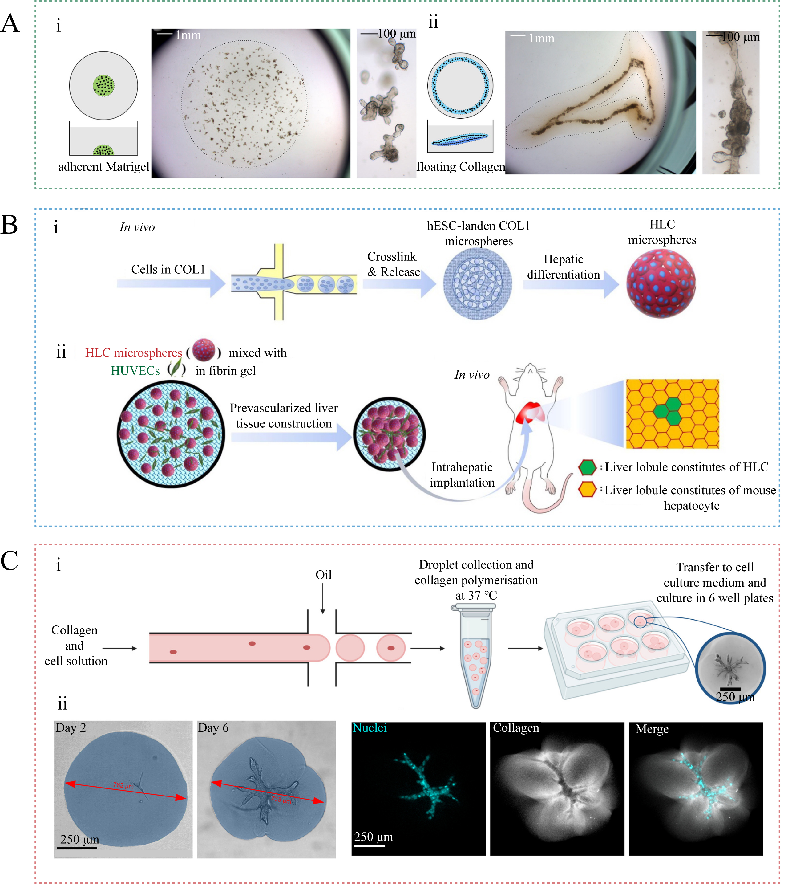
Figure 5. Collagen-based matrices and microgels for organoid and assembloid-oriented culture. (A) Comparison of small intestinal organoids cultured in traditional Matrigel and in floating type I collagen gel rings: (i) Organoids in traditional Matrigel mainly grow as single budding cysts. (ii) In floating collagen gel rings, the organoids self-organize into continuous macroscopic tubular structures that better mimic native intestinal architecture through mechanical tension-mediated morphogenesis. Figure 5A is adapted in part with permission from Ref.[110]. Copyright 2017, The Company of Biologists Ltd; (B) Schematic diagram of the preparation of HLC microspheres and the culture of liver organoids: (i) Microfluidic fabrication of collagen microspheres encapsulating hESCs and a schematic diagram of hESC differentiation into HLCs. (ii) The HLC microspheres were further assembled with human umbilical vein endothelial cells (HUVECs) to form prevascularized liver tissue (PLT), which was implanted into a mouse liver. Figure 5B is adapted in part from Ref.[111]. Reused under the terms of the Creative Commons Attribution License (CC BY); (C) Cell-laden collagen microgels for the culture of complex branched organoids. (i) Microfluidic fabrication of collagen microgel encapsulating pancreatic ductal adenocarcinoma cells and culture of complex branched organoids. (ii) Deformation of collagen microgels and formation of branched structures under the traction of organoids demonstrating the capacity of microgel confinement to guide organoid morphogenesis. Figure 5C is adapted in part from Ref.[112]. Reused under the terms of the Creative Commons Attribution License (CC BY). hESC: Human embryonic stem cell; HLC: hepatocyte-like cell; COL1: Collagen type I.

