fig4

Nanoplastics remodel the extracellular matrix mechanical microenvironment to activate hepatic stellate cells

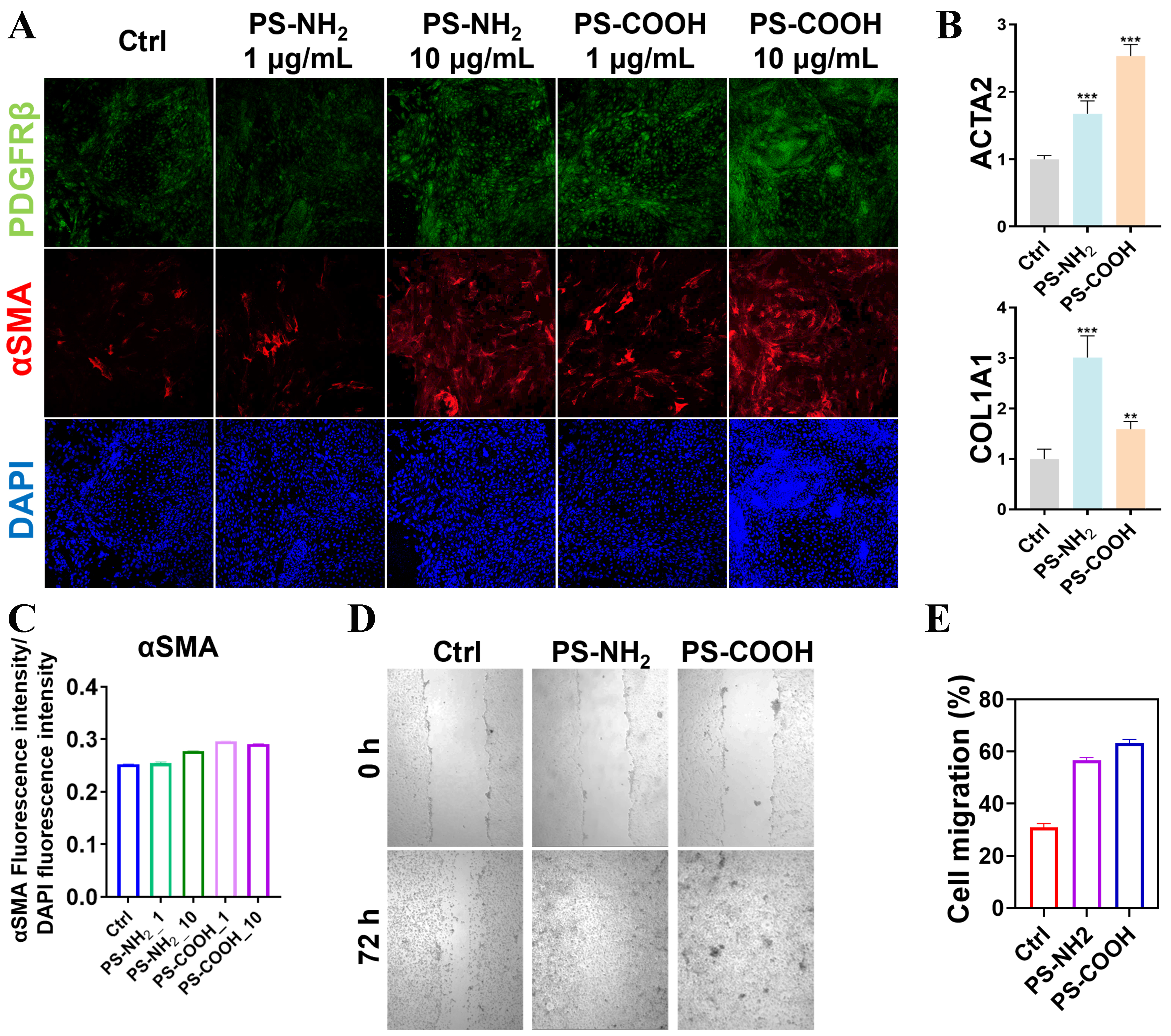
Figure 4. Regulation of HSC behavior by nanoplastics. (A) Immunofluorescence staining of HSC protein markers following treatment with different nanoplastics (PDGFRβ, α-SMA, and DAPI); (B) Quantitative PCR analysis of mechanotransduction-related genes in HSCs after nanoplastic exposure; (C) Comparison of α-SMA fluorescence staining intensity under different nanoplastic treatments; (D) Images of 72 h migration of HSCs after treatment with different nanoplastics; (E) Comparison of the 72 h migration capacity of HSCs after treatment with different nanoplastics. Values represent the mean of three independent biological replicates. **P ≤ 0.01, ***P ≤ 0.001 compared to the control group. HSC: Hepatic stellate cell; PDGFRβ: platelet-derived growth factor receptor beta; α-SMA: alpha-smooth muscle actin; DAPI: 4′,6-diamidino-2-phenylindole; PCR: polymerase chain reaction; PS-NH2: amino-functionalized polystyrene; PS-COOH: carboxyl-functionalized polystyrene; ACTA2: alpha-smooth muscle actin; COL1A1: collagen type I alpha 1 chain.

Journal of Environmental Exposure Assessment
ISSN 2771-5949 (Online)

Portico

All published articles are preserved here permanently:

https://www.portico.org/publishers/oae/

Portico

All published articles are preserved here permanently:

https://www.portico.org/publishers/oae/