



fig3
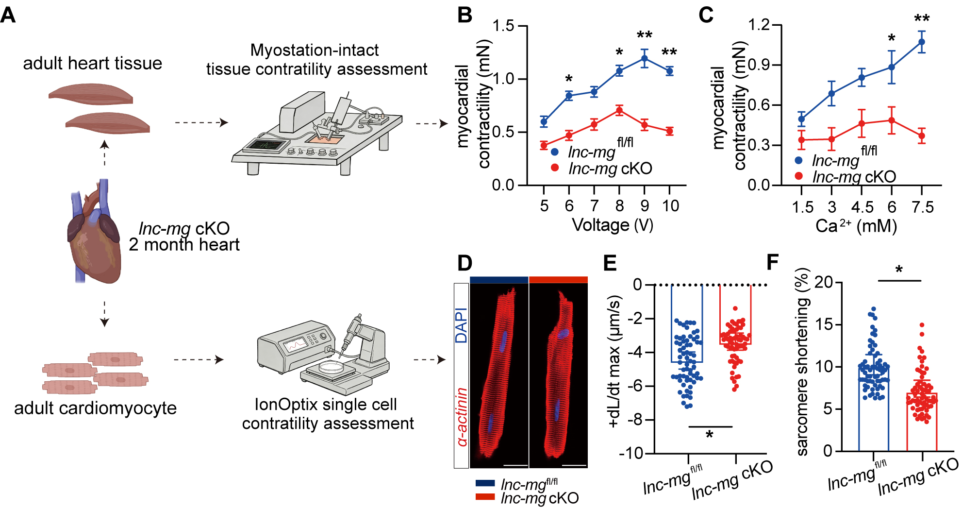
Figure 3. Lnc-mg deficiency impairs contractile function in myocardial tissue and isolated cardiomyocytes. (A) Schematic of myocardial tissue and single-cardiomyocyte contractility assessment using the Myostation-intact and IonOptix systems. Created in BioRender. Jia S, (2026) https://BioRender.com/0o3q3pu. (B and C) Contractile force of left ventricular myocardial strips isolated from 2-month-old lnc-mgfl/fl and lnc-mg cKO mice under graded electrical stimulation (5-10 V) (B) and increasing extracellular Ca2+ concentrations (1.5-7.5 mM) (C). Myocardial strips were obtained from six independent mice per group (n = 6 biological replicates per group), with the mouse serving as the experimental unit. Measurements obtained from the same myocardial preparation at different stimulation voltages or Ca2+ concentrations were treated as repeated measurements. (D) Representative immunofluorescence images of freshly isolated adult cardiomyocytes stained for α-actinin (red) and DAPI (blue), demonstrating the morphology of isolated rod-shaped cardiomyocytes before IonOptix analysis, Scale bar, 10 μm. (E and F) IonOptix analysis of maximum shortening velocity (+dL/dt max) (E) and sarcomere shortening (F) in isolated adult cardiomyocytes. The 60 points shown per group represent pooled cell-level measurements obtained from three independent mice (n = 3 biological replicates per group). Statistical comparisons were performed using nested t-tests, with cardiomyocytes nested within their mouse of origin. Data are presented as mean ± SEM. *P < 0.05; **P < 0.01.




