



fig2
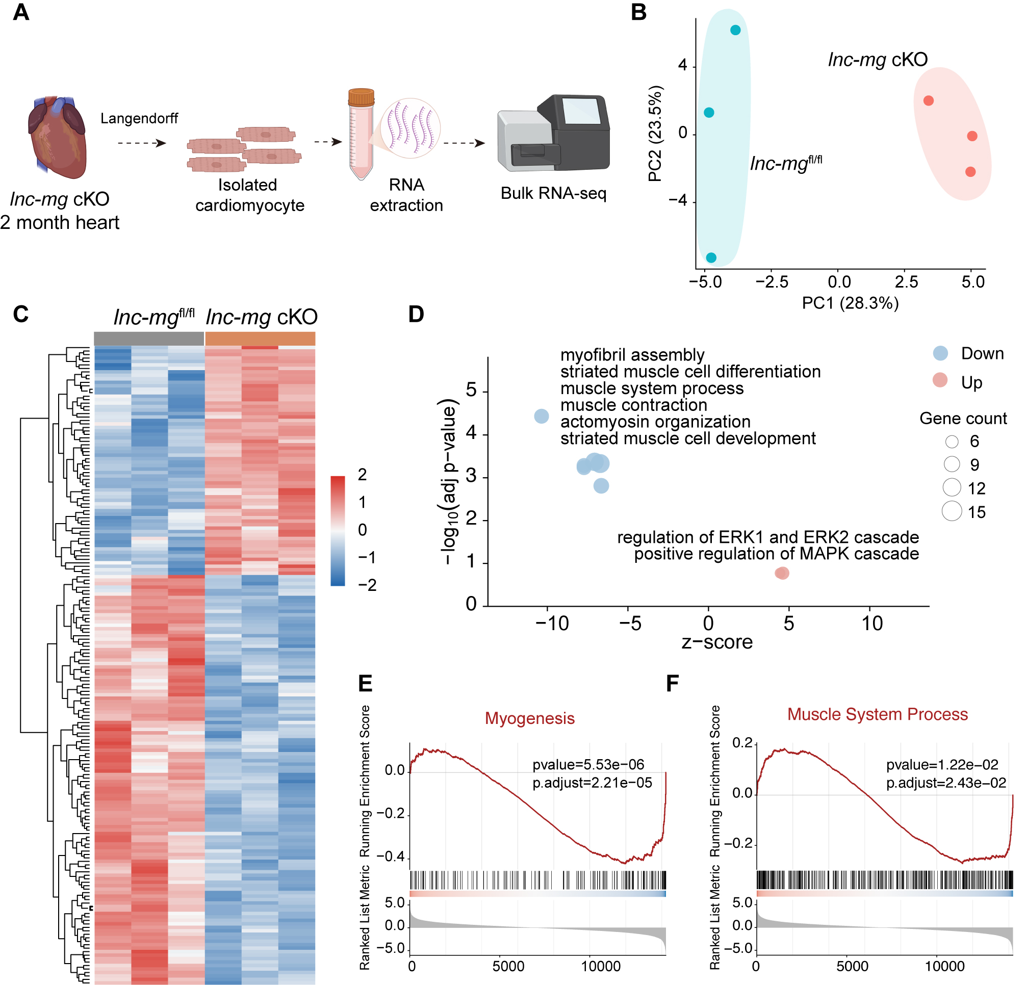
Figure 2. Transcriptomic analyses reveal suppression of contractile gene programs in lnc-mg-deficient hearts. (A) Schematic illustration of the experimental workflow. Myocardial tissues were collected from 2-month-old lnc-mgfl/fl and lnc-mg cKO mice, followed by RNA-seq. Created in BioRender. Jia S, (2026) https://BioRender.com/umio76p. (B) PCA of RNA-seq samples from lnc-mgfl/fl and lnc-mg cKO hearts. Three independent mice were analyzed per genotype (n = 3 biological replicates per group). (C) Heatmap showing the expression patterns of differentially expressed genes identified between lnc-mgfl/fl and lnc-mg cKO hearts. Gene expression values were row-scaled for visualization. (D) GO enrichment analysis of upregulated and downregulated genes. Bubble size represents gene count, and bubble color indicates upregulated or downregulated gene sets. (E and F) GSEA of the Myogenesis gene set (E) and the Muscle System Process gene set (F) using genes ranked according to DESeq2 statistics. All transcriptomic analyses were based on the same three independent biological replicates per group.




