

fig5
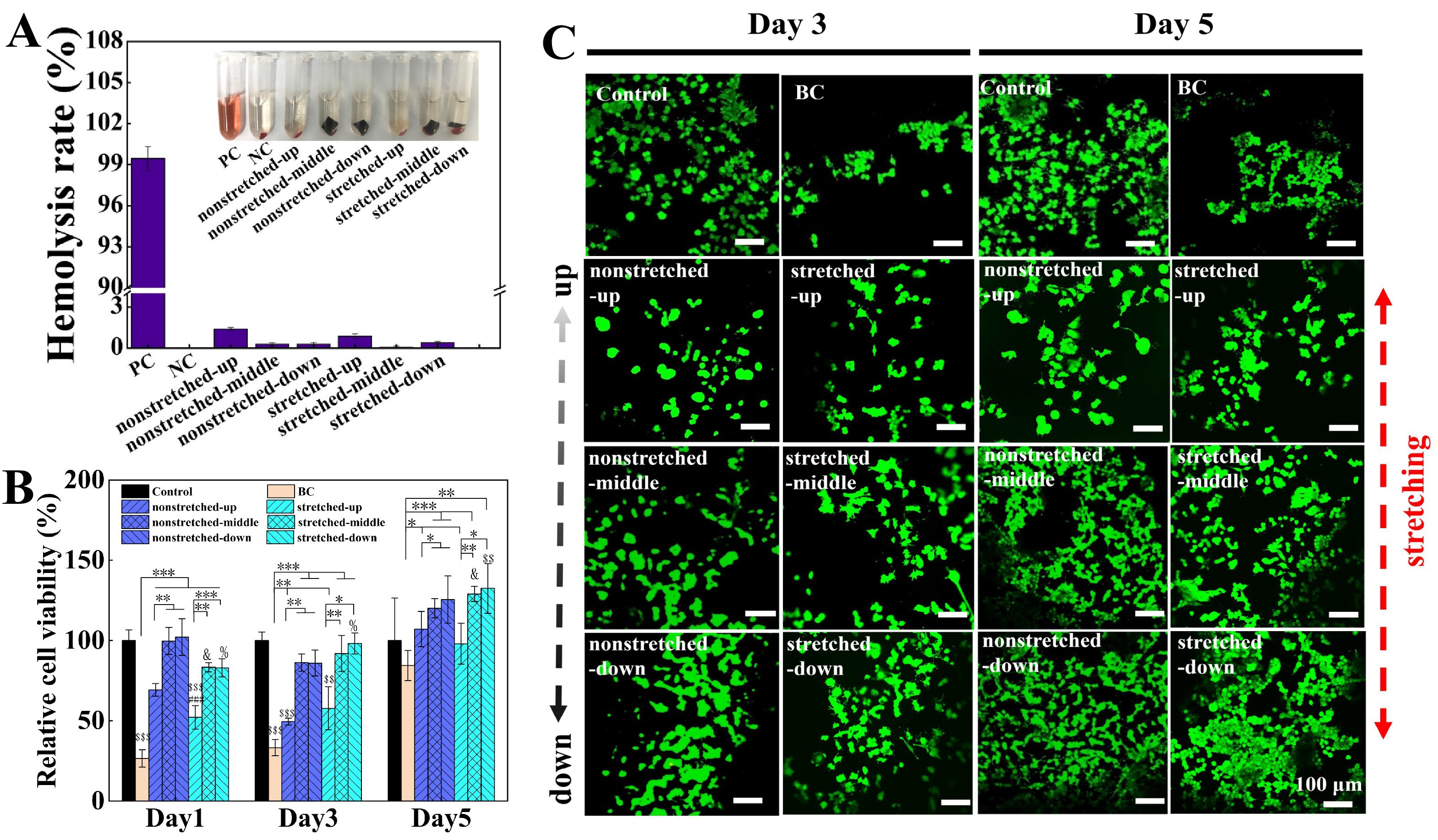
Figure 5. Biocompatibility of BC/PPy conductive gradient membranes and cell morphology observations. (A) Hemolysis test for non-stretched and stretched BC/PPy conductive gradient membranes. PC: positive control (distilled water); NC: negative control (normal saline), n = 3; (B and C) MTT assay and Live/Dead staining images of PC12 cells cultured on Control (petri dish), BC, non-stretched and stretched BC/PPy conductive gradient membranes. n = 3, *P < 0.05, **P < 0.01, ***P < 0.001, ##P < 0.01, # when compared with the non-stretched upper segment under the same conditions. &P < 0.05, & when compared with the non-stretched middle segment under the same condition. %P < 0.05, % when compared with the non-stretched lower segment under the same condition. $$P < 0.01, $$$P < 0.001, $ when compared with control group under the same condition. Scale bars are 100 μm. Error bars represent standard deviation (SD). BC: Bacterial cellulose; PPy: polypyrrole; MTT: 3- (4,5-dimethylthiazol-2-yl)-2,5-diphenyltetrazolium bromide.

