

fig3
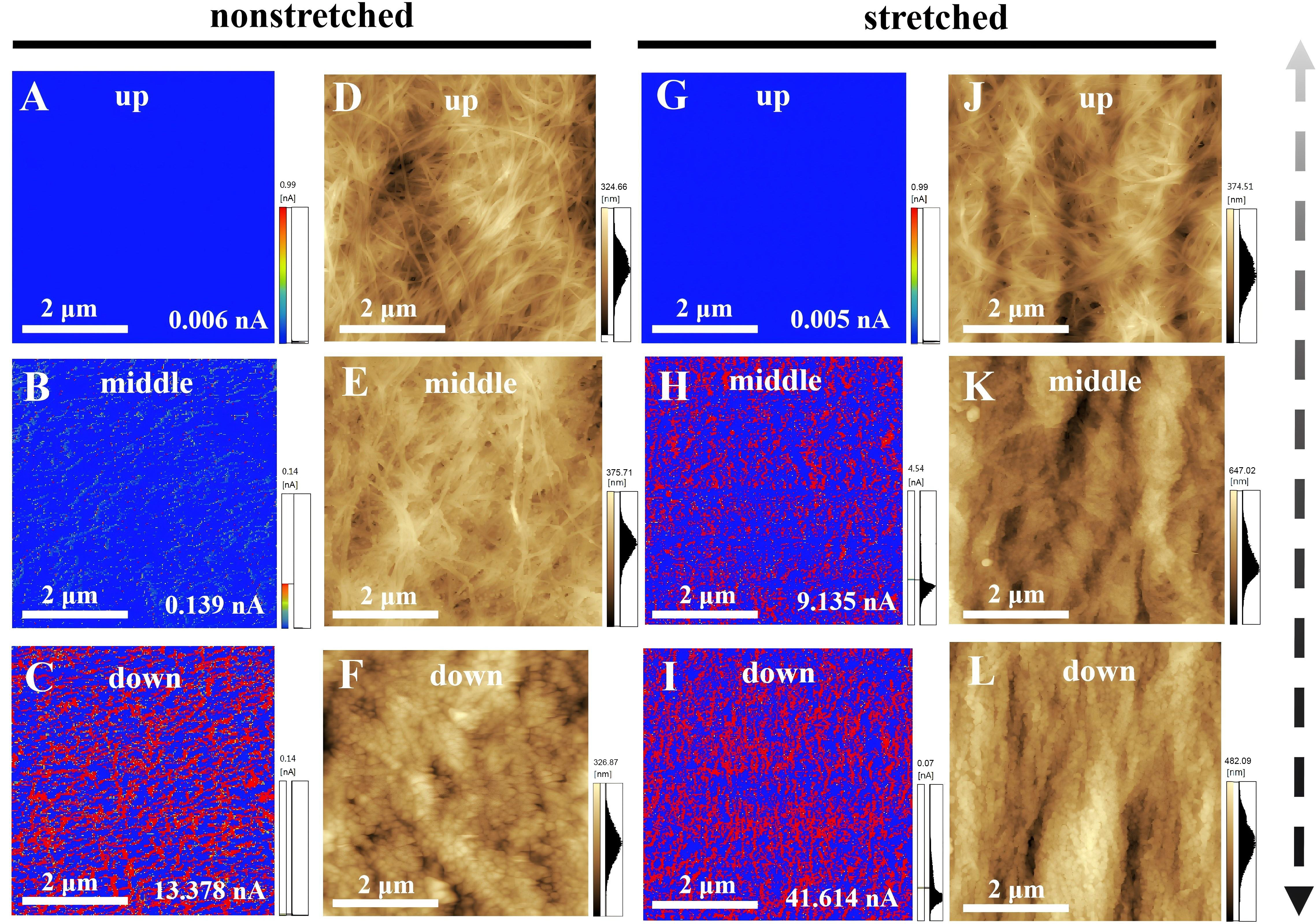
Figure 3. AFM images of non-stretched (A-F) and stretched (G-L) BC/PPy gradient conductive membranes. Current images are shown for non-stretched (A-C) and stretched (G-I) membranes, and height images for non-stretched (D-F) and stretched (J-L) membranes. Scan size: 5 μm × 5 μm. AFM: Atomic force microscopy; BC: bacterial cellulose; PPy: polypyrrole.

