





Plant-derived extracellular vesicles as natural nucleic acid carriers in gut-liver communication: implications for metabolic liver disease
 0
0 
Abstract
Plant-derived extracellular vesicles (PDEVs) are nano-sized vesicles released by plant cells that contain endogenous nucleic acids, including microRNAs (miRNAs), small interfering RNAs (siRNAs), transfer RNA fragments (tRFs), and mitochondrial DNA (mtDNA). These characteristics have led to growing interest in PDEVs as naturally occurring nanocarriers suitable for oral delivery. Three biogenesis routes have been described for PDEV formation, namely multivesicular body release, plasma membrane budding, and plasmodesmatal transit. However, whether PDEVs-associated nucleic acids withstand gastrointestinal digestion and subsequently regulate mammalian gene expression remains controversial. Such interactions are generally considered within the framework of cross-kingdom gene regulation. This review summarizes current evidence concerning the nucleic acid biology of PDEVs, including their biogenesis, cargo diversity, and isolation methods. Evidence concerning gastrointestinal stability, intestinal uptake, and movement toward the liver is subsequently evaluated, along with experimental support for cross-kingdom transfer and the methodological limitations that complicate its interpretation. PDEVs-associated nucleic acids have been proposed as candidate mediators of gut-liver axis signaling in inflammatory and metabolic contexts, but their causal contribution and intravesicular localization remain incompletely defined. This review also discusses engineering strategies and potential biomarker applications. PDEVs-mediated nucleic acid transfer shows promise but requires more rigorous methodological standards. Standardized isolation workflows, stringent contamination controls, and causal validation are needed to establish biological relevance and support clinical translation.
Keywords
INTRODUCTION
Extracellular vesicles (EVs) are membrane-delimited particles ranging from nano- to micro-scale sizes and are released by diverse cell types[1,2]. Their cargo includes proteins, RNAs, and DNA, mediating intercellular communication[3,4]. Plants also produce vesicle-like nanoparticles. Plant-derived extracellular vesicles (PDEVs), also known as exosome-like nanoparticles (ELNs) or plant-derived nanovesicles (PDNVs), have been isolated from edible plants and display size and morphological characteristics resembling those of mammalian EVs while carrying diverse molecular cargo[3-5].
PDEVs display favorable safety profiles and low immunogenicity[3,6-10]. However, most research focuses on PDEVs as drug delivery vehicles[3,4,11], whereas their role as natural nucleic acid carriers in cross-kingdom communication remains less explored. Oral administration studies indicate that PDEVs can withstand gastrointestinal digestion and may transport nucleic acid cargo toward the gut-liver axis and systemic circulation[2,4,10].
The ability of dietary plant nucleic acids to functionally regulate mammalian gene expression remains controversial. Several studies have reported regulatory effects of dietary plant miRNAs on mammalian gene expression[12-14], and specific plant miRNAs have been detected in animal plasma and tissues[15-17]. However, these observations have not been consistently reproduced, with multiple independent studies reporting conflicting results[17-19]. Furthermore, direct evidence that plant xenomiRs interact with the mammalian RNA interference machinery has not been established[20,21]. Accordingly, recent reviews have called for more rigorous methodological standards for detecting and interpreting dietary plant nucleic acids[15,20,21].
Dietary nucleic acids may be protected from degradation through packaging within vesicles or association with proteins and lipoproteins[3,22]. In experimental settings, PDEVs have been shown to withstand gastrointestinal exposure and to be taken up by intestinal epithelial and immune cells[23]. They have also been reported to enter portal and lymphatic circulation and reach the liver and other organs[24], although the relative contributions of these routes and the distinction between intact vesicles and released components remain incompletely defined. This vesicle-mediated model provides an alternative to the original “naked miRNA” hypothesis, but has not been systematically reviewed.
The gut-liver axis provides a route through which food-derived PDEVs have been proposed to influence systemic metabolism and liver disease. Plant-derived vesicles modulate intestinal barrier integrity, epithelial cell renewal, and gut microbiota composition, and then act on the liver via microbial metabolites and immune mediators[25,26]. Orally administered PDEVs protect against inflammatory bowel disease (IBD), colitis-associated cancer, and experimental liver injury[8,23,27-29]. Several reports have suggested that PDEVs, which carry small RNAs, contribute to these effects, although the RNA cargo’s specific contribution remains to be directly established[4,23,30].
These findings support a focused re-evaluation of PDEVs as nucleic acid reservoirs and delivery vehicles, particularly in relation to gut-liver communication. This review addresses five questions to provide a complementary perspective to existing PDEVs reviews: (i) What nucleic acids are carried by PDEVs? (ii) How are PDEVs and their cargo absorbed and transported following oral intake? (iii) What established and controversial roles do PDEVs-associated nucleic acids have in cross-kingdom gene regulation? (iv) How do PDEVs influence the gut-liver axis and metabolic liver diseases? (v) How can plant vesicles be engineered as oral nucleic acid nanocarriers, and how can circulating plant-derived nucleic acids be detected? These five questions are addressed sequentially in the main text: Section 1 addresses question (i), Section 2 addresses questions (ii) and (iii), Section 3 addresses question (iv), and Section 4 addresses question (v).
To facilitate interpretation of the evidence discussed below, three levels of evidence are distinguished: (i) studies demonstrating biological activity of PDEV preparations in experimental models; (ii) studies suggesting, through small RNA profiling, target prediction, or correlative readouts, that vesicle-encapsulated nucleic acids contribute to these effects; and (iii) studies providing causal evidence, such as loss-of-function or rescue experiments, that specific nucleic acids are responsible. Because PDEV preparations also contain proteins, lipids, and metabolites, biological effects observed with whole preparations do not by themselves establish nucleic acid-mediated mechanisms or demonstrate systemic cross-kingdom regulation in the absence of direct evidence.
PDEVs AS NUCLEIC ACID RESERVOIRS
Biogenesis and subtypes of PDEVs
The formation of plant-derived vesicles has been described through three routes. In the first, multivesicular bodies (MVBs) fuse with the plasma membrane and release intraluminal vesicles as exosome-like particles. In the second, outward budding of the plasma membrane directly generates microvesicle-like structures. A third route involves the transit of some vesicles through plasmodesmata to adjacent cells, representing a plant-specific route with no mammalian counterpart[5,31]. In most studies, PDEVs are isolated from fruit and vegetable juices by differential centrifugation, ultracentrifugation, or density gradient centrifugation[32,33]. The resulting vesicle preparations are heterogeneous and are commonly referred to as PDEVs, exosome-like nanoparticles (ELNs), plant-derived nanovesicles (PDNVs), or plant-derived exosome-like nanoparticles (PDENs)[1,3,34] [Figure 1].
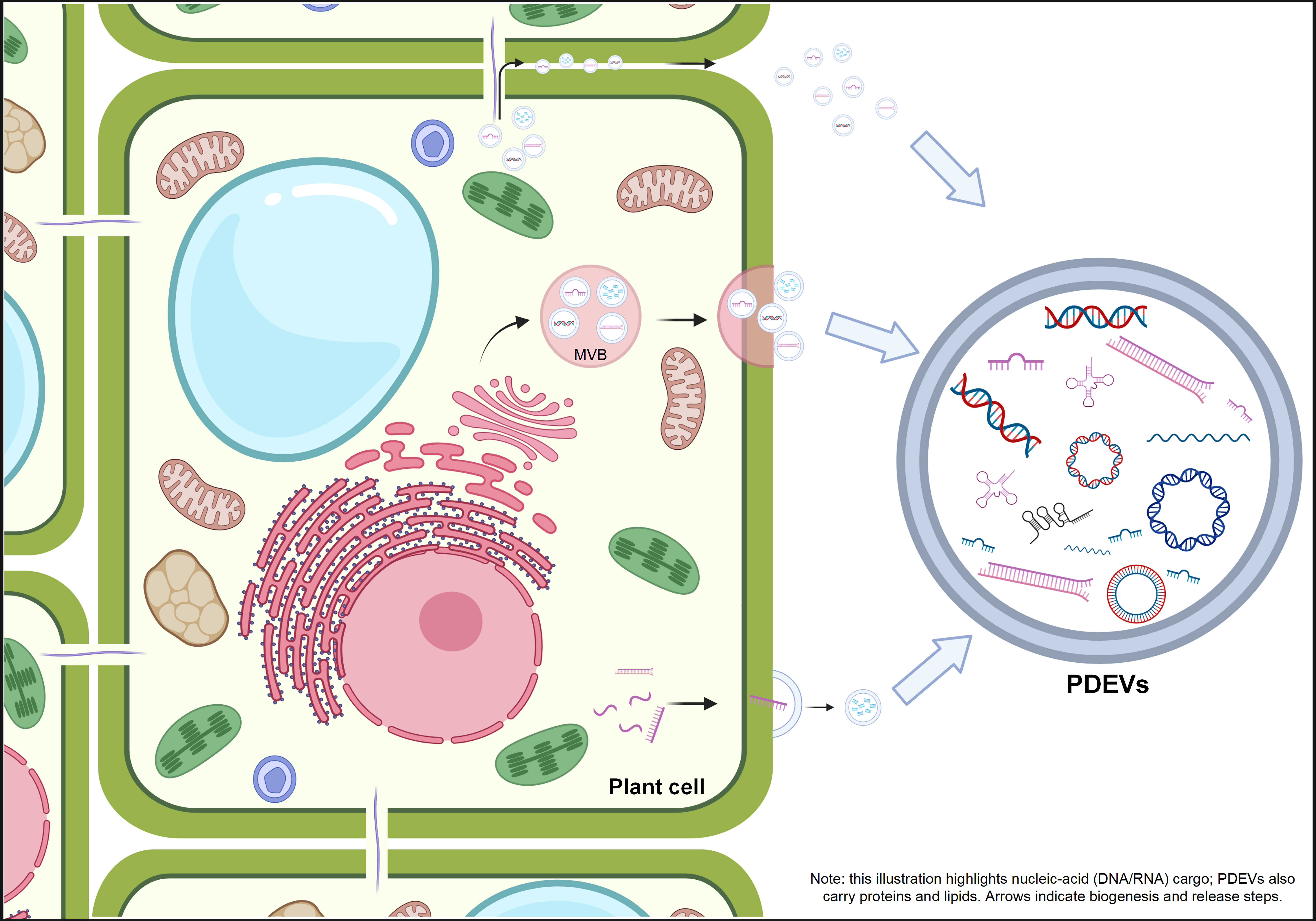
Figure 1. The formation and release of PDEVs from plant cells. This illustration depicts PDEV biogenesis and secretion pathways, with emphasis on nucleic acid cargo. MVBs fuse with the plasma membrane and release intraluminal vesicles. Alternatively, vesicles can be directly shed from the plasma membrane or transit through plasmodesmata to adjacent cells. Although PDEVs carry diverse cargo, including DNA, RNA, and protein, this illustration highlights their nucleic acid content. Created in BioRender. Ruting, C. (2026) https://BioRender.com/r47hcp8. PDEVs: Plant-derived extracellular vesicles; MVBs: multivesicular bodies; DNA: deoxyribonucleic acid; RNA: ribonucleic acid.
The resulting preparations generally contain vesicles 30 to 200 nm in diameter and display a cup-shaped morphology under transmission electron microscopy. However, the cup-shaped appearance may partly result from fixation and dehydration artifacts rather than represent an intrinsic structural feature. PDEV preparations are also enriched in phosphatidic acid, phosphatidylcholine, and phosphatidylserine[5]. Unlike mammalian EVs, which have well-established markers such as Cluster of Differentiation 9 (CD9), Cluster of Differentiation 63 (CD63), and Cluster of Differentiation 81 (CD81), PDEVs currently lack universally accepted protein markers[34]. PDEVs originate from diverse plant tissues (fruits, rhizomes, leaves), and their nucleic acid payload can vary with plant species, tissue origin, developmental stage, and environmental conditions, particularly pathogen infection, which has been associated with preferential loading of stress- and defense-related small RNAs into PDEVs[35-37]. The relative influence of other factors, such as abiotic stress and cultivation conditions, remains poorly characterized, and this variability is an underexplored determinant of their functional specificity[38].
Recent reviews have differentiated EVs isolated from the plant apoplast, the intercellular fluid occupying the extracellular spaces between plant cells, from nanovesicles obtained from juiced plant tissues[4]. A matrix-based taxonomy according to plant part and cargo type has also been proposed[39]. Some researchers define PDENs as nanovesicles from edible plants that meet fundamental EV criteria, highlighting their translational medical potential[11]. This review uses “PDEVs” as a collective term for plant vesicle preparations with EV-like characteristics and measurable biological activity. Narrower categories (ELNs, PDENs) are explicitly noted where study-specific data apply. From a nucleic acid perspective, biological and operational heterogeneity implies that distinct PDEV subpopulations may exhibit unique cargo preferences and targeting properties. MVB-derived vesicles may be enriched in specific small RNAs and messenger RNA (mRNA) destined for secretion[35,36]. Membrane-budded vesicles may carry different nucleic acid profiles[30]. The plasmodesmatal route, unique to plants, has received little study in this regard[5,31]. Therefore, linking PDEVs-associated nucleic acids to functional outcomes in mammalian hosts requires more precise isolation strategies and molecular markers.
Consistent with the Minimal Information for Studies of Extracellular Vesicles (MISEV) guidelines, the term “exosome” is reserved for vesicles for which endosomal biogenesis, intraluminal vesicle formation within MVBs, and subsequent release through fusion with the plasma membrane have been experimentally demonstrated. Because most plant-derived vesicle preparations are heterogeneous and their intracellular origin has not been verified, they should not be designated as “exosomes” unless endosomal biogenesis has been directly demonstrated. Accordingly, this review uses “PDEVs” or “ELNs” for plant vesicle preparations without demonstrated endosomal origin, whereas “exosome” is used only when the endosomal biogenesis of a specific vesicle population has been confirmed.
Methodological considerations for nucleic acid profiling in PDEVs
Accurate characterization of PDEV-associated nucleic acids depends strongly on the isolation and analytical methods employed. To date, most studies have relied on differential centrifugation and ultracentrifugation, sometimes combined with sucrose or iodixanol density gradients, to enrich vesicles from plant juices or homogenates[32,40]. However, these methods may co-isolate non-vesicular contaminants such as protein-RNA complexes, cell debris, and aggregated lipids, which can interfere with downstream nucleic acid analysis. Emerging techniques, including size exclusion chromatography and tangential flow filtration, offer higher purity and scalability, but their application in plant systems remains limited[5,11]. Different separation methods can substantially influence vesicle yield, size distribution, and cargo composition. Extraction conditions, particularly pH and clarification steps, can also significantly alter PDEVs’ characteristics[11] [Figure 2]. A systematic comparison of conventional herbal extracts with vesicles isolated from fresh plant tissue found that drying and alcohol-based extraction selectively depleted heat-labile and amphipathic constituents, including miRNAs, proteins, and lipids, whereas vesicles obtained from fresh material retained the native co-encapsulated cargo profile and associated multi-component activity[41]. This comparison is directly relevant to the interpretation of cross-kingdom studies because differences in reported small RNA content may arise from upstream processing of the source material as well as from biological variation among plant species, tissues, or physiological states.
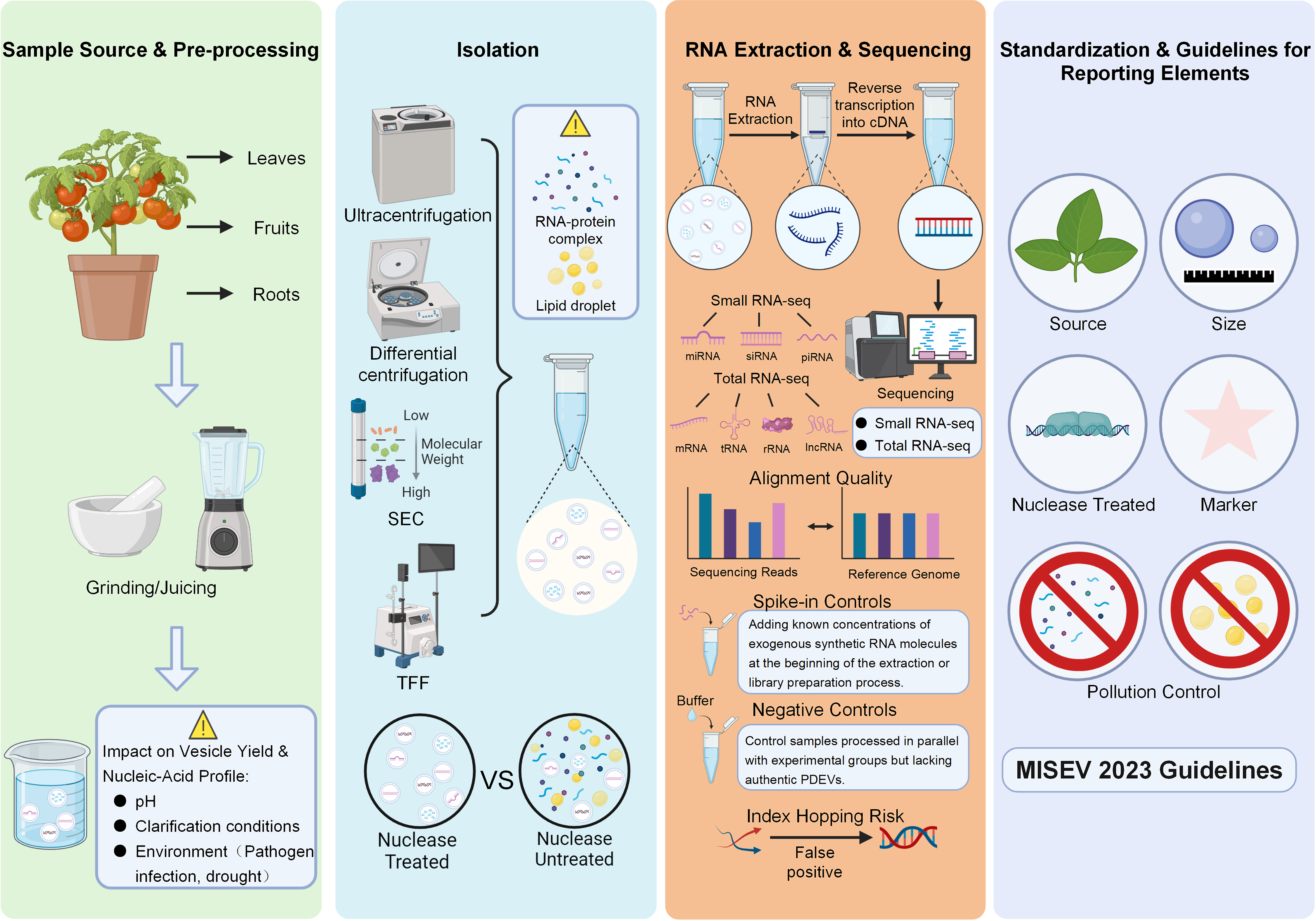
Figure 2. The PDEVs research workflow following MISEV 2023 guidelines. The diagram outlines four phases of PDEVs research: sample and pre-treatment; sequential separation (centrifugation, SEC, and TFF); RNA extraction and sequencing; and standardized reporting. This workflow is informed by MISEV 2023 guidelines to promote rigor, reproducibility, and transparency in PDEVs research. Created in BioRender. Ruting, C. (2026) https://BioRender.com/ufeeq9m. TFF: Tangential flow filtration; cDNA: complementary DNA; lncRNA: long non-coding RNA; miRNA: microRNA; MISEV: Minimal Information for Studies of Extracellular Vesicles; mRNA: messenger RNA; PDEVs: plant-derived extracellular vesicles; piRNA: PIWI-interacting RNA; RNA: ribonucleic acid; rRNA: ribosomal RNA; SEC: size exclusion chromatography; siRNA: small interfering RNA; tRNA: transfer RNA.
The distinction between starting materials is equally important. A minority of studies isolate PDEVs from apoplastic fluid, occupying the extracellular spaces of intact plant tissues, avoiding mechanical tissue disruption. Most studies use juice, pressed tissue, or homogenates, in which mechanical disruption of cell walls and membranes can introduce intracellular membranes, organellar material, and non-vesicular nucleic acids into vesicle-enriched preparations. These categories should therefore not be considered methodologically equivalent. This distinction is particularly important when interpreting nuclear, mitochondrial, or chloroplast DNA and long RNA species, because their detection in vesicle-enriched fractions may reflect organellar contamination rather than intravesicular cargo. Therefore, the cargo literature should be stratified according to starting material and isolation strategy, with evidence for intravesicular localization, such as nuclease protection assays, evaluated separately from findings from unfractionated preparations Table 1.
PDEVs’ nucleic acid cargo literature summary
| Plant source | Nucleic acid cargo identity | Starting material | Isolation/Purification method | Evidence for intravesicular localization | Known/Predicted mammalian targets | Key functional findings | Nucleic acids dependence | Ref. |
| Blueberry | Small RNA; miRNAs predicted to target CD36, SREBP-1c, FASN, ACC, PPARα/γ | Fresh blueberry fruit (Vaccinium corymbosum), washed and juiced | Differential centrifugation (1,000/3,000/10,000 g); 1.0 µm filtration; 8% PEG8000 precipitation | TEM (spherical morphology); DLS 150-250 nm (mean ~189.6 nm); zeta -2.52 mV; TLC (RNA/protein/lipid); DiO membrane dye uptake; DiR biodistribution | CD36, SREBP-1c, FASN, ACC, PPARα/γ (Predicted) | Ameliorated diet-induced fatty liver | Not tested | [8] |
| Grape | miRNAs associated with lipid metabolism | Fresh grapes, peeled, pressed at 4 °C, diluted in cold PBS | Differential centrifugation + sucrose-gradient ultracentrifugation | TEM 50-300 nm; DLS 380.5 ± 37.5 nm; zeta -26.3 ± 8.1 mV; lipidomics (PA 53.2%, PE 26.1%); RNase-resistant RNA; PKH26/DiR labeling; cryo-EM | LGR5, Wnt/β-catenin pathway genes | Intestinal stem cell induction; anti-colitis | Not tested | [9] |
| Panax notoginseng | miRNAs | Fresh roots/rhizomes of Panax notoginseng (plus 3 other medicinal plants), washed, homogenized in PBS, filtered through 200-mesh gauze | Sequential centrifugation (1,000/2,500/5,000/10,000 g); 0.22-µm filtration; 100 kDa ultrafiltration; 120,000 g for 1 h; 8%/30%/45%/60% sucrose gradient | TEM (cup-shaped); NTA ~94 nm, 7.57 × 10¹² particles/mL; zeta -8.80 mV; DiD-labeled confocal uptake | G3BP2 (partially validated: luciferase assay in vitro) | Cross-kingdom miRNA delivery restored neuronal function after ischemic injury | Partial | [13] |
| Polygoni multiflori radix | miRNAs | Fresh Polygoni Multiflori Radix, crushed, homogenized, and filtered | Differential/gradient centrifugation to remove cells and debris; ultracentrifugation enrichment | TEM (round/cup-shaped); NTA monodisperse, peak ~77 nm (30-120 nm); yield ~2.2 × 10⁹ particles/g; DiD-labeled uptake (caveolin/HSPG endocytosis) | SRD5A2, AR, Wnt/β-catenin genes (Predicted) | Cross-kingdom miRNA delivery promoted hair growth via androgen pathway modulation | Partial | [14] |
| Rice | miR-168a | N/A - no plant vesicle isolation; MIR168a from rice total RNA (TRIzol) | N/A - rice total RNA extraction; animal serum MVs pelleted at 110,000 g for 2 h | No plant vesicle evidence; MIR168a detected in serum MVs (110,000 g pellet; AGO2 IP; periodate oxidation) | LDLRAP1 (validated: luciferase, AGO2 IP, in vivo ASO rescue) | miR-168a detected in human serum; represses LDLRAP1 translation | Yes | [17] |
| Por Artemisia L. | Small RNAs | Whole Portulaca oleracea L., washed, chopped and homogenized | Sequential centrifugation (500-3,000 g 10-15 min; 3,000-10,000 g 20-40 min; 10,000-150,000 g 1.5-2 h); 8%/30%/45%/60% sucrose gradient at 150,000 g | DLS ~160 nm; zeta -31.4 mV; cryo-TEM; IRDye 800CW-labeled IVIS tracking (colonic targeting) | ZBTB7B (validated: WB, IHC) | Ameliorated DSS-induced colitis | Not tested | [28] |
| Artemisia | mtDNA | Fresh Artemisia annua L., washed, blended in PBS (1:2 g/mL) for 5 min | Sequential centrifugation (200/2,000/10,000 g); 150,000 g for 2 h; 15%/30%/45%/60% sucrose gradient (150,000 g for 2 h); 30% band washed ×3 in PBS, filtered | TEM (nano-scaled membrane-bound vesicles); Dil labeling | cGAS-STING pathway: MB21D1 (cGAS), TMEM173 (STING) | Medicinal plant-derived mtDNA via nanovesicles induced cGAS-STING pathway | Yes (DNA) | [42] |
| Plant miR159 (dietary origin) | miR159 | N/A - no plant vesicle isolation; synthetic miR159 mimics used in functional assays | N/A - human serum EVs by sequential centrifugation (500 g 5 min; 18,700 g 20 min; 110,000 g 70 min ×2) | No plant vesicle evidence; miR159 enriched in serum EV fraction; 2’-O-methylation (plant origin) confirmed by periodate oxidation | TCF7 (partially validated: TCF7 3’UTR luciferase assay) | Plant miR159 detectable in human serum; oral miR159 mimics inhibited xenograft tumor growth | Partial | [43] |
| Grape/Grapefruit/Ginger/Carrot (EPDENs) | Multiple miRNAs from mixed plant sources | Edible plant juices (grape, grapefruit, ginger, carrot), washed ×3 and juiced | Sequential centrifugation (1,000 g 10 min; 3,000 g 20 min; 10,000 g 40 min); 150,000 g for 90 min; 8%/15%/30%/45%/60% sucrose gradient (150,000 g for 120 min); bands at 8%/30% and 30%/45% interfaces | PKH26 membrane dye labeling; confocal uptake by F4/80+ macrophages and Lgr5+ intestinal stem cells after oral gavage | NFE2L2 (NRF2), HMOX1 (HO-1), NQO1, TGF-β, IL-10 (Predicted) | EPDENs taken up by intestinal macrophages; delivered miRNAs induced anti-inflammatory and antioxidant responses | Not tested | [44] |
| Tea | miRNAs | Fresh tea leaves (Camellia sinensis) | EVs isolated from tea (TEVs) by centrifugation-based protocol; RNase-treated D-TEVs generated for RNA-dependence assays | TEM imaging of TEVs/D-TEVs; small RNA-seq (miRNA enrichment); time-dependent macrophage uptake (flow cytometry); RNase digestion abolished activity | AKT1, IKBKB (validated: dual-luciferase) | Reprogrammed macrophage polarization; alleviated intestinal inflammation | Yes | [45] |
| Polygonatum sibiricum | miR166u | Polygonatum sibiricum rhizomes | Tangential flow filtration | TEM; NTA; miRNA-seq (miR166u-enriched) | CD11c (validated); downstream TLR4/AKT | miR166u-enriched ELNs improved intestinal barrier through TLR4/AKT signaling | Partial | [46] |
| Coptis chinensis Franch | miRNA-5106 | Fresh Coptis chinensis Franch., homogenized; juice collected | Sequential centrifugation (1,000 g 10 min; 3,000 g 20 min; 10,000 g 30 min); 0.22 µm filtration; 120,000 g for 60 min | TEM (cup-shaped); PKH26-labeled confocal internalization | Zinc transporter genes: Slc39a2 (validated: dual-luciferase); ZIP1-14, ZnT1-10 (predicted) | Delivered miRNA-5106 suppressed NETs by restoring zinc homeostasis | Partial | [47] |
| Plant miR8126 (dietary origin) | miR8126-3p, miR8126-5p | N/A - synthetic plant miRNA mimics (miR8126-3p/5p) | N/A - in vitro transfection of human HepG2 hepatocytes | N/A - no vesicle isolation or characterization | Metabolic genes: SREBF1, FASN, ACACA (Predicted) | Plant miR8126-3p/5p decreased lipid accumulation | Partial | [48] |
| Grapefruit | miRNAs associated with oxidative stress and metabolic pathways | Fresh grapefruit pulp (peeled), homogenized at 4 °C for 1 min; grapefruit-derived lipids | Differential centrifugation (2,000 g 20 min; 10,000 g 1 h); 150,000 g for 1.5 h; 8%/30%/45%/60% sucrose gradient (band 2 at 30%/45% interface); reconstituted GDNs | TEM; DLS 105.7-396.1 nm (mean ~210.8 nm); negative zeta; lipidomics (PE 45.5%, PC 28.5%); DiR/PKH26 tracking; gastric/intestinal enzyme stability; GDN nanovectors | Macrophage polarization: NOS2, ARG1, TNF-α, IL-6, IL-10 | Anti-inflammatory; targeted drug delivery to intestinal macrophages | Not tested | [2,29,49] |
| Ginger (curcumin-loaded GELNs) | Endogenous GELN miRNAs | Fresh ginger, washed ×3, soaked in ice water and juiced; ginger juice | Differential centrifugation (1,000/3,000/10,000 g); 140,000 g for 2.5 h; 5%-60% sucrose gradient (140,000 g for 3 h; 30%/45% interface); PEG 8000 precipitation + sucrose density gradient | TEM/AFM/DLS 168.2 ± 9.0 nm (PDI 0.21); zeta -17.6 ± 0.67 mV; PKH67-labeled CLSM; curcumin loading 94.0%, encapsulation 89.3% | N/A (curcumin cargo, not miRNA-mediated) | Curcumin-loaded GELNs alleviated ionizing radiation-induced intestinal damage via gut microbiota regulation | N/A | [50,51] |
| Arabidopsis | Small RNAs silencing virulence genes; mRNAs | Arabidopsis thaliana leaves (infected with Botrytis cinerea); AWF collected by vacuum infiltration | AWF filtered; differential centrifugation (~40,000 × g) then ultracentrifugation (100,000 × g, 60 min, 4 °C) to pellet EVs | TEM/confocal (EVs accumulate at infection sites; MVBs fuse with plasma membrane); nuclease-protection assay (sRNAs protected unless vesicles ruptured); uptake by B. cinerea | N/A (plant-fungal system, not plant-mammalian) | EV-encapsulated small RNAs silence fungal virulence genes; plant mRNAs move into fungal pathogens via EVs | Yes | [32,35,37,52] |
| Plant (multiple species) - bacterial EVs | Small RNAs | Arabidopsis thaliana leaves; AWF | AWF filtered; ultracentrifugation at 100,000-120,000 × g to pellet small EVs (exosomes) | TEM; NTA (EVs enriched in 10-30 nm nanoparticles); small RNA-seq (tiny RNAs 10-17 nt enriched) | N/A (plant-microbe system, not plant-mammalian) | Bidirectional sRNA exchange at plant-microbe interfaces; both vesicular and non-vesicular sRNA trafficking | Partial | [53,54] |
| Honeysuckle | miR2911 | Dried honeysuckle (Lonicera japonica) flowers | Soaked in PBS for 30 min, homogenized; cleared by centrifugation (10,000 rpm for 60 min, repeated); hollow-fiber filtration (200 nm pore, 750 kDa MWCO) hierarchical separation; HNVs = fractions ≤ 200 nm and > 750 kDa | Cryo-EM, TEM, DLS; MIR2911 from honeysuckle decoction is absorbed and self-assembles into host exosomes, directly targeting SARS-CoV-2 RNA | SARS-CoV-2 genomic RNA: ORF1ab | miR2911 orally absorbed; directly targets SARS-CoV-2 RNA | Partial | [55,56] |
| Ginseng | Exosomal miRNAs; miRNAs targeting KRAS-MAPK axis; miRNAs targeting inflammatory cytokines | Ginseng (Panax ginseng) root/material | Ginseng homogenized/extracted, cleared by low-speed centrifugation, vesicles pelleted by ultracentrifugation; characterized by DLS, NTA, TEM, Cryo-TEM | TEM/Cryo-TEM morphology; DLS (size/zeta); NTA (~6.93 × 10¹² particles); exosomal miRNA profiling | KRAS, MAPK1/3, TNF-α, IL-6, IL-1β (Predicted) | Ameliorated rheumatoid arthritis via KRAS-MAPK; suppressed colitis inflammatory cytokines | Partial | [57,58] |
| Citrus sinensis | SARS-CoV-2 spike mRNA | Fresh orange (Citrus sinensis) juice (plant-derived EVs, oEVs) | Juice filtered, centrifuged (4,000 × g, 30 min), ultracentrifuged (10,000 × g, 1 h; 100,000 × g, 2 h, 4 °C); exogenous mRNA encapsulated via osmotic stress with cationic proteins, washed (100,000 × g, 2 h) | TEM/NTA; mRNA protected from RNase and stable ≥ 1 year at room temp after lyophilization; oral gavage elicits humoral immunity | N/A (engineered mRNA cargo, not natural plant miRNA) | Oral/intranasal mRNA vaccine delivery by PDEV carriers | Yes (engineered) | [59,60] |
| Ginger | Multiple miRNAs enriched in inflammation/cancer-targeting profiles; miRNAs targeting Lactobacillus rhamnosus transcripts | Fresh ginger (Zingiber officinale) rhizome, juiced | Ginger juice sequentially centrifuged (1,000/3,000/10,000 g) then ultracentrifuged (150,000 × g, 2 h); sucrose-gradient purification (major band at 8%/30% interface) | TEM/NTA/DLS; membrane (PKH) labeling; miRNA-seq (~125 miRNAs); colon-targeted uptake by intestinal cells/macrophages; gut microbiota remodeling | NLRP3, TNF-α, IL-6, IL-1β (Predicted) | Anti-inflammatory; anti-colitis; liver protection against ALD; gut microbiota reshaping via miRNA transfer to L. rhamnosus | Partial | [2,6,23,61,62] |
A major limitation of current nucleic acid profiling is that small RNA sequencing and total RNA often do not distinguish vesicle-enclosed RNA from RNA associated with the outer vesicle surfaces[2,4,5]. Several studies have addressed this limitation by treating preparations with nucleases before RNA extraction to degrade unprotected nucleic acids. Nuclease resistance alone does not unequivocally demonstrate intravesicular localization: extravesicular RNA can be protected from nucleases by RNA-binding proteins or other macromolecular complexes, and nuclease digestion may be incomplete under suboptimal reaction conditions. More conclusive evidence is provided by comparing nuclease treatment performed with and without detergent-induced membrane disruption, or by single-vesicle approaches such as immunoelectron microscopy or flow-cytometric analysis of permeabilized vs. non-permeabilized vesicles. Studies reporting only nuclease resistance in the absence of such controls should be interpreted accordingly. When detecting low-abundance plant-derived RNA in mammalian samples, it is essential to include spike-in controls and negative controls, and to rigorously assess sequencing alignment quality, to minimize alignment artifacts and contamination risks[63].
Establishing standardized workflows and reporting guidelines for PDEVs nucleic acid analysis is therefore essential. Drawing on established recommendations for mammalian EVs, future studies should at minimum report plant species and tissue sources, detailed isolation procedures, vesicle characteristics such as size, morphology, and protein/lipid markers, RNA extraction and sequencing methods, and contamination-control measures, including nuclease sensitivity testing with and without membrane disruption[1,33,64,65]. The absence of standardized procedures continues to hinder cross-laboratory data comparison and complicates definitive conclusions regarding cross-kingdom nucleic acid transfer. Establishing a robust methodological foundation is essential to achieve two primary objectives: validating the functional roles of PDEVs-derived nucleic acids in diseases and developing these molecules as circulating biomarkers or therapeutic agents.
A critical yet frequently overlooked methodological challenge is the presence of plant-derived contaminants, particularly polyphenols and polysaccharides, which are abundant in plant tissues and can strongly interfere with RNA extraction and downstream sequencing[66]. Effective removal therefore draws on established purification strategies. Polyphenols oxidize rapidly during tissue homogenization, forming quinones that irreversibly bind to nucleic acids and inhibit reverse transcription and PCR amplification. Polysaccharides co-precipitate with RNA during conventional extraction procedures, reducing yield and purity while potentially interfering with library preparation and sequencing accuracy. Effective removal of these contaminants requires optimized protocols: polyvinylpyrrolidone (PVP) or polyethylene glycol (PEG) can be included during homogenization to adsorb polyphenols; cetyltrimethylammonium bromide (CTAB)-based extraction buffers with high-salt precipitation steps are effective for polysaccharide-rich tissues such as fruits and leaves; and commercial silica-column purification with DNase I and RNase A treatment removes contaminating nucleic acids, thereby reducing nucleic-acid-protein complexes. Future studies should explicitly report contamination control measures and validate RNA integrity and purity using both spectrophotometric (A260/A230 and A260/A280 ratios) and electrophoretic methods.
Intrinsic nucleic acid cargo: miRNAs and beyond
With these methodological caveats in mind, the nucleic acid species reported in PDEV preparations can be summarized and interpreted accordingly. High-throughput sequencing has consistently identified abundant nucleic acids in these preparations, particularly small RNAs, although the resulting profiles may also contain nucleic acids originating from co-isolated contaminants or from vesicle surfaces[35,67]. Plant miRNAs have been detected in vesicles derived from edible plants, including ginger, grapes, grapefruit, lemon, broccoli, and carrots, and their abundance profiles often differ significantly from those of the corresponding whole plant tissues[5,11,29,35,36] Table 1. Ginger-derived ELNs, for instance, are enriched in miRNAs predicted to target mammalian genes involved in inflammation and cancer, whereas grapefruit-derived ELNs contain miRNAs associated with oxidative stress and metabolic pathways[29]. Species- and tissue-specific small RNA signatures have been reported in vesicle-enriched preparations, although whether these “small RNA fingerprints” represent true intravesicular cargo, surface-associated RNA, or differential co-isolation of contaminants remains to be determined[36,37].
In addition to canonical miRNAs, PDEV preparations have been reported to contain diverse small RNA species, including small interfering RNAs (siRNAs), phased small interfering RNAs (phasiRNAs), and fragments derived from transfer RNA (tRNA) and ribosomal RNA (rRNA)[30,35-37,68]. The roles of these molecules in mammals remain largely unexplored, but their functions in antiviral defense, transposon silencing, and developmental regulation in plants make them potential candidates for cross-kingdom communication[15,16]. Furthermore, vesicle preparations frequently contain long RNAs, such as mRNA, long non-coding RNAs (lncRNAs), and circular RNAs, as well as DNA fragments of nuclear, mitochondrial, and chloroplast origin[2,4,5]. For example, analyses of PDEVs have identified mitochondrial and chloroplast DNA fragments in addition to small RNAs. Given the possibility of co-isolated organellar remnants, interpret these DNA species cautiously before considering them intravesicular cargo. If confirmed to be vesicle-associated, plant-derived DNA may contribute to immune modulation or serve as a source of cell-free DNA signals[42,69].
The nucleic acids in PDEVs are not simply a random representation of cellular components but may represent a “selective subset” enriched through specific molecular mechanisms. Multiple studies have shown that the RNA composition of PDEV preparations differs significantly from that of their source tissues, which has been interpreted as evidence of active sorting and loading; however, differential co-isolation of contaminants, degradation during processing, and methodological variation across studies could also contribute to such compositional differences[10,36,70]. Under environmental stresses such as pathogen infection, plants have been reported to exhibit “differential loading” in PDEVs, with preferential enrichment of small RNAs involved in stress response and defense[35-37]. These findings suggest that, once validated using methods that distinguish intravesicular from contaminating nucleic acids, the nucleic acid profile of PDEVs could serve as a plant-specific molecular “barcode,” with compositional features potentially reflecting plant species, tissue origin, and physiological status. From a translational medicine perspective, this raises two possibilities: first, specific plant vesicles could be selected or engineered to achieve desired nucleic acid payloads and targeted therapeutic effects; second, plant-derived nucleic acids detected in host circulation may carry information about dietary intake and the physiological state of the source plants, potentially serving as novel biomarkers for diet-related health assessment or disease diagnosis.
ORAL DELIVERY AND CROSS-KINGDOM GENE REGULATION OF PDEV NUCLEIC ACIDS
Gastrointestinal stability, uptake, and systemic transport of PDEVs
PDEVs administered orally must withstand gastric acid, bile salts, and digestive enzymes before reaching the intestinal mucosa. Multiple studies have demonstrated that ELNs from ginger maintain their size distribution and morphological integrity after exposure to simulated gastric and intestinal fluids[6,23,50,71]. Vesicles from grapefruit and blueberries similarly resist acidic and bile environments, a stability attributed to their lipid composition and nanostructure[8,29]. Most food-derived PDEVs have been reported to reach the small intestine intact or partially intact. This generalization is, however, not universal. Vesicles isolated from Lilium lancifolium remained stable in simulated salivary and gastric fluid but showed a propensity to aggregate under simulated intestinal conditions, indicating that gastrointestinal stability is vesicle- and compartment-specific rather than an intrinsic property of PDEVs[72]. The same study provided a useful structure-function control: intact vesicles outperformed mechanically disrupted vesicles with identical nominal composition, supporting a contribution of vesicular architecture to the observed biological activity[72]. Reports of gastrointestinal stability should therefore be interpreted in relation to the specific conditions of the stability assay and the structural state of the preparation that was actually tested.
In the intestinal lumen, PDEVs can interact with and be internalized by epithelial cells, immune cells, and intestinal microbiota. Ginger- and grapefruit-derived ELNs have been reported to enter epithelial cells through endocytosis and macropinocytosis[6,23,29]. Ginger-derived ELNs preferentially target colonic epithelial cells and lamina propria macrophages, where they inhibit the NOD-like receptor pyrin domain-containing 3 (NLRP3) inflammasome and reduce pro-inflammatory cytokines[23,73]. These ELNs can also be internalized by gut bacteria, potentially reshaping microbial metabolism and indirectly modulating host immunity[51,71,74]. Through such local interactions, PDEVs may influence barrier integrity, mucus composition, and microbiota-derived metabolites, with downstream effects on hepatic inflammation and metabolism through the portal circulation[25,26,75,76].
After intestinal uptake, PDEVs and their cargo may reach the liver and systemic circulation through two pathways: the portal vein system and the lymphatic network. The portal vein transports absorbed intestinal material directly to the liver[25]. Lipophilic particles such as chylomicrons enter systemic circulation via the lymphatic network and thoracic duct. Evidence suggests that a subset of PDEVs may utilize similar transport mechanisms [Figure 3].
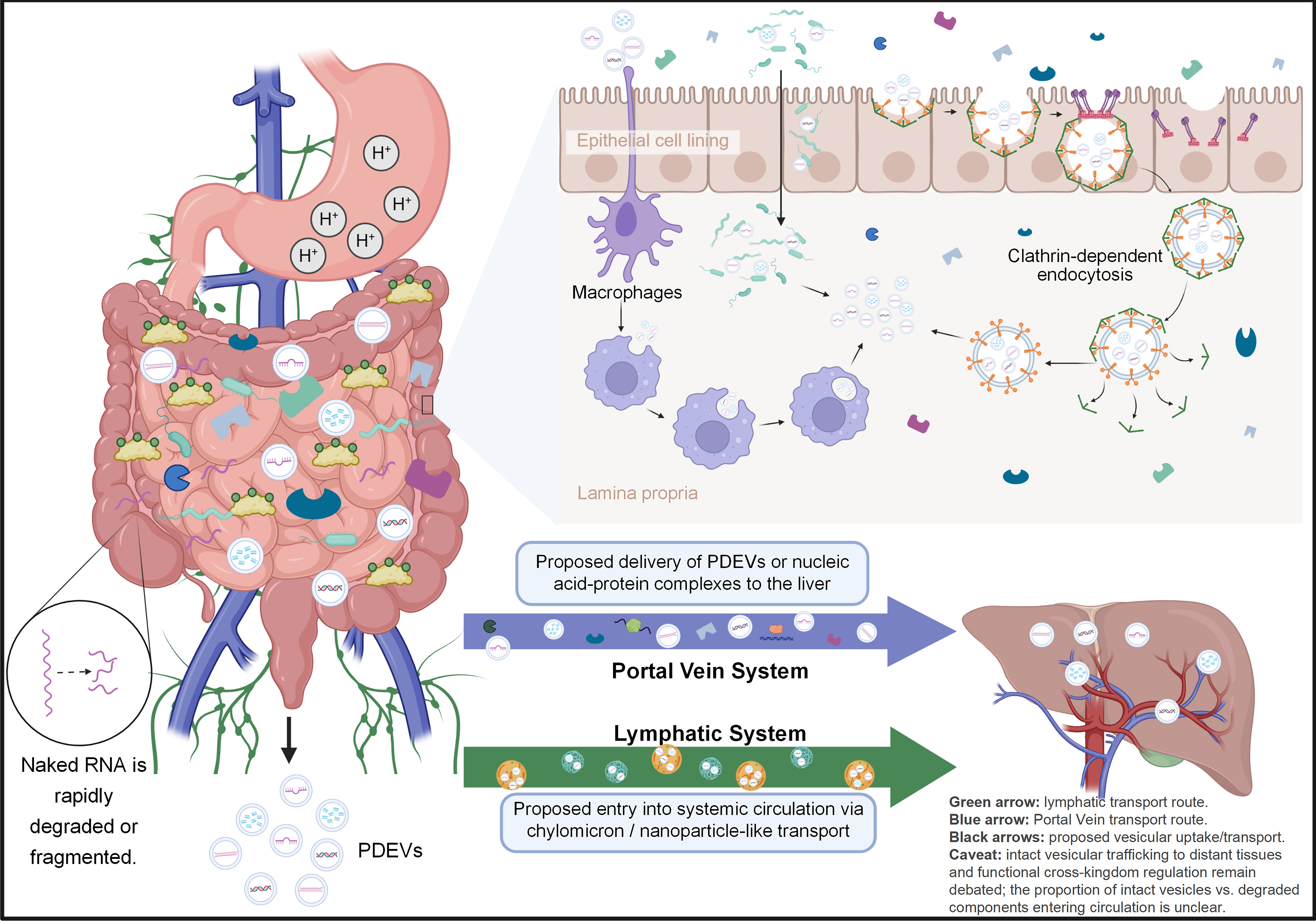
Figure 3. PDEV uptake and trafficking after oral administration. This diagram illustrates the proposed uptake and trafficking of PDEVs following oral administration. PDEVs can cross the intestinal epithelium and potentially enter the circulation through portal vein transport to the liver or through lymphatic transport followed by systemic distribution. PDEVs-associated nucleic acids may subsequently reach distant organs and potentially contribute to cross-kingdom regulation. Created in BioRender. Ruting, C. (2026) https://BioRender.com/pnlssbr. PDEVs: Plant-derived extracellular vesicles; RNA: ribonucleic acid.
Orally administered ginger-derived ELNs accumulated in the liver and protected against alcohol-induced liver injury[6]. Blueberry-derived ELNs ameliorated non-alcoholic fatty liver disease in mice and localize to hepatocytes and Kupffer cells, showing that PDEVs can reach both parenchymal and non-parenchymal liver cells[8]. Grapefruit-derived ELNs can deliver therapeutic agents to the liver and other organs, and their biodistribution is influenced by particle size, surface modification, and disease state[29,49].
These findings support a model in which a portion of orally administered PDEVs, or their released payloads, can be transported from the intestine to the liver and potentially to distant tissues. However, the relative contributions of the portal and lymphatic pathways, as well as the proportion of intact vesicles vs. degraded components entering the circulation, remain unclear. Further investigation of PDEV transport mechanisms will require advanced imaging techniques, tracer-based pharmacokinetic analyses, and synchronized sampling of the portal vein, lymph, and systemic circulation.
Cross-kingdom gene regulation: evidence and controversies
Whether dietary plant miRNAs can survive gastrointestinal digestion, enter the systemic circulation, and functionally regulate mammalian gene expression remains one of the most contentious questions in RNA biology. An earlier study reported that rice-derived miR168a was detectable in human serum and repressed LDLRAP1 translation by binding to its mRNA coding sequence[17]. In mice, dietary rice increased plasma LDL cholesterol, a phenotypic change considered consistent with cross-kingdom gene regulation[17]. However, these findings have been challenged. Dickinson et al. conducted a rigorous feeding study and failed to detect dietary plant miRNA uptake above background, attributing the earlier observations to technical artifacts, including cross-hybridization and environmental contamination[18]. These methodological concerns are substantial because trace-level detection of exogenous RNA is highly sensitive to experimental design, including RNA isolation, library preparation, hybridization stringency, sequencing depth, and bioinformatic filtering[18,77]. Reanalysis of public animal small RNA sequencing (sRNA-seq) datasets further suggested that apparent plant miRNA reads could reflect sequencing errors rather than biological uptake[77,78], including mis-mapping, incomplete reference databases, and index-hopping artifacts. Witwer and colleagues argued that dietary plant miRNAs reach extremely low systemic concentrations that are unlikely to be sufficient for functional regulation of mammalian gene expression[19]. These conflicting findings highlight the difficulty of distinguishing trace exogenous RNA from technical background and of defining reproducible criteria for functional cross-kingdom gene regulation.
The controversy has, however, stimulated methodological advances and mechanistic investigation. Plant miR159 has been detected in human serum and reported to target transcription factor 7 (TCF7), suppressing breast cancer cell proliferation; oral administration of synthetic miR159 mimics also significantly inhibited xenograft tumor growth in mice[43]. These findings support the biological plausibility of cross-kingdom RNA regulation, although synthetic miRNA alone does not establish dietary PDEVs-mediated delivery. Evidence from EV studies provides an additional mechanistic framework. Edible plant-derived exosome-like nanovesicles (EPDENs) from grape, grapefruit, ginger, and carrot are taken up by intestinal macrophages and have been reported to deliver miRNAs associated with anti-inflammatory and antioxidant responses[44]. Ginger-derived exosome-like nanoparticles (GELNs) are also taken up by Lactobacillus rhamnosus in a lipid-dependent manner, and GELN-associated miRNAs have been reported to target bacterial transcripts, suggesting a potential role for vesicle-mediated RNA transfer in host-microbiome interactions[61]. At the plant-microbe interface, Arabidopsis secretes EV-encapsulated small RNAs that silence virulence genes in the fungal pathogen Botrytis cinerea[35], consistent with earlier demonstrations of plant mRNA transfer to fungal pathogens and bidirectional sRNA exchange[52-54].
Several studies have further investigated the functional significance of dietary PDEVs-associated nucleic acids. A clinical pilot study reported accelerated viral clearance in patients consuming honeysuckle decoction, with miR2911 proposed as a candidate mediator; however, direct evidence that PDEVs-associated miR2911 reaches target tissues and mediates the observed effect remains to be established[55]. Tea EV-derived miRNAs have been reported to alter macrophage polarization and alleviate intestinal inflammation[45]. Ginseng exosomal miRNAs have been associated with amelioration of rheumatoid arthritis through the KRAS-MAPK axis[57]. Polygonatum sibiricum nanoparticle-enriched miR166u has been reported to improve intestinal barrier function through TLR4/AKT signaling[46]. Vesicle-mediated, microbiota-dependent protection against colitis has similarly been reported for Centella asiatica-derived exosomes carrying aof-miR396b and fve-miR396c-3p[79]. However, these findings encompass different experimental systems and levels of evidence, and not all demonstrate that native PDEVs-associated nucleic acids are the direct mediators of the observed biological effects. Important uncertainties therefore remain regarding the extent to which dietary miRNAs reach the body at biologically meaningful concentrations[19], the mechanism by which intact EV-associated cargo crosses the intestinal epithelium[44] and the potential for environmental contamination to confound trace-level detection, despite the use of periodate oxidation and standardized bioinformatic pipelines[18,77,80]. Thus, although vesicle-mediated delivery may facilitate cross-kingdom RNA transfer, the physiological relevance of dietary plant miRNAs in mammalian gene regulation remains actively debated[16,20,21].
Critics have emphasized that the absolute quantities of dietary plant miRNAs reaching the systemic circulation may be extremely low, potentially below the concentrations required for functional gene regulation in mammalian cells[18,19]. The field also lacks consensus regarding whether reported biological effects result from direct sequence-specific targeting or from non-specific responses, such as Toll-like receptor (TLR) activation by exogenous RNAs. Even when plant miRNAs are detected in circulation, functional repression in mammalian tissues remains uncertain. The number of plant miRNA molecules per vesicle may be insufficient for substantial RNA-induced silencing complex (RISC) loading, while targetsite accessibility and seedregion complementarity may further constrain repression efficacy. Quantitative data on tissue-specific uptake of dietary vesicles also remain limited[18,19,77,81]. In addition, studies administering high doses of plant-derived vesicles or synthetic miRNA mimics may not reflect exposure associated with normal dietary consumption. Resolving these issues will require standardized dose-response studies, rigorous contamination controls, and independent replication across diverse experimental models. Within this evidence framework, three distinct claims can be separated: (i) PDEV preparations exert biological effects at mucosal surfaces, particularly in the intestine; (ii) some of these effects may involve vesicular nucleic acids; and (iii) PDEV-associated nucleic acids mediate systemic cross-kingdom gene regulation in distant mammalian tissues. The first two claims are supported by a growing body of work, whereas the third remains controversial and has not reached a consistent conclusion.
FUNCTIONAL RELEVANCE ALONG GUT-LIVER AXIS
Immunomodulatory and anti-inflammatory effects
A growing body of evidence indicates that PDEVs exert immunomodulatory and anti-inflammatory effects in the intestine, although the contribution of vesicle-encapsulated small RNAs remains incompletely defined. In multiple murine models, edible ginger-derived ELNs alleviated acute and chronic colitis, promoted intestinal repair, and prevented colitis-associated tumorigenesis[23,73]. Native plant vesicles have also been investigated for anti-tumor immunotherapy. In an MC38 colorectal cancer model, oral administration of Scutellaria baicalensis-derived vesicles downregulated programmed death-ligand 1 (PD-L1) and increased intratumoural CD3+/CD8+ T cells; combined treatment with an anti-programmed cell death protein 1 (PD-1) antibody enhanced tumor suppression while reducing antibody-associated colonic toxicity[82]. Similarly, celery-derived PDEVs were reported to suppress PD-L1 expression and improve the chemotherapeutic efficacy of lung-cancer treatment[83], suggesting that PDEVs-associated immune-checkpoint modulation is not limited to colorectal cancer. Astragalus-derived PDEVs have been reported to repolarize tumor-associated macrophages toward a tumor-suppressive phenotype through Toll-like receptor 2 (TLR2) engagement, indicating that PDEVs can influence the tumor immune microenvironment[84]. These beneficial outcomes are associated with reduced production of pro-inflammatory cytokines, including tumor necrosis factor-alpha (TNF-α), interleukin-6 (IL-6), and interleukin-1 beta (IL-1β), in intestinal macrophages and epithelial cells, increased expression of anti-inflammatory cytokines, including interleukin-10 (IL-10) and interleukin-22 (IL-22)[28] and suppression of NLRP3 inflammasome activation[23,73]. Ginger-derived ELNs contain diverse miRNAs, several of which are predicted to target mammalian inflammatory signaling pathways. Recent studies have begun to experimentally validate specific miRNA-target interactions in intestinal immune cells[23,62] [Figure 4]. As a representative example, tea EV-derived small RNAs were examined in a colitis model: osa-miR166d-5p and gma-miR396a-3p were experimentally validated by dual-luciferase reporter assays to directly target the 3’ untranslated regions (3’UTRs) of AKT1 and IKBKB, respectively, attenuating AKT/nuclear factor kappa B (NF-κB) signaling and reprogramming intestinal macrophages toward an anti-inflammatory phenotype[45]. In another study, exosomes isolated from Centella asiatica selectively accumulated in inflamed colonic tissue after oral administration and contained miRNA cargo enriched in aof-miR396b and fve-miR396c-3p, which were investigated as regulators of Peak1 in intestinal immune cells[79]. Single-cell transcriptomic profiling, combined with fecal microbiota transfer into pseudo-germ-free recipients, indicated that both immune remodelling and restoration of the microbial community contributed to the therapeutic effect[79]. Further, RNA-removal experiments in which RNase treatment reduced the vesicle efficacy provide supportive, though not definitive, evidence that the observed anti-inflammatory effects involve vesicle-associated RNA[45].
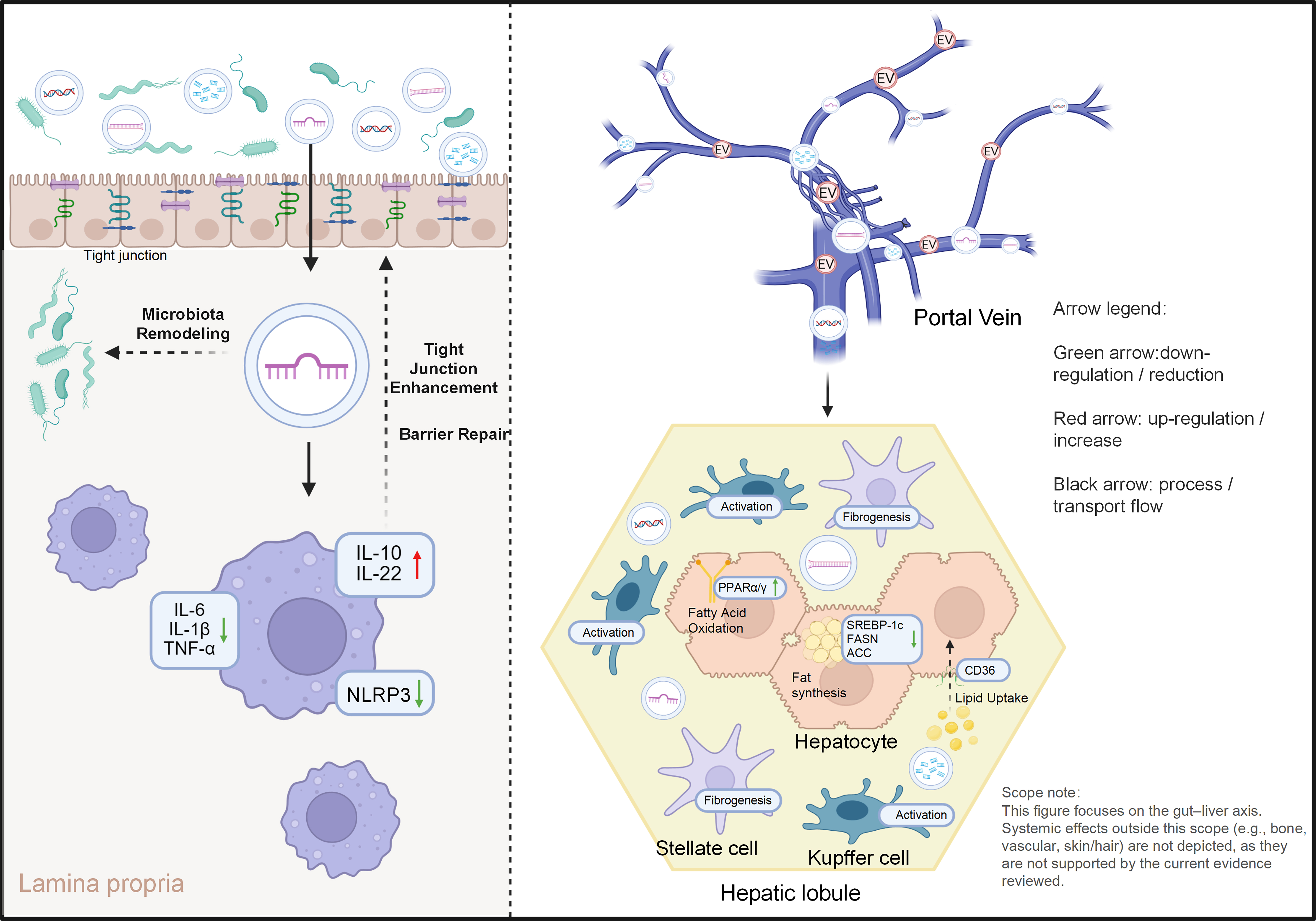
Figure 4. Functional roles of PDEVs-delivered nucleic acids along the gut-liver axis. The schematic summarizes the proposed roles of PDEV-carried nucleic acids in intestinal and hepatic pathways associated with inflammatory and metabolic liver diseases. In the intestine, PDEVs-derived miRNAs and other RNAs may act on epithelial cells and immune cells, reducing pro-inflammatory cytokine production and supporting barrier function. PDEVs may also modulate the gut microbiota and reduce endotoxin exposure. In the liver, PDEVs-associated nucleic acids may influence Kupffer cell activity, hepatic lipid metabolism, and fibrotic signaling in stellate cells. These effects may contribute to protection against IBD, MASLD, and liver fibrosis. Created in BioRender. Ruting, C. (2026) https://BioRender.com/9iamk5c. ACC: Acetyl-CoA carboxylase; CD36: cluster of differentiation 36; EV: extracellular vesicle; FASN: fatty acid synthase; IBD: inflammatory bowel disease; IL-1β: interleukin-1 beta; IL-6: interleukin-6; IL-10: interleukin-10; IL-22: interleukin-22; MASLD: metabolic dysfunction-associated steatotic liver disease; NLRP3: NOD-, LRR- and pyrin domain-containing protein 3; PDEVs: plant-derived extracellular vesicles; PPARα/γ: peroxisome proliferator-activated receptor alpha/gamma; SREBP-1c: sterol regulatory element-binding protein 1c; TNF-α: tumor necrosis factor alpha.
Mechanistic evidence has also been reported for Coptis chinensis-derived vesicle-like nanoparticles containing miR-5106. This miRNA was experimentally validated to target Slc39a2, which encodes the zinc transporter ZIP2, in intestinal tissues. Restoration of zinc homeostasis was associated with reduced neutrophil extracellular trap formation and alleviation of experimental colitis[47]. Such studies provide a relatively detailed mechanistic chain from a specific small RNA to a validated target and downstream phenotype. However, they represent a minority of the available literature. In most studies, the contribution of PDEVs-associated small RNAs remains based on cargo profiling, target prediction, or correlations with biological outcomes rather than direct causal testing[28,58].
ELNs derived from Portulaca oleracea also ameliorate dextran sulfate sodium (DSS)-induced colitis, restore tight junction integrity, and modulate the composition of the gut microbiota. Small RNAs within these vesicles are considered potential effector molecules[28]. Nanovesicles isolated from Rosa roxburghii Tratt (RNVs) similarly attenuated DSS-induced colitis in mice, restoring tight-junction-associated barrier readouts and increasing short-chain fatty acid output, while rebalancing CD25+Foxp3+ regulatory T cells against IL-17+CD4+ T cells in both splenic and peripheral compartments[85]. These findings broaden the immunological scope of PDEVs activity beyond the macrophage- and inflammasome-centered mechanisms described above. As in most studies summarized in this section, however, the responsible molecular species were not resolved, and the respective contributions of vesicular RNAs, lipids, and small molecules remain undetermined. Similarly, ginseng- and mushroom-derived vesicles demonstrated protective effects in models of colitis and acute liver injury, reducing inflammatory cell infiltration and improving tissue organization[58,86]. Honeysuckle-derived nanovesicles have also been reported to ameliorate DSS-induced colitis through remodeling of gut microbiota composition[87]. Oral garlic-derived nanoparticles further indicate that plant vesicles can interact with intestinal innate lymphocytes and induce IFNγ-producing γδ T cells, thus improving anti-tumor immunity[88]. These findings demonstrate reproducible biological activity of PDEV preparations at mucosal surfaces, with some evidence implicating vesicular small RNAs; causal validation of specific nucleic acids remains limited.
Given the anatomical and functional relationship between the intestine and liver, these immunomodulatory effects may have consequences along the gut-liver axis[11,24,89]. Two levels of evidence should be distinguished. Gut-liver axis activity refers here to studies reporting coordinated changes in intestinal barrier function, microbiota or microbial metabolites, and hepatic outcomes within the same experiment. Direct hepatoprotective effects refer primarily to hepatic outcomes and do not by themselves establish an axis-mediated mechanism. For studies meeting the former definition, PDEVs-induced enhancement of intestinal barrier function and modulation of microbiota-derived signaling molecules have been proposed to reduce hepatic exposure to endotoxins and pro-inflammatory metabolites; however, coordinated gut-liver measurements have been reported in only a limited number of studies[59]. PDEVs-derived small RNAs may also interact with hepatic immune cells, including Kupffer cells and infiltrating monocytes, but direct experimental evidence remains limited[24,25] Table 2.
Animal/cell studies of PDEVs in gut disease models
| Plant source | Formulation type | Model | Main observation index | Nucleic acid dependence | Ref. |
| Grape | ELNs | DSS-induced colitis | Lgr5+ intestinal stem cells - tissue repair and barrier restoration | Not tested | [9] |
| Portulaca oleracea L. | ELNs | DSS-induced colitis | Intestinal barrier (ZO-1, occludin) -barrier repair and immune modulation | Not tested | [28] |
| Grapefruit | ELNs | DSS-induced colitis | Macrophage polarization (M1/M2) -anti-inflammatory reprogramming | Not tested | [29] |
| Plant-derived nanovesicles | ELNs | Colorectal cancer | PD-L1/CD8+ T cell -tumor growth inhibition, anti-PD-L1 response | Yes | [71] |
| Ginger | GELNs | Radiation-induced intestinal damage | Intestinal barrier and microbiota -injury repair and microbial restoration | N/A | [51] |
| Grape/Grapefruit/Ginger/Carrot | EPDENs | Intestinal inflammation | Inflammatory cytokines and miRNA target genes -intestinal inflammation | Not tested | [44] |
| Ginger | GELNs | DSS-induced colitis | Gut microbiota -bacteria-mediated anti-inflammatory effect | Yes | [61] |
| Tea | EVs | DSS-induced colitis | Reprogramming macrophages -alleviated intestinal inflammation | Yes | [45] |
| Polygonatum sibiricum | ELNs | DSS-induced colitis | Intestinal barrier (ZO-1, occludin, claudin) -barrier protection | Partial | [46] |
| Ginseng | ELNs | DSS-induced colitis | TNF-α, IL-6, IL-1β and macrophage infiltration -colonic inflammation | Not tested | [58] |
| Coptis chinensis Franch | ELN-like nanoparticles | Colitis | NETs formation and zinc homeostasis -colonic inflammation | Partial | [47] |
| Momordica charantia L. | EVs | Ulcerative Colitis | Intestinal barrier and microbiota -inflammation and metabolic balance | Not tested | [90] |
| Ginger | ELNs | DSS-induced colitis | Intestinal inflammation and immune effectors -enteritis protection | Not tested | [23,62,73] |
Metabolic regulation and protection against MASLD/MASH
Metabolic dysfunction-associated steatotic liver disease (MASLD) and MASH represent prototypical gut-liver axis disorders, with their pathogenesis closely linked to dietary factors, intestinal barrier dysfunction, microbial dysbiosis, and abnormalities in hepatic lipid metabolism[91-96]. Following oral intake, food-derived PDEVs, particularly those from plants, are taken up by intestinal epithelial and immune cells. They also interact with the gut microbiota , modulating microbial composition and metabolite production[8,24]. These interactions have been associated with improved intestinal barrier integrity. Increased expression of tight junction proteins, together with an enhanced mucus layer, has been observed, consequently reducing gut-derived endotoxin entering the portal circulation[24,26]. These barrier-related readouts were obtained largely from intestinal models or following oral administration; only a minority of studies have demonstrated coordinated hepatic changes within the same experimental system.
PDEVs modulate hepatic lipid metabolism in experimental models; small RNA target predictions implicate vesicular nucleic acids as potential mediators, but their causal contribution remains to be validated. At the cellular level, dietary micronutrients and hormones, including selenium and 17β-oestradiol, directly influence lipid accumulation in hepatocyte models of MASLD[97]. Small RNAs enriched in blueberry, citrus, and grape vesicles are predicted to target mammalian regulators of lipid uptake [e.g., Cluster of Differentiation 36 (CD36)], lipogenesis [sterol regulatory element-binding protein-1c (SREBP-1c), fatty acid synthase (FASN), acetyl-CoA carboxylase (ACC)], and nuclear receptors [peroxisome proliferator-activated receptor alpha/gamma (PPARα/γ)][8,98]. To date, none of these predicted lipid-related interactions has been experimentally validated in the context of vesicle delivery. The closest evidence comes from in vitro studies using synthetic mimics: transfection of plant miR8126-3p and miR8126-5p into a human hepatocyte model of steatosis downregulated the predicted targets QKI and MAPKAPK2 and decreased expression of lipogenic genes including FASN, reducing lipid accumulation[48]. Complementary in vivo evidence comes from Opuntia Milpa Alta-derived vesicles, which homed to inflamed colonic tissue after oral administration and reprogrammed host fatty acid metabolism, with coordinated changes in Acsl1, Acaca, and Fabp4a expression alongside restoration of short-chain fatty acid-producing taxa[99]. The absorbed mediators in that study were identified as phenolic constituents rather than vesicular nucleic acids, and the metabolic changes were documented in the colon rather than the liver. This example illustrates a recurring interpretive boundary: PDEV preparations can demonstrably reprogram host lipid metabolism in vivo, but such phenotypes should not be attributed to vesicular RNA in the absence of direct loss-of-function evidence. The study also used synthetic miRNA mimics rather than vesicle-delivered RNA, and the translational relevance to dietary PDEV uptake remains uncertain. Blueberry-derived ELNs alleviated diet-induced fatty liver in mice by reducing mitochondrial oxidative stress, improving hepatic lipid metabolism, and suppressing pro-inflammatory cytokine production[8]. Similarly, ginger-derived ELNs protected against alcohol-induced liver injury, reduced hepatic steatosis, and modulated gene expression related to lipid oxidation and antioxidant defense. Extending this pattern to a MASH model, terpenoid-enriched Curcuma wenyujin-derived nanovesicles attenuated high-fat-diet-induced hepatic steatosis, serum Alanine aminotransferase (ALT) and Aspartate aminotransferase (AST), and circulating triglycerides and total cholesterol, while suppressing Kupffer-cell/macrophage IL-6, IL-1β, and TNF-α, downregulating lipogenic FASN, and increasing Peroxisome proliferator-activated receptor gamma coactivator 1-alpha (PGC1α)/Microsomal triglyceride transfer protein (MTTP) expression[100]. These hepatic phenotypes were demonstrated using whole PDEV preparations, and none of the liver disease studies compiled in Table 3 met the criteria for unequivocal nucleic acid dependence, such as RNase treatment, specific miRNA depletion/knockdown, anti-miR inhibition, or rescue experiments. The contribution of nucleic acids to hepatic lipid metabolism therefore remains an open question, and miRNA mimics, antagomirs, and loss-of-function or rescue experiments in hepatocyte and MASLD/MASH models are needed to establish causality.
Animal/cell studies of PDEVs in liver disease models
| Plant source | Formulation type | Model | Main observation index | Nucleic acid dependence | Ref. |
| Ginger | GELNs | ALD | NF-κB (p65) - reduced hepatic inflammation and oxidative stress | No | [6] |
| Blueberry | ELNs | MASLD | Lipid metabolism and oxidative stress | Not tested | [8] |
| Shiitake mushroom | ELNs | ALI | Hepatic inflammatory response | Not tested | [86] |
| Honeysuckle | ELNs | ALF | TLR4/NF-κB -ALF | Not tested | [56] |
| Lycium barbarum L. (goji) | ELNs | ALI | Gut microbiota and metabolites -ameliorated alcohol-induced liver injury | Not tested | [101] |
| Morus nigra L. | LNPs from leaves | HCC | Tumor cell signaling -anti-tumor effect in HCC | Not tested | [102] |
Evidence involving both sides of the gut-liver axis within a single experiment remains limited. The available studies provide the most direct evidence for PDEVs’ activity across the axis. Pomegranate-derived ELNs reduced binge alcohol-induced leaky gut and concurrent liver injury through the TLR4/NF-κB pathway[103]. Lycium barbarum ELNs improved acute liver injury while also reshaping the intestinal flora, supporting a gut-liver axis mechanism[101]. Honeysuckle-derived ELNs have also been reported to ameliorate metabolic-associated fatty liver disease by modulating gut microbiota and their metabolites[104]. In these studies, changes in intestinal barrier function or microbiota were accompanied by hepatic outcomes within the same experimental system. The studies compiled in Table 3 were assessed primarily according to hepatic outcomes. Throughout this review, we therefore refer to these findings as direct hepatoprotective effects rather than gut-liver axis activity, and their nucleic acid dependence remains to be determined.
Antifibrotic and tissue-regenerative implications
Beyond their effects on steatosis and inflammation, PDEVs also exhibit antifibrotic properties. Studies have reported that vesicles isolated from ginger, grapes, and medicinal plants attenuate liver fibrosis in experimental models, reduce collagen deposition, and downregulate key fibrotic markers such as alpha-smooth muscle actin (α-SMA) and transforming growth factor-β (TGF-β)[103,105,106]. These vesicles can also reduce oxidative stress and inflammatory responses, both of which are important drivers of fibrosis progression[6,107]. The antioxidant effects of PDEVs may also involve regulation of ferroptosis. American ginseng-derived ELNs mitigate oxidative injury, at least in part, by inhibiting glutathione peroxidase 4 (GPX4)-mediated ferroptosis, extending the reported cytoprotective effects of plant vesicles beyond the liver[108]. Mushroom-derived vesicles have been reported to protect against acute liver injury by enhancing hepatocyte survival and suppressing inflammatory pathways[86,109]. PDEV preparations also show regenerative potential in extrahepatic tissues[7,11,110,111]. Beyond internal organs, PDEVs are being investigated for skin applications such as facial aesthetics, where they have been reviewed for anti-scarring, anti-aging, and anti-pigmentation activities[112]. These applications lie outside the gut-liver axis that is central to this review but illustrate the broader translational interest in PDEVs. Inula japonica Thunb.-derived ELNs provide another example of this extrahepatic potential: oral administration attenuated lipopolysaccharide (LPS)-induced acute lung injury, while 16S rRNA profiling and untargeted metabolomics showed restoration of gut microbial homeostasis, including Lachnospiraceae_NK4A136_group, Roseburia and Lactobacillus, together with coordinated changes in eicosanoid and butyrate metabolism and activation of the Aryl hydrocarbon receptor (AhR)-Cytochrome P450 family 1 subfamily A member 1 (CYP1A1) axis[113]. These findings suggest that PDEVs-mediated effects may extend beyond the gut-liver axis, although whether nucleic acids contribute to these extrahepatic effects remains to be determined.
ENGINEERING STRATEGIES AND APPLICATIONS
Engineering strategies for PDEVs-mediated nucleic acid delivery
The studies discussed in this section concern engineered PDEVs-based delivery systems. Evidence that engineered PDEVs can serve as vehicles for exogenous cargo does not constitute evidence that naturally occurring nucleic acids in dietary PDEVs mediate cross-kingdom regulation of host genes; these two concepts are based on different experimental foundations and are therefore treated separately here.
Loading exogenous nucleic acids into PDEVs relies on two main strategies. Post-isolation methods include electroporation, sonication, extrusion, and transfection techniques[59,60,114]. Early studies demonstrated that nanocarriers assembled from grapefruit-derived lipids could encapsulate siRNA, DNA expression vectors, and proteins, achieving functional delivery both in vitro and in vivo. These lipid-based nanoparticles are engineered formulations derived from plant components rather than native PDEV preparations[49]. Acerola-derived ELNs, isolated from fruit juice as native plant EVs, encapsulated exogenous miRNA by simple incubation and protected it against RNase, acid, and base treatments; oral administration delivered small RNA to both the small intestine and liver, achieving target gene suppression in vivo within 24 h[115]. These findings provide direct evidence that native plant-derived vesicles can serve as oral carriers for nucleic acid delivery to the gut-liver axis. Drug-loaded nanocarriers have enhanced therapeutic efficacy while reducing toxicity in colitis models; however, plant-derived EV formulations such as Momordica charantia EVs[90] and Coptis chinensis EV-like nanoparticles[47] carry endogenous cargo rather than exogenous drug payloads, and their therapeutic effects were attributed to these native components. Similar benefits were reported for non-plant EV platforms, including bacterial[116] and milk-derived EVs[117], support the general concept of EV-mediated drug delivery but were not obtained with plant-derived systems. The plant biofactory strategy offers an alternative approach in which therapeutic miRNA or siRNA expressed in plant tissues is naturally incorporated into vesicles during biogenesis; to date, however, this concept rests on feasibility studies of plant-derived production of RNA-loaded EVs, and its validation in PDEVs remains incomplete.
PDEVs confer advantages for targeting the intestine and liver. Oral administration delivers high local concentrations to the intestine, while vesicles or their cargo can reach the liver via the portal vein[24,29,102]. To enhance targeting, surface ligands can be introduced to promote preferential binding to receptors on epithelial or immune cells; this has been demonstrated with grapefruit-derived nanovesicles[29] and ginger-derived ELNs loaded with curcumin[51]. Ligand-mediated targeting validated with synthetic nanoparticles or liposomes, such as anionic nanoparticles that enhance intestinal permeability[118] and RGD-modified liposomal formulations for oral delivery[119], supports the general feasibility of this strategy, although these platforms are not plant-derived. Hybrid nanoparticles coated with ginger-derived ELNs exemplify this approach[6]. In MASLD/MASH, PDEV formulations carrying miRNA mimics targeting SREBP-1c or CD36 can be combined with anti-inflammatory miRNAs[5,98]. For colitis-associated cancers, PDEVs carrying tumor-suppressor miRNAs target both intestinal epithelial cells and hepatic metastatic niches[120-122].
PDEVs offer several potential advantages over mammalian EVs. First, PDEVs can be produced at scale from agricultural by-products[123-125]. A temporary immersion bioreactor (TIB) system has recently been developed for controlled, large-scale production of PDEVs from food and medicine homology plants[126]. Second, derived from edible plant sources, PDEVs exhibit favorable safety and immunogenicity profiles[5,77]. Third, PDEVs are inherently suited for oral administration and gut-liver targeting[24,75]. However, PDEVs face significant limitations, including batch-to-batch variability influenced by plant species, growth conditions, tissue origin, and extraction methods[11,64,127,128]. The absence of universally accepted markers complicates quality control. Moreover, the loading efficiency of exogenous nucleic acids is unstable and difficult to quantify precisely. The regulation of therapeutic drugs based on plant vesicles is still in its infancy. Establishing robust methodologies, analytical techniques, and regulatory systems is essential for clinical translation.
Applications and detection of plant-derived nucleic acids as potential circulating biomarkers
Detecting plant-derived nucleic acids in blood requires sensitive, highly specific analytical methods. Conventional quantitative polymerase chain reaction (qPCR) and digital polymerase chain reaction (dPCR) have been used to quantify specific plant miRNAs in plasma or serum[19,80]. High-throughput small RNA sequencing provides a broader profile of circulating RNAs but requires stringent bioinformatic filtering[66]. Pre-analytical variables substantially affect the results[24,75]. Enrichment of EV-associated nucleic acids may improve detection sensitivity but can also introduce selection bias. Nuclease protection assays can provide supportive evidence for vesicle association but do not, by themselves, establish intravesicular RNA localization, because extravesicular RNA may be protected from nuclease degradation by RNA-binding proteins. Conclusions regarding intravesicular cargo are strengthened when nuclease treatment is combined with membrane-disruption controls or direct vesicle-imaging approaches[4,5]. Emerging technologies may further improve the detection and characterization of low-abundance plant nucleic acids. However, robust standard operating procedures (SOPs) and inter-laboratory ring trials are needed to ensure reproducible, biologically relevant detection results.
Circulating plant-derived nucleic acids have two potential applications. As dietary biomarkers, unique plant miRNA or small RNA sequence signatures may indicate recent food intake[129]. As therapeutic indicators, detecting vesicle-associated nucleic acids in plasma may provide pharmacokinetic evidence of PDEV absorption. However, detection of plant nucleic acids in plasma does not, by itself, demonstrate uptake of intact PDEVs, because plant-derived small RNAs may also circulate in non-vesicular forms, such as free RNA or RNA associated with RNA-binding proteins or lipoproteins[19]. When combined with measurements of host-derived extracellular vesicles and cell-free DNA, these analyses may help clarify the relative contributions of different sources to changes in total circulating nucleic acids.
Circulating plant-derived nucleic acids may also serve as pharmacodynamic indicators. Temporal changes in specific plant miRNA levels in plasma have been suggested to correlate with changes in host tissue gene expression[129]. In MASLD models, small RNAs enriched in blueberry, citrus, and grape vesicles have been predicted to target mammalian lipid regulators such as PPARγ and SREBP-1c. However, no study to date has simultaneously detected circulating PDEV-derived miRNAs and evaluated their relationship with lipid profiles in vivo. The closest evidence comes from in vitro studies using synthetic miRNA mimics: transfection of plant miR8126-3p and miR8126-5p into a human hepatocyte model of steatosis downregulated lipogenic genes and reduced lipid accumulation[48]. These experiments, however, used synthetic mimics rather than vesicle-delivered RNA. Thus, the potential biomarker value of simultaneously profiling circulating PDEVs-derived miRNAs and lipid profiles in MASLD models remains to be demonstrated. Integrating these measurements with multi-omics analyses could facilitate system-level modeling of PDEVs’ effects[24,25], and, if validated in prospective cohorts, may help identify responder subpopulations and inform dose optimization. Ultimately, well-designed clinical studies are required to determine whether circulating plant-derived nucleic acids can serve as reliable biomarkers in clinical practice.
Challenges and future directions
Methodological heterogeneity remains a major obstacle to nucleic acid research on PDEVs. Differences in plant cultivation, tissue processing, vesicle isolation, characterization, and nucleic acid profiling can influence reported outcomes. Unified terminology and standardized experimental protocols are therefore needed[38,41,130]. PDEVs-related studies should explicitly report plant species, growth conditions, extraction methods, and vesicle characterization procedures. Establishing minimum information standards for PDEVs nucleic acid research would substantially improve the reproducibility and comparability of findings. Multicenter studies based on shared plant materials and harmonized procedures could further generate benchmark datasets.
Standardized reporting should also account for plant-specific contaminants that can compromise nucleic acid analysis. Polyphenol and polysaccharide contents vary substantially among plant species, tissues, and developmental stages, necessitating tailored extraction protocols rather than one-size-fits-all approaches. The effects of these contaminants on downstream nucleic acid analyses, including inhibition of reverse transcription, reduced sequencing depth, and false-positive alignment artifacts, should be systematically quantified and reported. Developing reference materials and spike-in controls derived from plant matrices with defined polyphenol and polysaccharide profiles could facilitate inter-laboratory benchmarking and quality control. Such advances are essential to move PDEVs nucleic acid research beyond descriptive observations toward rigorous, reproducible, and clinically translatable findings.
Controversies surrounding dietary plant miRNAs and cross-kingdom regulation have highlighted the need for more definitive experimental approaches. Rigorous studies incorporating gene “barcode” labeling of plant miRNAs are needed to establish their origin and track their fate[35-37]. When combined with appropriate membrane-disruption controls, nuclease protection assays and advanced single-vesicle imaging techniques can help distinguish vesicle-encapsulated nucleic acids from contaminants. In the absence of such controls, nuclease resistance alone should be interpreted cautiously. Functional validation is also essential, particularly through experiments showing loss of a biological effect following removal of a specific plant RNA and restoration after its reintroduction. Multi-center replication and pre-registration of key experiments would further strengthen the evidence base. PDEVs therefore provide a useful experimental system for investigating the potential mechanisms of cross-kingdom nucleic acid biology.
PDEVs may also contribute to the development of precision nutrition and gut-liver axis therapies. Distinct molecular profiles among plant vesicles could be matched to specific individual phenotypes. Through personalized dietary interventions, specific nucleic acid payloads might eventually be delivered to modulate intestinal and hepatic signaling pathways[56,131]. Engineered PDEVs have been proposed as potential oral RNA therapeutics, either alone or in combination with other treatments, for MASLD/MASH, IBD, liver fibrosis, and hepatocellular carcinoma[9,103]. Successful translation of nucleic acid-based therapies will also require alignment with regulatory requirements. PDEVs products may be classified as foods, supplements, biological products, or advanced therapeutic medicinal products[10,132]. Key challenges include defining quality attributes and establishing appropriate manufacturing practice processes. Preclinical studies, including oral administration in colitis models[29] and systemic administration in tumor models[49], have reported generally favorable safety and tolerability profiles for PDEV preparations; however, clinical experience with orally administered PDEV-based therapeutics remains limited. Collaborative efforts among EV researchers, nutrition scientists, clinicians, and regulatory agencies will be important for the responsible and scientifically rigorous development of plant vesicles as natural nucleic acid delivery vehicles.
CONCLUSION
PDEVs represent natural nanocarriers that connect dietary intake with extracellular vesicle biology and the potential for cross-kingdom nucleic acid communication, with the gut-liver axis representing an important context for their study. Their intrinsic properties, including edibility, low immunogenicity, scalability, and endogenous nucleic acid cargo, such as miRNAs and siRNAs, are associated with biological effects involving gut barrier integrity, hepatic metabolism, and inflammatory responses, highlighting their potential for the management of MASLD, IBD, and liver fibrosis.
This review highlights that PDEVs can exert biological effects along the gut-liver axis, and that their passage from the gastrointestinal tract to the liver through portal and lymphatic routes provides a plausible basis for systemic activity. At the same time, current evidence is largely centered on the biological activity of whole PDEV preparations and on correlative or predictive associations involving their nucleic acid cargo. Definitive causal evidence, including loss-of-function or rescue experiments demonstrating that specific plant nucleic acids are required for the observed effects, remains limited. Uncertainty also persists regarding the intravesicular localization of these nucleic acids and the methodological robustness of their detection. PDEVs-associated nucleic acids should therefore be considered promising candidate mediators, but their causal contribution to the observed biological effects remains incompletely established. The activity of whole PDEV preparations may also reflect contributions from proteins, lipids, and other vesicle components. Other limitations include methodological heterogeneity in PDEVs isolation and nucleic acid profiling, which compromises reproducibility, ongoing controversies regarding the functional relevance of cross-kingdom nucleic acids, and batch-to-batch variability in PDEVs preparations that may hinder translational development.
Future research should prioritize standardized approaches for PDEVs characterization, definitive experimental validation of nucleic acid-mediated effects along the gut-liver axis, and engineering strategies for targeted PDEVs delivery. Addressing these challenges could help clarify the role of PDEVs at the interface of nutritional science and precision medicine and support their potential development as next-generation nutraceuticals and nucleic acid-based therapeutics for metabolic and inflammatory disorders.
DECLARATIONS
Acknowledgments
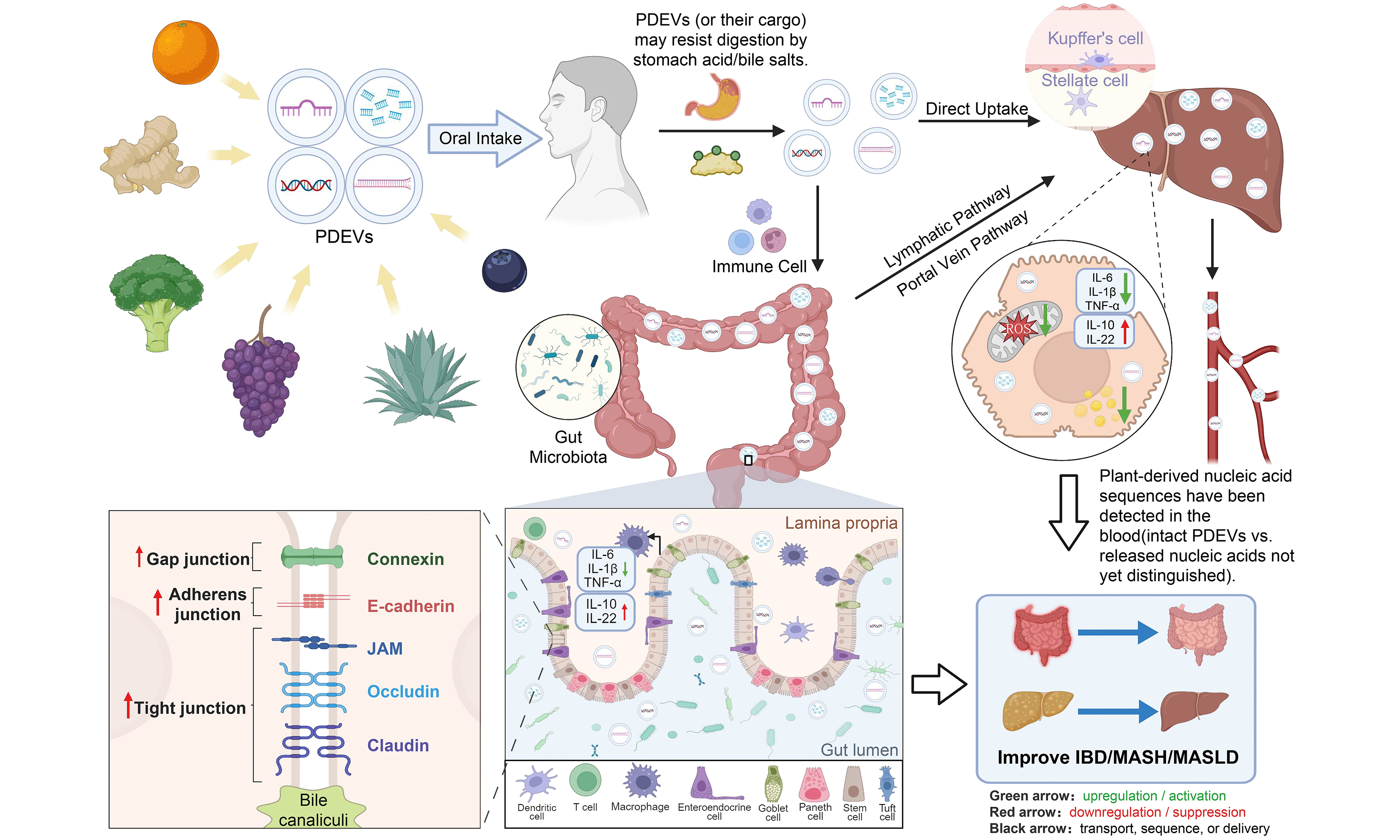
The Graphical Abstract was created in BioRender. Ruting, C. (2026) https://BioRender.com/vd5ibir.
Authors’ contributions
Conducted the literature search and data collection: Cheng R
Drafted the manuscript: Cheng R, Liang R
Critically revised the manuscript for important intellectual content: Feng J, Zhong Z, You Z
Availability of data and materials
Not applicable.
AI and AI-assisted tools statement
During the preparation of this manuscript, the AI tool WorkBuddy (version 5.5.6, released 2026-09-10) was used to enhance readability and language expression. The tool did not influence the study design, data collection, analysis, interpretation, or the scientific content of the work. All authors take full responsibility for the accuracy, integrity, and final content of the manuscript.
Financial support and sponsorship
This work was financially supported by the National Natural Science Foundation of China (No. 82204759), the Shenzhen Science and Technology Program (No. JCYJ20240813113239051), and the Macao Science and Technology Development Fund (FDCT 0047/2025/ITP1, 0123/2022/A, and 0008/2023/RIC).
Conflicts of interest
Zhong Z is a Guest Editor of the Special Topic “Plant-Derived Vesicles: From Fundamental Biology to Precision Therapeutics” in the journal Extracellular Vesicles and Circulating Nucleic Acids and serves as an Editorial Board Member. Zhong Z was not involved in any steps of the editorial process for this manuscript, including reviewer selection, manuscript handling, or decision-making. The other authors declare no conflicts of interest.
Ethical approval and consent to participate
Not applicable.
Consent for publication
Not applicable.
Copyright
© The Author(s) 2026.
REFERENCES
1. Welsh JA, Goberdhan DCI, O’Driscoll L, et al. ; MISEV Consortium. Minimal information for studies of extracellular vesicles (MISEV2023): from basic to advanced approaches. J Extracell Vesicles. 2024;13:e12404.
2. Sarasati A, Syahruddin MH, Nuryanti A, et al. Plant-derived exosome-like nanoparticles for biomedical applications and regenerative therapy. Biomedicines. 2023;11:1053.
3. Lo KJ, Wang MH, Ho CT, Pan MH. Plant-derived extracellular vesicles: a new revolutionization of modern healthy diets and biomedical applications. J Agric Food Chem. 2024;72:2853-78.
4. Kameli N, Dragojlovic-Kerkache A, Savelkoul P, Stassen FR. Plant-derived extracellular vesicles: current findings, challenges, and future applications. Membranes. 2021;11:411.
5. Huang D, Chen J, Zhao M, et al. Plant-derived extracellular vesicles: composition, function and clinical potential. J Transl Med. 2025;23:1065.
6. Zhuang X, Deng ZB, Mu J, et al. Ginger-derived nanoparticles protect against alcohol-induced liver damage. J Extracell Vesicles. 2015;4:28713.
7. Di Raimo R, Mizzoni D, Aloi A, et al. Antioxidant effect of a plant-derived extracellular vesicles’ mix on human skin fibroblasts: induction of a reparative process. Antioxidants. 2024;13:1373.
8. Zhao WJ, Bian YP, Wang QH, et al. Blueberry-derived exosomes-like nanoparticles ameliorate nonalcoholic fatty liver disease by attenuating mitochondrial oxidative stress. Acta Pharmacol Sin. 2022;43:645-58.
9. Ju S, Mu J, Dokland T, et al. Grape exosome-like nanoparticles induce intestinal stem cells and protect mice from DSS-induced colitis. Mol Ther. 2013;21:1345-57.
10. Zeng YB, Deng X, Shen LS, et al. Advances in plant-derived extracellular vesicles: isolation, composition, and biological functions. Food Funct. 2024;15:11319-41.
11. Liu H, Dong T, Dong C, et al. Plant-derived exosome-like nanovesicles: a novel therapeutic perspective for skin diseases. J Nanobiotechnology. 2025;23:640.
12. Shi L, Guo C, Fang M, Yang Y, Yin F, Shen Y. Cross-kingdom regulation of plant microRNAs: potential application in crop improvement and human disease therapeutics. Front Plant Sci. 2024;15:1512047.
13. Yu Y, Tan N, Xu Z, et al. Cross-kingdom miRNA delivery by Panax notoginseng-derived extracellular-like nanoparticles vesicles restores neuronal function after ischemic injury. J Nanobiotechnology. 2026;24:242.
14. Li S, Zhang Y, Liu X, et al. Cross-kingdom delivery and putative gene modulation of androgen pathways by plant-derived exosome-like nanoparticles from polygoni multiflori radix promotes hair growth via miRNA cargo. Pharmacol Res. 2025;222:108033.
15. Jia M, He J, Bai W, et al. Cross-kingdom regulation by dietary plant miRNAs: an evidence-based review with recent updates. Food Funct. 2021;12:9549-62.
16. Yang L, Feng H. Cross-kingdom regulation by plant-derived miRNAs in mammalian systems. Animal Model Exp Med. 2023;6:518-25.
17. Zhang L, Hou D, Chen X, et al. Exogenous plant MIR168a specifically targets mammalian LDLRAP1: evidence of cross-kingdom regulation by microRNA. Cell Res. 2012;22:107-26.
18. Dickinson B, Zhang Y, Petrick JS, Heck G, Ivashuta S, Marshall WS. Lack of detectable oral bioavailability of plant microRNAs after feeding in mice. Nat Biotechnol. 2013;31:965-7.
19. Witwer KW, McAlexander MA, Queen SE, Adams RJ. Real-time quantitative PCR and droplet digital PCR for plant miRNAs in mammalian blood provide little evidence for general uptake of dietary miRNAs: limited evidence for general uptake of dietary plant xenomiRs. RNA Biol. 2013;10:1080-6.
20. DeLucas M, Sánchez J, Palou A, Serra F. The impact of diet on miRNA regulation and its implications for health: a systematic review. Nutrients. 2024;16:770.
21. Tambaro F, Gallicchio C, Orlando S, Carnevale S, Muscaritoli M. The conundrum of XenomiRs and human health. Adv Nutr. 2025;16:100510.
22. Nunes A, Zhang T, Mu X, Robbins PD. Therapeutic application of extracellular vesicles in human diseases. Mol Ther. 2025;33:2243-51.
23. Zhang M, Viennois E, Prasad M, et al. Edible ginger-derived nanoparticles: a novel therapeutic approach for the prevention and treatment of inflammatory bowel disease and colitis-associated cancer. Biomaterials. 2016;101:321-40.
24. Zhang S, Wang Q, Tan DEL, et al. Gut-liver axis: potential mechanisms of action of food-derived extracellular vesicles. J Extracell Vesicles. 2024;13:e12466.
25. Wu Q, Kan J, Fu C, et al. Insights into the unique roles of extracellular vesicles for gut health modulation: Mechanisms, challenges, and perspectives. Curr Res Microb Sci. 2024;7:100301.
26. Chang Z, Xu X, Xu Y, et al. Potential effects of functional foods and dietary supplements on metabolic-associated fatty liver disease and the underlying mechanisms: a narrative review with a focus on the modulation of the gut microbiota. J Agric Food Chem. 2025;73:29247-80.
27. Fang X, Feng J, Zhu X, Feng D, Zheng L. Plant-derived vesicle-like nanoparticles: a new tool for inflammatory bowel disease and colitis-associated cancer treatment. Mol Ther. 2024;32:890-909.
28. Zhu MZ, Xu HM, Liang YJ, et al. Edible exosome-like nanoparticles from portulaca oleracea L mitigate DSS-induced colitis via facilitating double-positive CD4+CD8+T cells expansion. J Nanobiotechnology. 2023;21:309.
29. Wang B, Zhuang X, Deng ZB, et al. Targeted drug delivery to intestinal macrophages by bioactive nanovesicles released from grapefruit. Mol Ther. 2014;22:522-34.
30. Cai Q, Halilovic L, Shi T, et al. Extracellular vesicles: cross-organismal RNA trafficking in plants, microbes, and mammalian cells. Extracell Vesicles Circ Nucl Acids. 2023;4:262-82.
31. Rome S. Biological properties of plant-derived extracellular vesicles. Food Funct. 2019;10:529-38.
32. Huang Y, Wang S, Cai Q, Jin H. Effective methods for isolation and purification of extracellular vesicles from plants. J Integr Plant Biol. 2021;63:2020-30.
33. Woith E, Guerriero G, Hausman JF, et al. Plant extracellular vesicles and nanovesicles: focus on secondary metabolites, proteins and lipids with perspectives on their potential and sources. Int J Mol Sci. 2021;22:3719.
34. Jeppesen DK, Zhang Q, Franklin JL, Coffey RJ. Extracellular vesicles and nanoparticles: emerging complexities. Trends Cell Biol. 2023;33:667-81.
35. Cai Q, Qiao L, Wang M, et al. Plants send small RNAs in extracellular vesicles to fungal pathogen to silence virulence genes. Science. 2018;360:1126-9.
36. He B, Cai Q, Qiao L, et al. RNA-binding proteins contribute to small RNA loading in plant extracellular vesicles. Nat Plants. 2021;7:342-52.
37. Baldrich P, Rutter BD, Karimi HZ, Podicheti R, Meyers BC, Innes RW. Plant extracellular vesicles contain diverse small RNA species and are enriched in 10- to 17-nucleotide “Tiny” RNAs. Plant Cell. 2019;31:315-24.
38. Pinedo M, de la Canal L, de Marcos Lousa C. A call for Rigor and standardization in plant extracellular vesicle research. J Extracell Vesicles. 2021;10:e12048.
39. Nueraihemaiti N, Dilimulati D, Baishan A, et al. Advances in plant-derived extracellular vesicle extraction methods and pharmacological effects. Biology. 2025;14:377.
40. Woith E, Melzig MF. Extracellular vesicles from fresh and dried plants-simultaneous purification and visualization using gel electrophoresis. Int J Mol Sci. 2019;20:357.
41. Han X, Zheng W, Sun Z, et al. Plant-derived exosomes: unveiling the similarities and disparities between conventional extract and innovative form. Phytomedicine. 2025;145:157087.
42. Liu J, Xiang J, Jin C, et al. Medicinal plant-derived mtDNA via nanovesicles induces the cGAS-STING pathway to remold tumor-associated macrophages for tumor regression. J Nanobiotechnology. 2023;21:78.
43. Chin AR, Fong MY, Somlo G, et al. Cross-kingdom inhibition of breast cancer growth by plant miR159. Cell Res. 2016;26:217-28.
44. Mu J, Zhuang X, Wang Q, et al. Interspecies communication between plant and mouse gut host cells through edible plant derived exosome-like nanoparticles. Mol Nutr Food Res. 2014;58:1561-73.
45. Luo T, Hou L, Cao Y, et al. Tea extracellular vesicle-derived microRNAs contribute to alleviate intestinal inflammation by reprogramming macrophages. J Agric Food Chem. 2025;73:6745-57.
46. Wei C, Chen Y, Chen J, et al. miR166u -enriched Polygonatum sibiricum exosome-like nanoparticles alleviate colitis by improving intestinal barrier through the TLR4/AKT pathway. Int J Biol Macromol. 2025;318:144802.
47. Yang Y, Yang L, Deng H, et al. Coptis chinensis-derived extracellular vesicle-like nanoparticles delivered miRNA-5106 suppresses NETs by restoring zinc homeostasis to alleviate colitis. J Nanobiotechnology. 2025;23:444.
48. Díez-Sainz E, Aranaz P, Amri EZ, Riezu-Boj JI, Lorente-Cebrián S, Milagro FI. Plant miR8126-3p and miR8126-5p decrease lipid accumulation through modulation of metabolic genes in a human hepatocyte model that mimics steatosis. Int J Mol Sci. 2024;25:1721.
49. Wang Q, Zhuang X, Mu J, et al. Delivery of therapeutic agents by nanoparticles made of grapefruit-derived lipids. Nat Commun. 2013;4:1867.
50. Huang S, Zhang M, Li X, et al. Formulation, characterization, and evaluation of curcumin-loaded ginger-derived nanovesicles for anti-colitis activity. J Pharm Anal. 2024;14:101014.
51. Zhang X, Cui Q, Yin L, et al. Ginger-derived vesicle-like nanoparticles loaded with curcumin to alleviate ionizing radiation-induced intestinal damage via gut microbiota regulation. Gut Microbes. 2025;17:2531210.
52. Wang S, He B, Wu H, et al. Plant mRNAs move into a fungal pathogen via extracellular vesicles to reduce infection. Cell Host Microbe. 2024;32:93-105.e6.
53. Ravet A, Zervudacki J, Singla-Rastogi M, et al. Vesicular and non-vesicular extracellular small RNAs direct gene silencing in a plant-interacting bacterium. Nat Commun. 2025;16:3533.
54. Cai Q, He B, Weiberg A, Buck AH, Jin H. Small RNAs and extracellular vesicles: New mechanisms of cross-species communication and innovative tools for disease control. PLoS Pathog. 2019;15:e1008090.
55. Zhou LK, Zhou Z, Jiang XM, et al. Absorbed plant MIR2911 in honeysuckle decoction inhibits SARS-CoV-2 replication and accelerates the negative conversion of infected patients. Cell Discov. 2020;6:54.
56. Li P, Tang Y, Chen Y, et al. Honeysuckle-derived exosome-like nanovesicles protect against acute liver failure by modulating gut microbiota. Int J Nanomedicine. 2025;20:12975-92.
57. Wang X, Liu H, Fan H, et al. Ginseng exosomal miRNA ameliorates rheumatoid arthritis by mediating KRAS-MAPK signaling. Int Immunopharmacol. 2025;161:115046.
58. Kim J, Zhang S, Zhu Y, Wang R, Wang J. Amelioration of colitis progression by ginseng-derived exosome-like nanoparticles through suppression of inflammatory cytokines. J Ginseng Res. 2023;47:627-37.
59. Pomatto MAC, Gai C, Negro F, et al. Oral delivery of mRNA vaccine by plant-derived extracellular vesicle carriers. Cells. 2023;12:1826.
60. Pomatto MAC, Gai C, Negro F, et al. Plant-derived extracellular vesicles as a delivery platform for RNA-based vaccine: feasibility study of an oral and intranasal SARS-CoV-2 vaccine. Pharmaceutics. 2023;15:974.
61. Teng Y, Ren Y, Sayed M, et al. Plant-derived exosomal microRNAs shape the gut microbiota. Cell Host Microbe. 2018;24:637-52.e8.
62. Yan L, Cao Y, Hou L, et al. Ginger exosome-like nanoparticle-derived miRNA therapeutics: A strategic inhibitor of intestinal inflammation. J Adv Res. 2025;69:1-15.
63. Lukasik A, Brzozowska I, Zielenkiewicz U, Zielenkiewicz P. Detection of plant miRNAs abundance in human breast milk. Int J Mol Sci. 2017;19:37.
64. Zhang F, Liang X, Liu H, Anayyat U, Yang Z, Wang X. Advancements and challenges of plant-derived extracellular vesicles in anti-cancer strategies and drug delivery. Curr Drug Deliv. 2025;22:921-34.
65. Théry C, Witwer KW, Aikawa E, et al. Minimal information for studies of extracellular vesicles 2018 (MISEV2018): a position statement of the International Society for Extracellular Vesicles and update of the MISEV2014 guidelines. J Extracell Vesicles. 2018;7:1535750.
66. Heintz-Buschart A, Yusuf D, Kaysen A, et al. Small RNA profiling of low biomass samples: identification and removal of contaminants. BMC Biol. 2018;16:52.
67. Lang C, Lin HT, Wu C, Alavi M. In Silico analysis of the sequence and structure of plant microRNAs packaged in extracellular vesicles. Comput Biol Chem. 2022;101:107771.
68. Liu Y, Teng C, Xia R, Meyers BC. PhasiRNAs in plants: their biogenesis, genic sources, and roles in stress responses, development, and reproduction. Plant Cell. 2020;32:3059-80.
69. Chen X, Xing X, Lin S, et al. Plant-derived nanovesicles: harnessing nature’s power for tissue protection and repair. J Nanobiotechnology. 2023;21:445.
70. Urzì O, Gasparro R, Ganji NR, Alessandro R, Raimondo S. Plant-RNA in extracellular vesicles: the secret of cross-kingdom communication. Membranes. 2022;12:352.
71. Teng Y, Luo C, Qiu X, et al. Plant-nanoparticles enhance anti-PD-L1 efficacy by shaping human commensal microbiota metabolites. Nat Commun. 2025;16:1295.
72. Li N, Zhong Z, Wen C, et al. Structural integrity facilitates multi-layer modulation of the brain-metabolome-microbiota network by Lilium lancifolium-derived vesicles in depression. J Nanobiotechnol 2026.
73. Bahri F, Mansoori M, Vafaei S, et al. A comprehensive review on ginger-derived exosome-like nanoparticles as feasible therapeutic nano-agents against diseases. Mater Adv 2024;5:1846-67.
74. Han Y, Guo X, Ji Z, et al. Colon health benefits of plant-derived exosome-like nanoparticles via modulating gut microbiota and immunity. Crit Rev Food Sci Nutr. 2025;65:7718-38.
75. Turner NP. Food-derived extracellular vesicles in the human gastrointestinal tract: Opportunities for personalised nutrition and targeted therapeutics. J Extracell Biol. 2024;3:e154.
76. Hsu CL, Schnabl B. The gut-liver axis and gut microbiota in health and liver disease. Nat Rev Microbiol. 2023;21:719-33.
77. Zhang Y, Wiggins BE, Lawrence C, Petrick J, Ivashuta S, Heck G. Analysis of plant-derived miRNAs in animal small RNA datasets. BMC Genomics. 2012;13:381.
78. Tosar JP, Rovira C, Naya H, Cayota A. Mining of public sequencing databases supports a non-dietary origin for putative foreign miRNAs: underestimated effects of contamination in NGS. RNA. 2014;20:754-7.
79. Shi R, Tan W, Jin H, et al. MicroRNA-enriched plant-derived exosomes alleviate colitis by modulating systemic immunity, metabolic homeostasis, and gut microbiota. Adv Sci. 2025;12:e05921.
80. Liang H, Zhang S, Fu Z, et al. Effective detection and quantification of dietetically absorbed plant microRNAs in human plasma. J Nutr Biochem. 2015;26:505-12.
81. Tosar JP, Gámbaro F, Sanguinetti J, Bonilla B, Witwer KW, Cayota A. Assessment of small RNA sorting into different extracellular fractions revealed by high-throughput sequencing of breast cell lines. Nucleic Acids Res. 2015;43:5601-16.
82. Cui D, Qiao W, Chen W, et al. Anti-tumor immunotherapy of Scutellaria baicalensis-derived vesicles on immune checkpoint modulation in colorectal cancer. Aging Dis. 2026;Epub ahead of print.
83. Lu X, Yu Z, Wang J, et al. The unexpected PD-L1 suppression function of celery-derived extracellular vesicles improves lung cancer chemotherapy efficacy. Extracell Vesicles Circ Nucl Acids. 2024;5:597-613.
84. Han X, Yang Y, Lu Q, et al. Astragalus-derived nano-agonist potentiates chemotherapy by reducing tumor-suppressive macrophages. Acta Pharm Sin B. 2026;16:3934-52.
85. Wang Y, Zeng Y, Liu G, et al. Rosa roxburghii ‐derived exosome‐like nanovesicles alleviate ulcerative colitis based on Th17/Treg immune balance and gut microbiota modulation. Food Frontiers. 2026;7:e70284.
86. Liu B, Lu Y, Chen X, et al. Protective role of shiitake mushroom-derived exosome-like nanoparticles in D-Galactosamine and lipopolysaccharide-induced acute liver injury in mice. Nutrients. 2020;12:477.
87. Wang Y, Zhou Y, Wu Q, et al. Honeysuckle-derived nanovesicles regulate gut microbiota for the treatment of inflammatory bowel disease. Adv Sci. 2025;12:e05208.
88. Xu J, Yu Y, Zhang Y, et al. Oral administration of garlic-derived nanoparticles improves cancer immunotherapy by inducing intestinal IFNγ-producing γδ T cells. Nat Nanotechnol. 2024;19:1569-78.
89. Li J, Luo T, Wang D, et al. Therapeutic application and potential mechanism of plant-derived extracellular vesicles in inflammatory bowel disease. J Adv Res. 2025;68:63-74.
90. Gao B, Huang X, Fu J, et al. Oral administration of Momordica charantia-derived extracellular vesicles alleviates ulcerative colitis through comprehensive renovation of the intestinal microenvironment. J Nanobiotechnology. 2025;23:261.
91. Guo F, Huang R-S, Li R-H, et al. Sichuan dark tea with a medicine-food-homology formula synergistically alleviates alcohol-associated gut microbiota disturbance, liver steatosis and death in mice. Food Med Homol. 2026;3:9420128.
92. Gao Y. Inflammation and gut microbiota in the alcoholic liver disease. Food Med Homol. 2024;1:9420020.
93. Rinella ME, Lazarus JV, Ratziu V, et al. ; NAFLD Nomenclature consensus group. A multisociety Delphi consensus statement on new fatty liver disease nomenclature. J Hepatol. 2023;79:1542-56.
94. Friedman SL, Neuschwander-Tetri BA, Rinella M, Sanyal AJ. Mechanisms of NAFLD development and therapeutic strategies. Nat Med. 2018;24:908-22.
95. Eslam M, Newsome PN, Sarin SK, et al. A new definition for metabolic dysfunction-associated fatty liver disease: an international expert consensus statement. J Hepatol. 2020;73:202-9.
96. Cao L, Wu Y, Liu K, et al. Cornus officinalis vinegar alters the gut microbiota, regulating lipid droplet changes in nonalcoholic fatty liver disease model mice. Food Med Homol. 2024;1:9420002.
97. Walsh SK, Pettigrew K, Mezzani I, Alaswad I, Bermano G. Role of selenium and 17 β oestradiol in modulating lipid accumulation in in vitro models of obesity and NAFLD. Food Med Homol. 2025;2:9420056.
98. Zou Z, Liu X, Yu J, et al. se in mice.Nuclear miR-204-3p mitigates metabolic dysfunction-associated steatotic liver disea. J Hepatol. 2024;80:834-45.
99. Zhou M, Shi R, Wu H, et al. Opuntia Milpa Alta-derived vesicles mitigate ulcerative colitis by regulating fatty acid metabolism. J Future Foods. 2026;Epub ahead of print.
100. Liu L, Niu J, Sun Y, et al. Terpenoid-enriched Curcuma wenyujin nanovesicles for suppressing inflammation and restoring lipid homeostasis in MASH. Extracell Vesicles Circ Nucl Acids. 2026;7:1010-28.
101. Guo L, Ding Q, Duan W, et al. Goji-derived exosomes-like nanoparticles ameliorate alcohol-induced acute liver injury by modulating gut microbiota and metabolites. Food Med Homol. 2026;3:9420081.
102. Gao Q, Chen N, Li B, et al. Natural lipid nanoparticles extracted from Morus nigra L. leaves for targeted treatment of hepatocellular carcinoma via the oral route. J Nanobiotechnology. 2024;22:4.
103. Kim JS, Kim DH, Gil MC, et al. Pomegranate-derived exosome-like nanovesicles alleviate binge alcohol-induced leaky gut and liver injury. J Med Food. 2023;Epub ahead of print.
104. Li P, Wu Q, Zhou Y, et al. Honeysuckle-derived exosome-like nanovesicles ameliorate metabolic-associated fatty liver disease by modulating gut microbiota and its metabolites. Nano Res. 2025;18:94907986.
105. Zheng Y, Wang T, Zhang J, et al. Plant-derived nanovesicles: a promising frontier in tissue repair and antiaging. J Agric Food Chem. 2025;73:13159-77.
107. Xie Q, Gu J, Sun Y, et al. Therapeutic potential of ginger exosome-like nanoparticles for alleviating periodontitis-induced tissue damage. Int J Nanomedicine. 2024;19:11941-56.
108. Liu T, Wang H, Wang R, et al. American ginseng-derived extracellular vesicle-like nanoparticles (AGELNs) mitigate doxorubicin-induced cardiotoxicity by inhibiting GPX4-mediated ferroptosis. Phytomedicine. 2026;152:157861.
109. Feng J, Xiu Q, Huang Y, Troyer Z, Li B, Zheng L. Plant-derived vesicle-like nanoparticles as promising biotherapeutic tools: present and future. Adv Mater. 2023;35:e2207826.
110. Savcı Y, Kırbaş OK, Bozkurt BT, et al. Grapefruit-derived extracellular vesicles as a promising cell-free therapeutic tool for wound healing. Food Funct. 2021;12:5144-56.
111. Hao Y, Yang Q, Zhang H, Bai C, Liu X, Gao Y. Ginger-derived extracellular vesicles: a natural solution for alopecia. Curr Drug Deliv. 2026;23:111-24.
112. Yang N, Wang Y, Liu H, Li T, Lu M, Lu R. Plant-derived extracellular vesicles in facial aesthetics. Extracell Vesicles Circ Nucl Acids. 2025;6:651-68.
113. Tang Y, Li P, Chen Y, et al. A natural derivative from exosome of Inula japonica Thunb.: Preventing acute lung injury through the gut microbiota-metabolites axis. Colloids Surf B Biointerfaces. 2026;264:115654.
114. Rabienezhad Ganji N. Functional characterization of mammalian and plant extracellular vesicles as systems for cancer treatment. 2024. Available from https://tesidottorato.depositolegale.it/handle/20.500.14242/126876. [accessed 13 October 2026].
115. Umezu T, Takanashi M, Murakami Y, et al. Acerola exosome-like nanovesicles to systemically deliver nucleic acid medicine via oral administration. Mol Ther Methods Clin Dev. 2021;21:199-208.
116. Chen J, Wei C, Pan C, et al. Genetically engineered Bacillus subtilis-derived extracellular vesicles Alleviates Ulcerative colitis by restoring intestinal barrier and regulating gut microbiota. Chemical Engineering Journal. 2025;512:162727.
117. Jing R, Zhang L, Li R, et al. Milk-derived extracellular vesicles functionalized with anti-tumour necrosis factor-α nanobody and anti-microbial peptide alleviate ulcerative colitis in mice. J Extracell Vesicles. 2024;13:e12462.
118. Lamson NG, Berger A, Fein KC, Whitehead KA. Anionic nanoparticles enable the oral delivery of proteins by enhancing intestinal permeability. Nat Biomed Eng. 2020;4:84-96.
119. Song Z, Lin Y, Zhang X, et al. Cyclic RGD peptide-modified liposomal drug delivery system for targeted oral apatinib administration: enhanced cellular uptake and improved therapeutic effects. Int J Nanomedicine. 2017;12:1941-58.
120. Zhang W, Fu X, Xie J, Pan H, Han W, Huang W. miR-26a attenuates colitis and colitis-associated cancer by targeting the multiple intestinal inflammatory pathways. Mol Ther Nucleic Acids. 2021;24:264-73.
121. Garo LP, Ajay AK, Fujiwara M, et al. MicroRNA-146a limits tumorigenic inflammation in colorectal cancer. Nat Commun. 2021;12:2419.
122. Cai Y, Zhang L, Zhang Y, Lu R. Plant-derived exosomes as a drug-delivery approach for the treatment of inflammatory bowel disease and colitis-associated cancer. Pharmaceutics. 2022;14:822.
123. Xu XH, Yuan TJ, Dad HA, et al. Plant exosomes as novel nanoplatforms for microRNA transfer stimulate neural differentiation of stem cells in vitro and in vivo. Nano Lett. 2021;21:8151-9.
124. Calzoni E, Bertoldi A, Cusumano G, Buratta S, Urbanelli L, Emiliani C. Plant-derived extracellular vesicles: natural nanocarriers for biotechnological drugs. Processes. 2024;12:2938.
125. Fang Z, Liu K. Plant-derived extracellular vesicles as oral drug delivery carriers. J Control Release. 2022;350:389-400.
126. Shu F, Yang N, Yu F, Luo P, Chen G, Chen J. Temporary immersion bioreactor system: an excellent strategy for preparing high-quality controlled extracellular vesicles from food and medicine homology plants. Food Med Homol. 2025;2:9420123.
127. Zhao B, Lin H, Jiang X, et al. Exosome-like nanoparticles derived from fruits, vegetables, and herbs: innovative strategies of therapeutic and drug delivery. Theranostics. 2024;14:4598-621.
128. Mun JG, Song DH, Kee JY, Han Y. Recent advances in the isolation strategies of plant-derived exosomes and their therapeutic applications. Curr Issues Mol Biol. 2025;47:144.
129. Liu YC, Chen WL, Kung WH, Huang HD. Plant miRNAs found in human circulating system provide evidences of cross kingdom RNAi. BMC Genomics. 2017;18:112.
130. Langellotto MD, Rassu G, Serri C, Demartis S, Giunchedi P, Gavini E. Plant-derived extracellular vesicles: a synergetic combination of a drug delivery system and a source of natural bioactive compounds. Drug Deliv Transl Res. 2025;15:831-45.
131. Wu C, Li J, Huang K, et al. Advances in preparation and engineering of plant-derived extracellular vesicles for nutrition intervention. Food Chem. 2024;457:140199.
Cite This Article
 Open Access
How to Cite
Download Citation
Export Citation File:
Type of Import
Tips on Downloading Citation
Citation Manager File Format
Type of Import
Direct Import: When the Direct Import option is selected (the default state), a dialogue box will give you the option to Save or Open the downloaded citation data. Choosing Open will either launch your citation manager or give you a choice of applications with which to use the metadata. The Save option saves the file locally for later use.
Indirect Import: When the Indirect Import option is selected, the metadata is displayed and may be copied and pasted as needed.
About This Article
Special Topic
Copyright

Data & Comments
Data
0










Comments
Comments must be written in English. Spam, offensive content, impersonation, and private information will not be permitted. If any comment is reported and identified as inappropriate content by OAE staff, the comment will be removed without notice. If you have any queries or need any help, please contact us at support@oaepublish.com.