


fig2
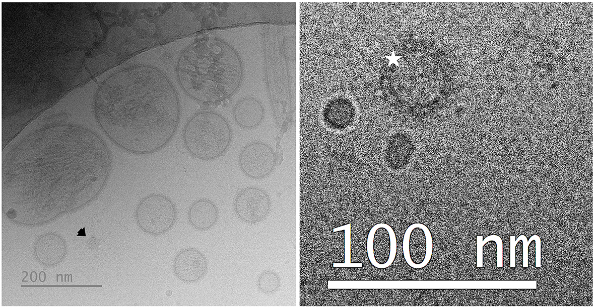
Figure 2. Representative cryogenic electron microscopy images of porcine seminal EVs. The left image shows a population of seminal EVs heterogeneous in size, shape, and electron density, with particles surrounded by a distinguishable membrane, as well as a non-vesicular extracellular particle (arrow). The right image shows a seminal EV surrounded by a protein corona (star). EVs: Extracellular vesicles.


