fig2
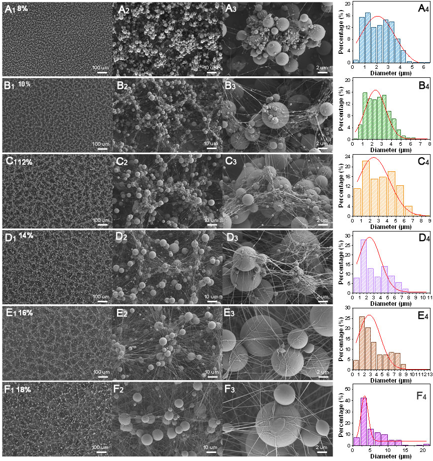
Figure 2. Microstructure morphology of the electrospun mats and the size distribution data of the spherical particles obtained with polymer solid content ranging from 8% to 18%. (A1-F3) SEM images of films prepared at 8%, 10%, 12%, 14%, 16% and 18% solid content, respectively; (A4-F4) Histogram of the corresponding particle size distribution. SEM: Scanning electron microscopy.