

fig11
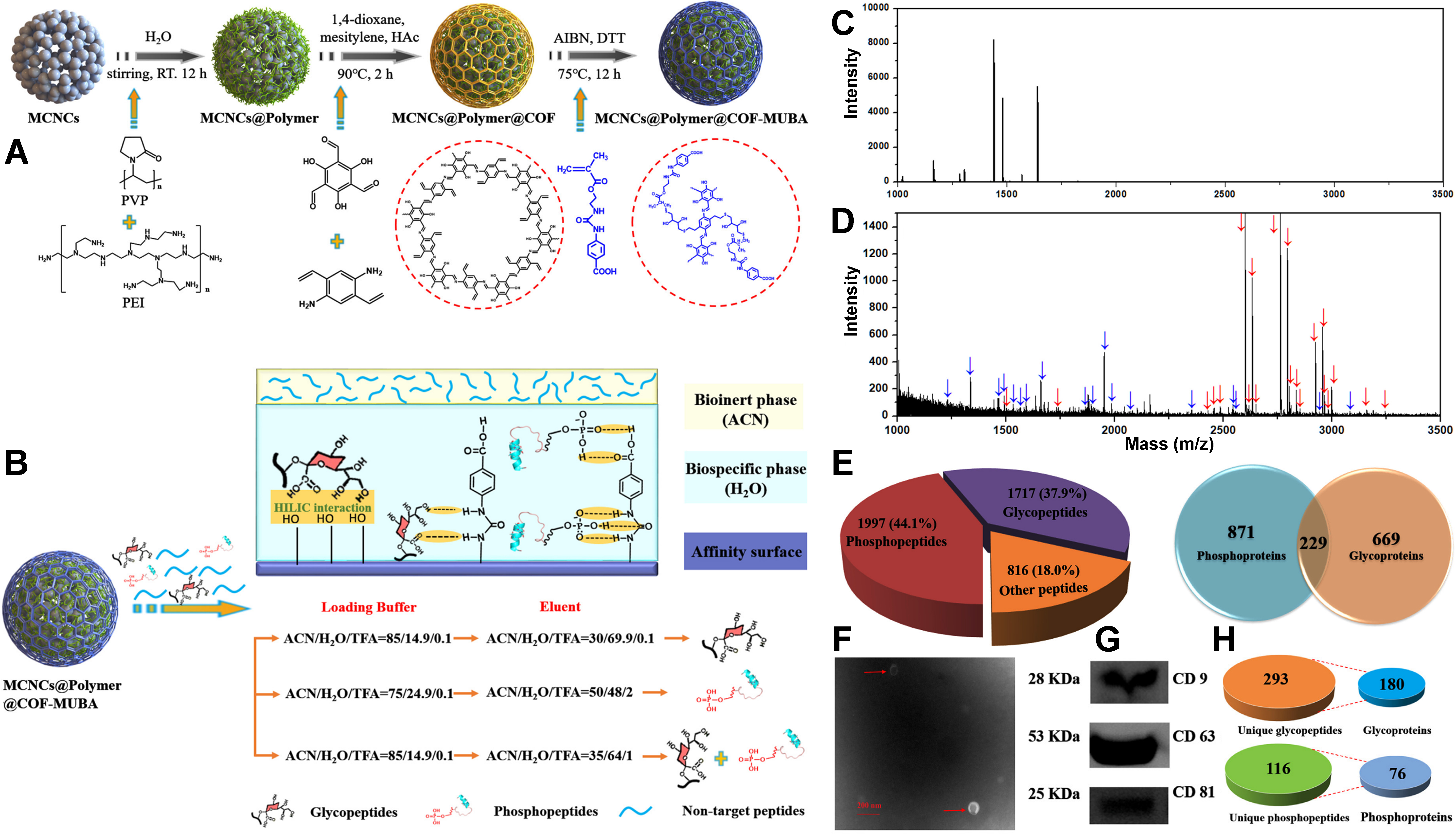
Figure 11. (A) Schematic representation of the synthesis of MCNC@polymer@COF-MUBA nanospheres; (B) Schematic diagram of the binding modes and controllable selective enrichment of the nanospheres for phosphopeptides and glycopeptides; (C) MALDI-TOF MS of peptide mixtures of tryptic digests of IgG, α-casein, and BSA with a molar ratio of 1:1:500 direct analysis; (D) MALDI-TOF MS of peptide mixtures of tryptic digests of IgG, α-casein, and BSA with a molar ratio of 1:1:1,000 after enrichment with the nanospheres. Glycopeptides are marked with red arrows and phosphopeptides are marked with blue arrows (For interpretation of the references to color in this Figure legend, the reader is referred to the Web version of this article); (E) The numbers of glycopeptides (glycoproteins) and phosphopeptides (phosphoproteins) identified using the nanospheres from mouse liver digests; (F) TEM negative staining analysis of the obtained exosomes; (G) Western blot analysis of the proteins collected from exosomes and three exosome markers (CD-9, CD-63, and CD81) were used; (H) Numbers of glycopeptides (glycoproteins) and phosphopeptides (phosphoproteins) in the exosome lysate identified using the material. (A-H) are reprinted with permission from Ref.[138], Copyright © 2021 by Elsevier. MCNC: Magnetic colloidal nanocrystalline cluster; COF: covalent organic framework; MUBA: 4-(3-(2-(methacryloyloxy)ethyl)-ureido)benzoic acid; MALDI-TOF MS: matrix-assisted laser desorption/ionization time-of-flight mass spectrometry; IgG: immunoglobulin G; BSA: bovine serum albumin; TEM: transmission electron microscope.