fig3
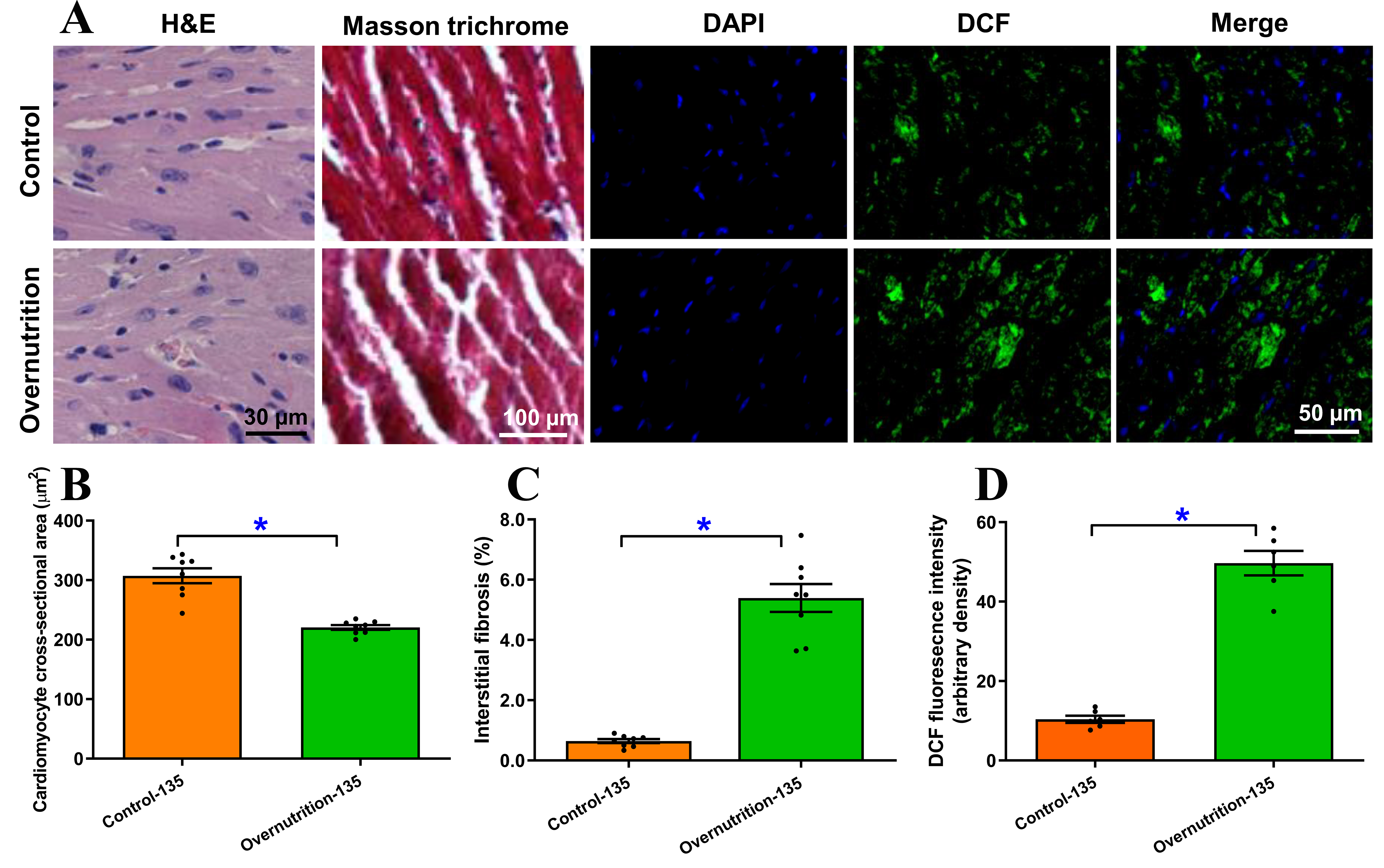
Figure 3. Morphological and free radical accumulation of fetal (D135) hearts from ewes receiving either control or overnutrition diet (150% NRC recommendation) for 135 days. (A) Representative micrographs depicting H&E staining, Masson Trichrome staining and DCF staining from fetal hearts from ewes receiving control or overnutrition diet. DCF staining includes DAPI (nucleus), DCF and merged images in 3 respective columns; (B) Pooled cardiomyocyte cross-sectional area from H&E staining; (C) Pooled myocardial interstitial fibrosis from Masson Trichrome staining; and (D) Pooled myocardial free radical accumulation from DCF staining. Mean ± SEM, *P < 0.05 between groups; n = 8 (B and C) or 6 (D) fetuses per group.