

Obesity-induced mesenchymal stem cell dysfunction: from molecular mechanisms to therapeutic strategies
 0
0 Abstract
Obesity is a systemic driver of cellular dysfunction that reshapes tissue biology well beyond energy storage, extending its influence to the regenerative machinery of the body. Mesenchymal stem/stromal cells (MSCs) support tissue repair through immunomodulatory, angiogenic, and paracrine functions, and accumulating evidence indicates that obesity impairs these functions within its metabolic milieu. This review integrates the mitochondrial, epigenetic, epitranscriptomic, and extracellular vesicle (EV) mechanisms by which obesity reprograms MSCs, traces the functional and organ-level consequences, and appraises rescue strategies against the strength and translational maturity of the supporting evidence. In the nutrient-excess environment of obesity, lipotoxicity, oxidative stress, and low-grade inflammation converge to erode mitochondrial integrity and bioenergetic homeostasis. Mitochondrial dysfunction is reinforced by dysregulated adenosine monophosphate-activated protein kinase (AMPK)-mechanistic target of rapamycin (mTOR) signaling, defective autophagy and mitophagy, and perturbed nicotinamide adenine dinucleotide (NAD+)-sirtuin homeostasis. These abnormalities intersect with epigenetic and epitranscriptomic remodeling, including altered chromatin regulation and non-coding ribonucleic acid signaling, as well as changes in EV cargo. Collectively, these interconnected processes reduce MSC stemness and metabolic adaptability, promote senescence and inflammatory phenotypes, and compromise reparative function. Consequently, obesity-compromised MSCs exhibit diminished immunomodulatory, angiogenic, and regenerative potential, which may limit their protective function across organs such as the heart, liver, and kidney and constrain their use as an autologous therapeutic. We discuss evolving therapeutic strategies, including metabolic reprogramming, mitochondrial protection, NAD+ restoration, senolytic approaches, and MSC- or EV-based interventions, and outline the translational gaps that remain before these approaches can benefit metabolically compromised patients.
Keywords
INTRODUCTION
Obesity has become a global pandemic. Between 1990 and 2022, the worldwide prevalence of adult obesity more than doubled, and projections indicate continued increases through 2040, with the burden shifting progressively toward lower-income countries[1,2]. This trajectory is expected to substantially increase the global burden of adiposity-related multimorbidity[3]. Beyond population trends, growing evidence indicates profound effects of obesity at the tissue and cellular levels, including on stem cell biology and regenerative potential[4]. Several mechanisms have been implicated in how obesity interferes with the reparative functions of circulating and tissue-resident stem cells. Disruption of stem cell niches by obesity-associated tissue and cellular changes, including chronic metabolic stress, low-grade inflammation, oxidative stress, and altered nutrient signaling, has been postulated to reduce stem cell proliferation and differentiation[5]. Consequently, individuals with obesity may have inadequate self-healing capacity and impaired recovery from insults. In addition, this functional deficiency of mesenchymal stem/stromal cells (MSCs) impedes their potential as a regenerative, autologous, cell-based strategy in individuals with obesity.
Throughout this review, we define “MSCs” according to the minimal criteria proposed by the International Society for Cellular Therapy (ISCT). These include: plastic adherence under standard culture conditions; expression of CD73, CD90, and CD105 with the absence of specified hematopoietic and endothelial markers; and trilineage differentiation in vitro[6]. Because these are population-level criteria rather than markers of a single-cell identity, and because MSC phenotype and function vary by tissue of origin, we specify the source of MSCs wherever the primary reports allow. Most human data on obesity-induced MSC dysfunction derive from adipose tissue-derived MSCs (AT-MSCs), typically harvested from abdominal subcutaneous fat, whereas bone marrow-derived MSCs (BM-MSCs) and MSCs from other depots may respond differently to the obese milieu.
The reports comparing AT- vs. BM-MSCs are, however, occasionally inconsistent. Donor-matched comparisons usually show tissue-specific differentiation bias, with AT-MSCs favoring adipogenesis and exhibiting higher proliferative rates, and BM-MSCs favoring osteogenesis and chondrogenesis, alongside substantial inter-donor variability[7,8]. Reported differences in immunomodulatory potency are less consistent. Studies have found AT-MSCs more potent, BM-MSCs to be superior, or the two broadly equivalent. These discrepancies may partly reflect donor matching, passage number, and assay design[9]. We therefore treat tissue source as requiring explicit statement; unless a source is specified, statements about “obese-MSCs” should be read as pertaining mainly to adipose tissue-derived cells.
Prior reviews have often addressed obesity-induced MSC impairment through a single mechanistic lens. The goal of the present review is integration: we connect the self-amplifying nicotinamide adenine dinucleotide (NAD+)-sirtuin-epigenetic loop to epitranscriptomic regulation and to extracellular vesicle (EV) cargo remodeling, trace these mechanisms through to organ-level functional deficits, and appraise candidate rescue strategies according to the experimental model and level of evidence supporting each. We also highlight voids in the current literature, including the reversibility of the obese-MSC phenotype and the near-absence of clinical data. Interpreting this literature requires attention to species, tissue source, and how obesity is defined. This review thus provides an overview of the multi-layered impact of the obese microenvironment on MSC function and considers the challenges to restoring their reparative capacity.
MECHANISMS RESPONSIBLE FOR OBESITY-INDUCED IMPAIRMENT OF MSCS
Metabolic dysregulation and mitochondrial dysfunction
Obesity-induced lipotoxicity impairs MSC function through palmitate accumulation, inducing endoplasmic reticulum (ER) stress, mitochondrial depolarization, apoptosis, and elevated reactive oxygen species (ROS). Excess palmitate forms toxic lipid intermediates (ceramides and diacylglycerols)[10], activates ER stress pathways including upregulation of activating transcription factor-4 (ATF4) and C/EBP homologous protein (CHOP)[11], and disrupts ER calcium homeostasis[12]. Palmitate also stimulates p38 mitogen-activated protein kinase (MAPK) phosphorylation, a central mediator of ER stress[13]. Palmitate-driven mitochondrial dysfunction lowers membrane potential[10,12] and triggers apoptosis via CHOP-mediated ER stress and mitochondrial pathways involving depolarization, calcium overload, and caspase-3/7 activation[12,14,15]. Concurrent nuclear factor kappa-B (NF-κB) and p38 activation amplifies pro-inflammatory mediators, including interleukin (IL)-6, vascular endothelial growth factor (VEGF), and monocyte chemoattractant protein-1 (MCP-1), further exacerbating apoptotic signaling[16].
MSCs require intact mitochondrial integrity and bioenergetics to orchestrate self-renewal, differentiation, and regeneration[17]. MSCs isolated from individuals with obesity (“obese-MSCs”) exhibit mitochondrial swelling, reduced membrane potential and adenosine triphosphate (ATP) levels, and elevated ROS[18]. These human findings are consistent with, and were first defined in, controlled animal models, allowing mechanisms to be dissected without the confounders of human cohorts[18,19]. Palmitate overload enhances mitochondrial β-oxidation, overwhelming the Krebs cycle and respiratory chain, thereby increasing mitochondrial superoxide production[18,19]. Several mechanisms may contribute to mitochondrial depolarization in obese-MSCs. These include lipotoxic injury by free fatty acids and ceramides, ER stress-mediated calcium overload, and impaired electron transport chain function due to structural damage[10,12,20]. Consequently, studies revealed increased mitochondrial superoxide production and loss of mitochondrial membrane potential[18,19].
Obese-MSCs also display impaired mitochondrial respiration, with reduced cytochrome-c oxidase (COX-IV) activity and ATP production[19,21]. Basal and maximal respiration, as well as spare respiratory capacity, are diminished, indicating chronic bioenergetic insufficiency[22]. Obesity further shifts mitochondrial dynamics toward increased fission and reduced fusion[23,24]. In swine metabolic-syndrome MSCs, the pro-fission GTPase dynamin-related protein-1 (DRP1) is upregulated, and the pro-fusion protein mitofusin-2 (MFN2) is reduced, yielding fragmented, low-volume mitochondria with disrupted three-dimensional networks[25]. In human adipose-MSCs, integrated hydroxymethylation and transcriptomic profiling have likewise identified coordinated alterations in nuclear-encoded mitochondrial genes governing ATP production, redox balance, and fatty acid metabolism[18]. Although data on DRP1/MFN2 specifically in obese human MSCs remain limited, structural defects, including cristae loss and reduced matrix density, impair ATP generation, promote ROS accumulation, and further compromise MSC bioenergetics[19,21].
Molecular pathways implicated in the impact of obesity on MSC mitochondria
MitomiR-181a as a candidate mediator of obesity-induced MSC dysfunction
One mechanism by which obesity may modulate MSC mitochondrial function involves micro-RNA (miR)-181a, a mitochondria-targeting miRNA (mitomiR) implicated in mitochondrial biogenesis, oxidative phosphorylation, and stress responses. In MSCs, miR-181a restrains proliferation and attenuates immunosuppressive capacity by targeting transforming growth factor-β receptor signaling[26]. It also targets mitochondrial transcription factor-A (TFAM) and electron transport chain components, and its downregulation is protective in models of mitochondrial disease, suggesting that sustained elevation would impair respiration and promote inflammation[27]. Direct evidence that obesity elevates miR-181a within MSCs is, however, lacking: obese swine AT-MSCs upregulate a small set of miRNAs predicted to target mitochondrial genes, in parallel with cristae remodeling and increased mitochondrial oxidative stress[19], but the specific contribution of miR-181a in obese MSCs remains unresolved. Any consequent disruption of mitochondrial-derived peptide (MDP) production, including humanin and the mitochondrial open-reading-frame of the 12S rRNA type-c (MOTS-c), is likewise an extrapolation from the broader literature on oxidative stress and autophagy in metabolically compromised MSCs[28].
MOTS-c expression in obese MSCs
MDPs are bioactive peptides encoded within mitochondrial DNA that regulate metabolism, stress responses, and cell survival. The MDP MOTS-c enhances insulin sensitivity, supports fatty acid β-oxidation, and mitigates metabolic dysfunction. It also activates the folate-5-aminoimidazole-4-carboxamide ribonucleotide (AICAR)-adenosine monophosphate (AMP)-activated protein kinase (AMPK) signaling axis to enhance mitochondrial bioenergetics, antioxidant defenses, and cellular stress adaptation[29]. The literature reports divergent obesity-associated patterns: serum MOTS-c is reduced in children with obesity and linked to endothelial dysfunction[30], whereas in adults it correlates with insulin sensitivity only in lean individuals[31]. We observed increased systemic MOTS-c levels in adults with obesity, indicating context- and tissue-dependent regulation[32]. Consistent with context-dependent regulation, Sequeira et al. reported that plasma MOTS-c concentrations were positively associated with android and liver fat in people without diabetes[33]. Whether MOTS-c expression is altered within obese-MSCs themselves remains to be determined.
AMPK-mTOR dysregulation and autophagy deficiency
AMPK, a cellular energy sensor that maintains homeostasis, is activated by cellular stress and glucose deprivation. In obesity, nutrient excess suppresses AMPK despite cellular dysfunction, creating a paradoxical signaling impairment. Reduced AMPK activity in obese-MSCs decreases fatty acid oxidation, increases lipid accumulation, and disrupts energy metabolism[34]. AMPK normally inhibits mechanistic target of rapamycin (mTOR) complex-1 (mTORC1) by phosphorylating the regulatory-associated protein of mTOR (RAPTOR) and activating tuberous sclerosis complex-2 (TSC2). However, in obesity, nutrient overload and AMPK suppression favor chronic mTORC1 activation[34]. Elevated mTORC1 subsequently promotes lipogenesis, exacerbating lipotoxicity[35], and concurrently inhibits autophagy, leading to lipid accumulation, insulin resistance, and mitochondrial dysfunction[35,36]. Accumulated lipids further repress AMPK and enhance mTORC1 signaling, reinforcing a positive feedback loop of metabolic impairment. Obesity-associated epigenetic alterations in mitochondrial genes involved in ATP generation, redox balance, and fatty acid metabolism add another layer of persistent dysregulation[18] [Figure 1]. Furthermore, MSCs from obese mice exhibit reduced cardiolipin levels, impairing LC3-cardiolipin interaction and disrupting cardiolipin-dependent mitophagy; this defect limits clearance of damaged mitochondria and diminishes their capacity for intercellular mitochondrial transfer[37].
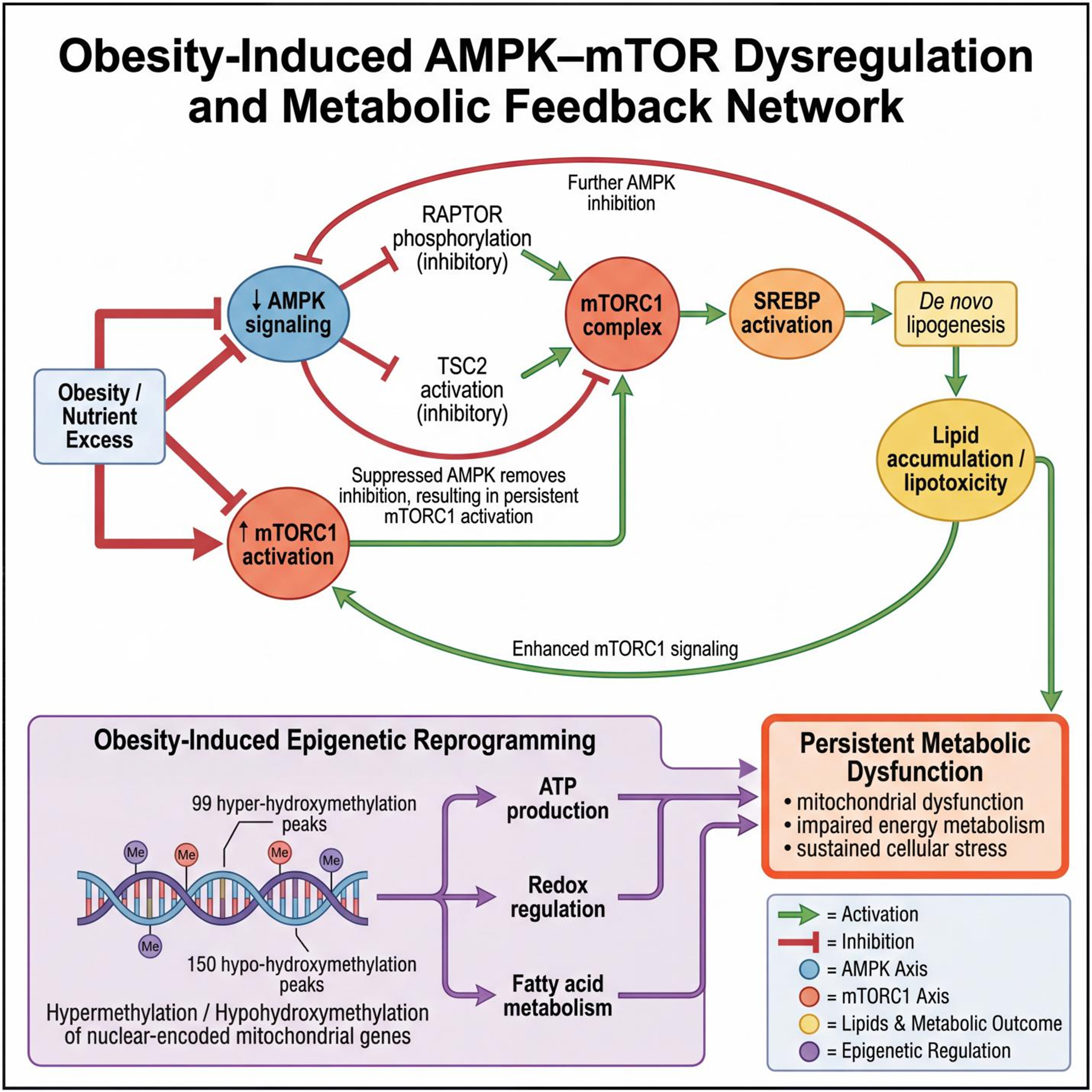
Figure 1. Obesity-induced AMPK-mTOR dysregulation and metabolic feedback network. Obesity and nutrient excess suppress AMPK signaling, releasing its inhibitory effect on the mTORC1 via reduced RAPTOR phosphorylation and diminished TSC2 activation. Sustained mTORC1 activation promotes SREBP-mediated de novo lipogenesis and lipid accumulation, ultimately producing lipotoxicity. Accumulated lipids further reinforce mTORC1 signaling, establishing a feed-forward loop. Concurrently, obesity induces epigenetic reprogramming of nuclear-encoded mitochondrial genes, characterized by altered hydroxymethylation patterns that disrupt ATP production, redox balance, and fatty acid metabolism. Together, these processes drive persistent metabolic dysfunction, including mitochondrial impairment, defective energy metabolism, and sustained cellular stress. The figure was created in BioRender [Elmaraezy, A. (2026) https://BioRender.com/k91vl7g] and refined using AI-assisted image-refinement tools (Figurelabs and Nano Banana Pro). AMPK: Adenosine monophosphate-activated protein kinase; mTOR: mechanistic target of rapamycin; mTORC1: mechanistic target of rapamycin complex-1; RAPTOR: regulatory-associated protein of mTOR; TSC2: tuberous sclerosis complex-2; SREBP: sterol regulatory element-binding protein; ATP: adenosine triphosphate.
Collectively, AMPK inhibition, mTORC1 hyperactivation, defective autophagy, and impaired mitophagy reduce stress resilience and stemness in obese-MSCs [Figure 1], reflected by downregulation of the pluripotency factors OCT4, NANOG, SOX2, and KLF4[38]. Notably, cellular stress in MSCs from donors with a body mass index (BMI) ≥ 30 kg/m2 resists melatonin and tauroursodeoxycholic acid (TUDCA), suggesting durable and potentially irreversible disruption of the AMPK-mTOR-autophagy axis at advanced stages[15].
Self-amplifying NAD+ depletion
Obesity-driven NAD+ depletion and sirtuin suppression create a self-reinforcing metabolic-epigenetic dysfunction. Nicotinamide N-methyltransferase (NNMT) upregulation reduces NAD+ and S-adenosylmethionine (SAM), impairing energy metabolism and methylation[39]. Concurrently, miR-34a targets sirtuin-1 (SIRT1) and nicotinamide phosphoribosyltransferase (NAMPT), diminishing sirtuin activity and NAD+ salvage. Reduced SIRT1 hyperacetylates peroxisome proliferator-activated receptor-γ coactivator-1α (PGC-1α), disrupting mitochondrial biogenesis[40]. The resulting mitochondrial dysfunction elevates ROS, activates poly(ADP-ribose) polymerases (PARPs), and accelerates NAD+ loss, while reduced ATP further limits NAD+ biosynthesis, perpetuating dysfunction in obese-MSCs[41] [Figure 2].
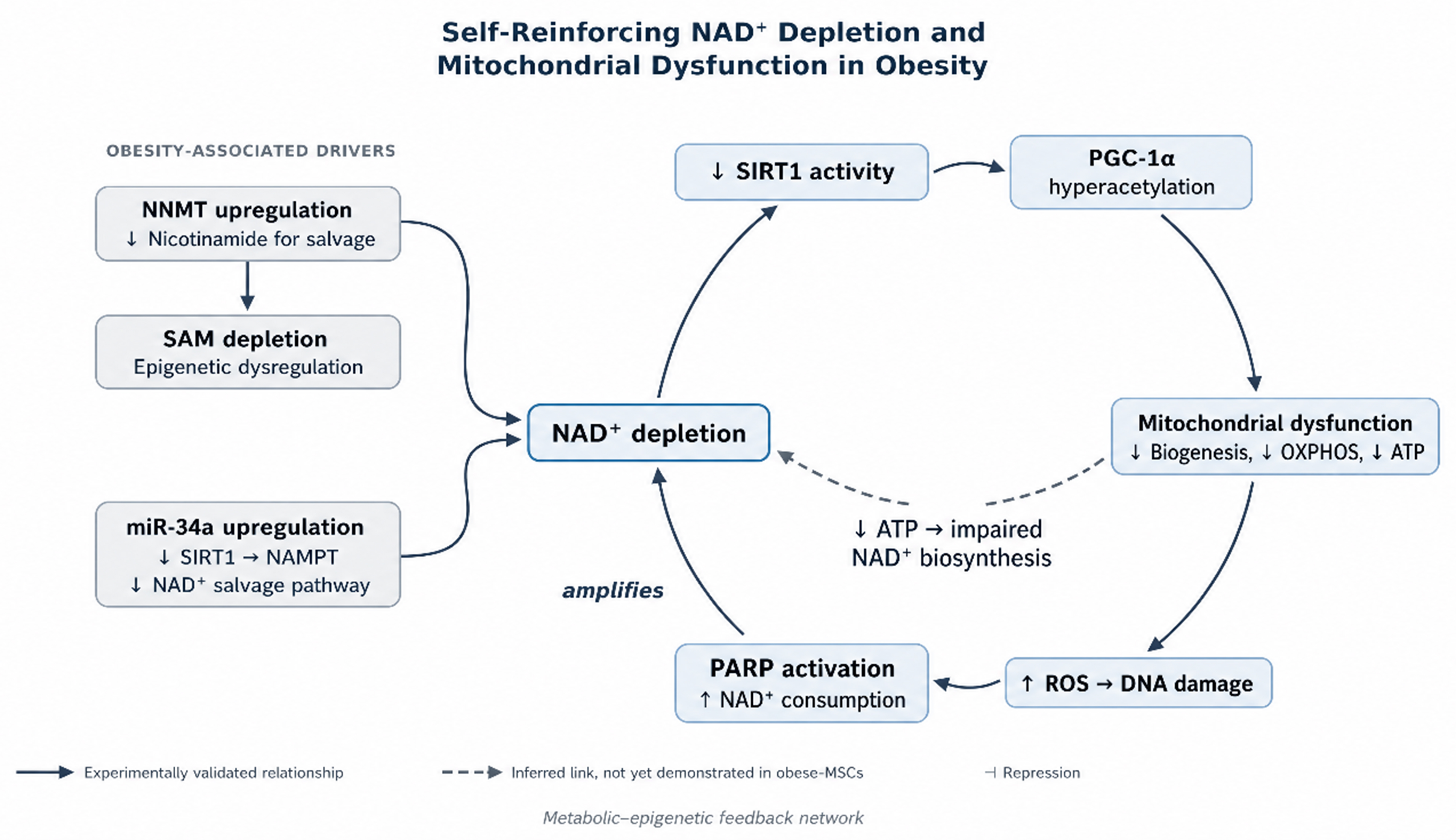
Figure 2. Self-reinforcing NAD+ depletion drives mitochondrial dysfunction in obesity. Obesity-associated drivers feed a self-amplifying cycle. Upregulated NNMT diverts nicotinamide from NAD+ salvage and consumes SAM, whose depletion impairs methylation and contributes to epigenetic dysregulation. In parallel, increased miR-34a represses SIRT1 and NAMPT, further impairing salvage. Within the cycle, reduced NAD+ diminishes SIRT1 activity, causing PGC-1α hyperacetylation and mitochondrial dysfunction, with impaired biogenesis, reduced OXPHOS, and ATP depletion. The resulting ROS production and DNA damage activate PARP, which consumes NAD+ and closes the loop. Solid arrows denote experimentally validated relationships, and the single dashed arrow denotes an inferred link (ATP depletion limiting NAD+ biosynthesis) not yet demonstrated in obese MSCs. The figure was created in BioRender [Elmaraezy, A. (2026) https://BioRender.com/sa8japm] and refined using AI-assisted image-refinement tools (Figurelabs and Nano Banana Pro). NAD+: Nicotinamide adenine dinucleotide; NNMT: nicotinamide N-methyltransferase; SAM: S-adenosylmethionine; miR: micro-RNA; SIRT1: sirtuin-1; NAMPT: nicotinamide phosphoribosyltransferase; PGC-1α: peroxisome proliferator-activated receptor-γ coactivator-1α; OXPHOS: oxidative phosphorylation; ATP: adenosine triphosphate; ROS: reactive oxygen species; PARP: poly(ADP-ribose) polymerase; MSCs: mesenchymal stem/stromal cells.
Epigenetic modifications in obese-MSCs
Environmental effects on cellular gene expression are often driven by epigenetic alterations, which are key drivers of the phenotypic and functional impairment of MSCs in obesity. Obesity reshapes the 5-hydroxymethylcytosine (5hmC) landscape, reducing MSC anti-inflammatory capacity[42]. Our work showed that these epigenetic shifts correlate with elevated inflammatory cytokines, particularly IL-1β, which amplifies inflammation and suppresses human MSC immunoregulation[4]. Obese-MSCs also exhibit disrupted NAD+ homeostasis and reduced methyl-donor availability, impairing the SAM cycle and leading to global hypomethylation with locus-specific hypermethylation[43].
Integrated hydroxymethylated DNA immunoprecipitation sequencing and mRNA sequencing of human abdominal subcutaneous AT-MSCs from subjects with obesity identified 99 hyper-hydroxymethylated and 150 hypo-hydroxymethylated peaks in nuclear-encoded mitochondrial genes, affecting metabolic regulators linked to redox balance, fatty acid oxidation, ATP transport, and mitochondrial biogenesis, with concordant transcriptional changes in a subset of overlapping genes[18]. Importantly, in swine obese-MSCs, the global hydroxymethylation and histone methylation changes induced by obesity were only partially corrected by the epigenetic modulator vitamin C, foreshadowing the concept of metabolic memory[42]. Additionally, hypermethylation of stemness genes corresponds with reduced SOX2 and KLF4 expression[38], suggesting that epigenetic silencing of the stemness program may promote a more differentiated, less stem-like cellular phenotype.
Obesity also drives miRNA dysregulation in MSCs, with increased miR-34a and miR-155 reinforcing SIRT1 suppression, inflammation, and reduced regenerative capacity[44,45]. miR-34a directly targets SIRT1 and indirectly lowers NAD+ via NAMPT inhibition, disrupting the NAD+/SIRT1 axis, impairing mitochondrial biogenesis, reducing membrane potential, and accelerating senescence[40,46-48]. Obesity-induced NF-κB activation elevates miR-155, establishing a feed-forward pro-inflammatory loop[49]. miR-155 suppresses peroxisome proliferator-activated receptor-γ (PPARγ) and glucose transporter type-4 (GLUT4), impairing adipogenic differentiation and insulin sensitivity, and increases ROS by repressing antioxidant regulators through CCAAT/enhancer-binding protein-β (C/EBPβ) targeting[49,50].
Taken together, miR-181a, miR-34a, and miR-155 form an integrated obesity-associated regulatory network linking mitochondrial dysfunction to epigenetic regulation. We discuss mitomiR-181a in the mitochondrial section because its principal targets, including TFAM and electron transport chain components, are mitochondrial.
Obese AT-MSCs exhibit leptin-associated histone remodeling, with elevated chromatin modifiers, including CREBBP/EP300 acetyltransferases and KDM6A/KDM6B demethylases, correlating with leptin levels[51]. Leptin exposure in vitro similarly increases their transcription, indicating nutrient-responsive chromatin modulation. This heightened chromatin-modifying activity promotes inflammatory gene activation while repressing tissue-repair programs[51]. Exposure of adipose-derived MSCs to plasma from obese donors induces cell-cycle arrest, increases senescence-associated β-galactosidase activity, and reduces mitochondrial respiration[52]. Activation of p38 MAPK and NF-κB precedes these secretory changes, suggesting that circulating mediators can impose the dysfunctional phenotype.
Epitranscriptomic regulation and RNA-binding proteins
Besides DNA methylation, hydroxymethylation, and histone modification, an epitranscriptomic layer, principally N6-methyladenosine (m6A) methylation of RNA, has emerged as a regulator of both adipose biology and MSC fate, and involves distinct classes of RNA-binding “writer”, “eraser”, and “reader” proteins. The methyltransferase-like (METTL) complex METTL3/METTL14 installs m6A, the demethylases fat mass and obesity-associated protein (FTO) and AlkB homolog-5 (ALKBH5) remove it, and reader proteins including YTH domain-containing family protein-2 (YTHDF2) and the insulin-like growth factor-2 messenger RNA-binding protein (IGF2BP) family determine transcript stability and translation. This machinery is itself nutritionally responsive in fat: METTL3/METTL14 expression varies with feeding, insulin, and high-fat-diet exposure[53]. In obesity, increased m6A modification of transcripts encoding β-adrenergic receptors and lipolytic proteins reduces their translation. The same layer intersects directly with the autophagy defects described earlier: when the demethylase FTO is depleted, transcripts for two core autophagy proteins accumulate the mark, are captured by the reader YTHDF2, and are degraded, so that autophagosome formation and adipogenic differentiation both fall[54]. Because FTO is the archetypal obesity-associated locus, this places an obesity-linked gene upstream of the autophagy machinery that fails in obese-MSCs.
In MSCs, this regulation affects the adipo-osteogenic balance that obesity skews. Removing the methyltransferase from murine BM-MSCs shifts lineage allocation, with reduced bone formation and expanded marrow adiposity; restoring or overexpressing it reinstates the balance, mediated at least in part through altered translation of a receptor governing MSC lineage choice[55]. A parallel study traced the osteogenic effect to autophagy induction, with IGF2BP-family reader proteins recognizing the methylated transcript of a core autophagy regulator and stabilizing it, a concrete illustration of how RNA-binding proteins, rather than the mark alone, determine the functional readout[56]. These studies were performed largely in models of osteoporosis and aging rather than obesity per se. Nevertheless, their targets, autophagy, adipo-osteogenic balance, and β-adrenergic/lipolytic signaling, overlap with pathways disrupted in obese-MSCs. Direct m6A profiling of MSCs from donors with obesity has, to our knowledge, not yet been reported, and should be considered a priority gap. The epitranscriptome is therefore a plausible but largely unexplored contributor.
Whether these epigenetic changes are durable is an actively contested question. One body of work supports persistence. Cellular stress in MSCs from donors with advanced obesity is refractory to agents that relieve it at earlier stages, such as melatonin and TUDCA[15]. Single-nucleus profiling of human and murine fat has shown that a substantial fraction of obesity-induced transcriptional differences survives major weight reduction, and that chromatin-level marks in murine adipocytes outlast the weight, priming the cells for exaggerated responses on re-exposure to a high-fat diet[57]. In women studied after bariatric surgery, the composition of the stromal-vascular fraction and the behavior of their adipose stem cells had not returned to the pattern seen in never-obese controls[58]. A longitudinal study sampled adipose-MSCs from the same donors before and after surgical weight loss. Respiration and proliferation recovered and several inflammatory transcripts fell, yet the cells remained unable to suppress macrophage activation effectively, indicating incomplete functional recovery[59]. In contrast, another body of work suggests recovery. In a large spatially resolved atlas of human subcutaneous fat, the senescent burden that accumulates in precursor, metabolic, and vascular populations fell markedly after weight loss, although macrophage activation did not fully normalize and not every obesity-associated abnormality resolved[60]. Current evidence therefore suggests that obesity-induced MSC dysfunction is partially reversible. The degree of recovery likely depends on the phenotype examined, the severity and duration of obesity, and the experimental readout used. This variability may influence the optimal timing of autologous cell-based interventions. The variable persistence of these abnormalities may, in part, reflect obesity-associated epigenetic remodeling, particularly in the context of aging. Consistent with this possibility, aging and obesity synergistically modify the epigenome, through overlapping hypomethylation of regulators including MAPT, NR3C2, APP, and CTNNB1, and hypermethylation of FOXO3 and CCND1[43].
Obesity alters the characteristics of MSC-derived EVs
MSC-derived EVs, lipid-bound nanoparticles secreted by most cell types, mediate reparative functions through genetic and protein cargo that modulate recipient-cell pathways[61]. Consistent with current field standards, EV nomenclature, isolation, and characterization in the studies discussed here are interpreted in light of the International Society for Extracellular Vesicles (ISEV) Minimal Information for Studies of Extracellular Vesicles (MISEV2023) framework. We use the generic term “EVs” except where a primary study defined a specific subpopulation[62]. Obesity profoundly alters EV biology, modifying their physical features and molecular content and thereby reducing their reparative capacity[63,64]. We showed that EVs from adipose-derived MSCs of obese pigs were smaller than those from lean pigs, suggesting changes in EV biogenesis[65]. High-throughput miRNA sequencing further revealed extensive remodeling of human obese-EV cargo, including upregulation of pro-inflammatory and pro-apoptotic miRNAs and downregulation of miRNAs associated with cell-cycle progression, angiogenesis, and Wnt-mediated regeneration[64]. In addition, obese-EVs from pigs with metabolic syndrome failed to enhance endothelial tube formation or boost human umbilical vein endothelial cell (HUVEC) migration, reflecting impaired angiogenic activity[66] linked to loss of pro-angiogenic miRNAs and enrichment of miRNAs targeting angiogenic pathways[64].
FUNCTIONAL CONSEQUENCES OF OBESE-MSC DYSFUNCTION
Human obese-MSCs show reduced engraftment owing to diminished proliferation and increased senescence, marked by elevated p16, p53, IL-6, and MCP-1 expression[67]. BMI correlates with p16/p21-mediated cell-cycle arrest, imposing cellular stress that limits the feasibility of autologous MSC therapy[15]. BM-MSC from high-fat-diet-fed mice also show reduced expression of VEGF-A and basic fibroblast growth factor (bFGF), resulting in impaired pro-angiogenic properties[68]. Notably, co-incubation with non-obese MSCs restored VEGF expression and tube formation that were blunted in injured HUVECs[67].
Obese-MSCs exhibit impaired immunomodulation with a shift toward pro-inflammatory activity. Whereas lean human MSCs promote an anti-inflammatory macrophage phenotype, this capacity is blunted in MSCs from obese donors, which are less effective at activating M2 polarization and suppressing lymphocyte proliferation, and which show increased expression of inflammatory markers and inflammasome activation[69]. In high-fat-diet-induced obesity, visceral adipose MSCs activate the IL-1 and granzyme-A pathways, reversing their immunoregulatory effects on leukocyte activation, inflammation, and chemokine signaling[70]. Moreover, obesity reverses the anti-fibrotic activity of MSCs toward pro-fibrotic signaling, accompanied by impaired differentiation and altered paracrine signaling. The senescence-associated secretory phenotype of obese-MSCs includes pro-fibrotic cytokines that promote extracellular matrix deposition and tissue scarring[70].
The diminished reparative potency of obese-MSCs has been demonstrated in several organ systems.
a) Kidney repair
Functionally, human obese-MSCs fail to protect against renal ischemia-reperfusion injury or to ameliorate chronic ischemic injury, as they develop cellular senescence[71] and blunted immunomodulatory function[69], while elevated IL-1β expression shifts their function from therapeutic to pathogenic[4]. Similarly, porcine obese-EVs fail to restore renal blood flow, glomerular filtration rate, mitochondrial integrity, and oxidative balance, or to reduce tubular injury and fibrosis in renal artery stenosis models[66,72]. EVs derived from obese adipose-tissue MSCs show impaired reparative activity, failing to suppress inflammation and exhibiting reduced anti-apoptotic and MAPK-modulatory effects compared with EVs from lean MSCs; obesity-associated alterations in EV miRNA cargo also involve regulators of Wnt and MAPK signaling[64].
b) Cardiac repair
Obesity markedly attenuates the cardioprotective efficacy of adipose-derived MSCs in vivo. In a murine model of renovascular hypertension (RVH), obese-MSCs only modestly reduced left ventricular (LV) mass, failed to correct systemic hemodynamics, and exacerbated LV systolic dysfunction, indicating a deleterious effect on contractile performance. These functional outcomes were mirrored by tissue-level responses: obese-MSCs displayed a pro-inflammatory phenotype that failed to mitigate oxidative stress and myocardial structural injury[73] [Figure 3]. Similarly, obese-MSC-EVs fail to prevent cardiac hypertrophy, exerting minimal effects on LV wall thickening and cardiomyocyte hypertrophy and reflecting a compromised cardioprotective phenotype. Notably, obese-EVs modestly reduced myocardial fibrosis, indicating selective preservation of anti-fibrotic signaling pathways[74].
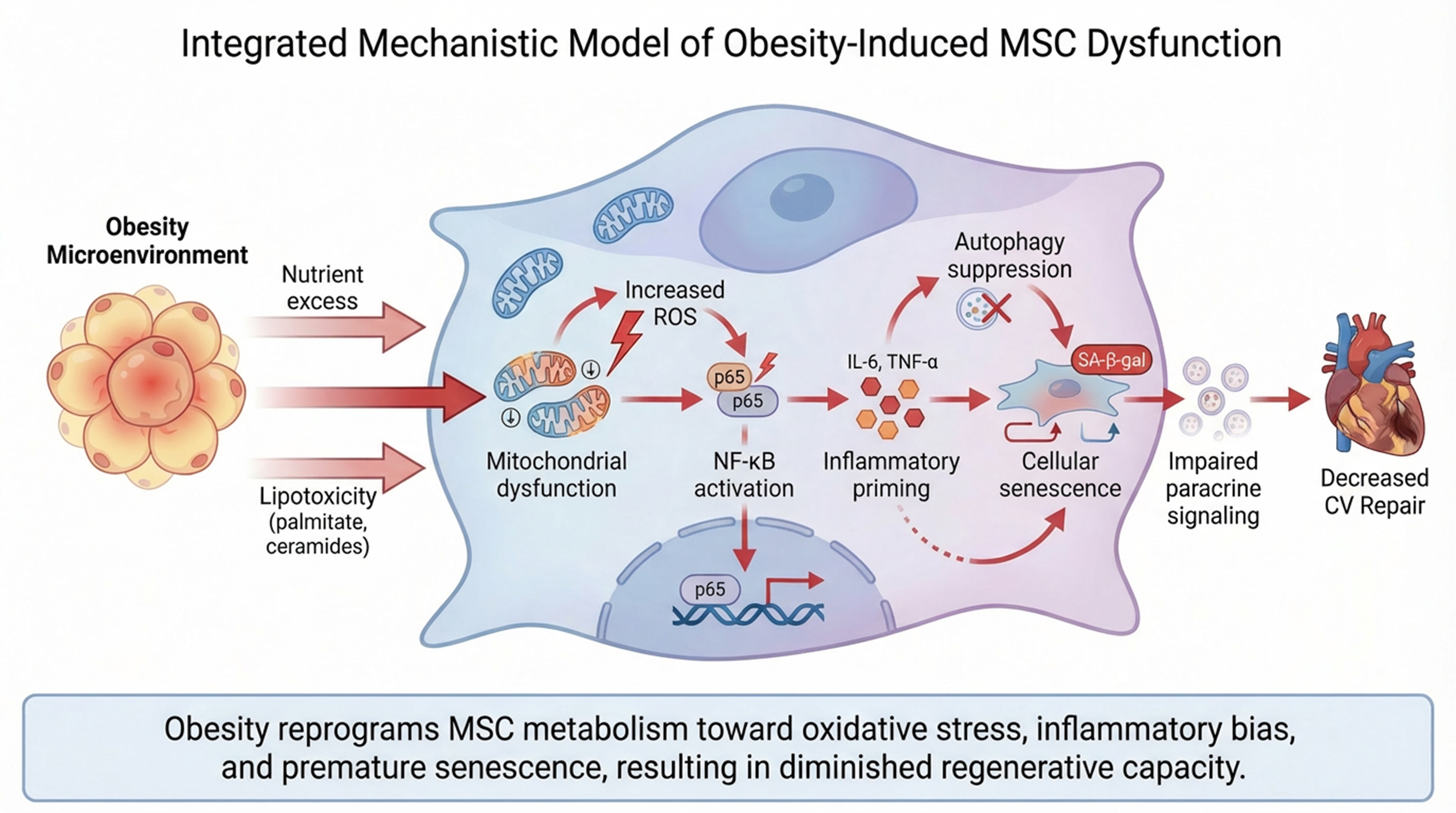
Figure 3. Integrated mechanistic model of obesity-induced MSC dysfunction. In the obese microenvironment, nutrient excess and lipotoxic mediators (e.g., palmitate and ceramides) induce mitochondrial dysfunction and increased ROS production in MSCs. Elevated oxidative stress activates NF-κB signaling, promoting inflammatory priming characterized by increased cytokine production (e.g., IL-6 and TNF-α). Concurrent suppression of autophagy contributes to the accumulation of damaged organelles and further oxidative stress. These processes drive cellular senescence, indicated by SA-β-gal activity, and impair MSC paracrine signaling capacity. Collectively, obesity reprograms MSCs toward a pro-inflammatory, senescent phenotype with diminished regenerative potential. Solid arrows denote experimentally supported relationships; the dashed arrow denotes an inferred link. The figure was created in BioRender [Elmaraezy, A. (2026) https://BioRender.com/mzjqngm] and refined using AI-assisted image-refinement tools (Figurelabs and Nano Banana Pro). MSC: Mesenchymal stem/stromal cell; ROS: reactive oxygen species; NF-κB: nuclear factor kappa-B; IL: interleukin; TNF-α: tumor necrosis factor-α; SA-β-gal: senescence-associated β-galactosidase; CV: cardiovascular.
Obese-MSCs show impaired paracrine signaling marked by increased pro-inflammatory activity and reduced antioxidant responses, consistent with obesity-associated senescence and metabolic dysfunction. As a result, RVH mice treated with obese-MSCs retain a heightened oxidative burden and show only partial reversal of adverse cardiac remodeling, whereas lean-MSC therapy restores redox homeostasis and supports more effective cardiac protection[73] [Figures 3 and 4].
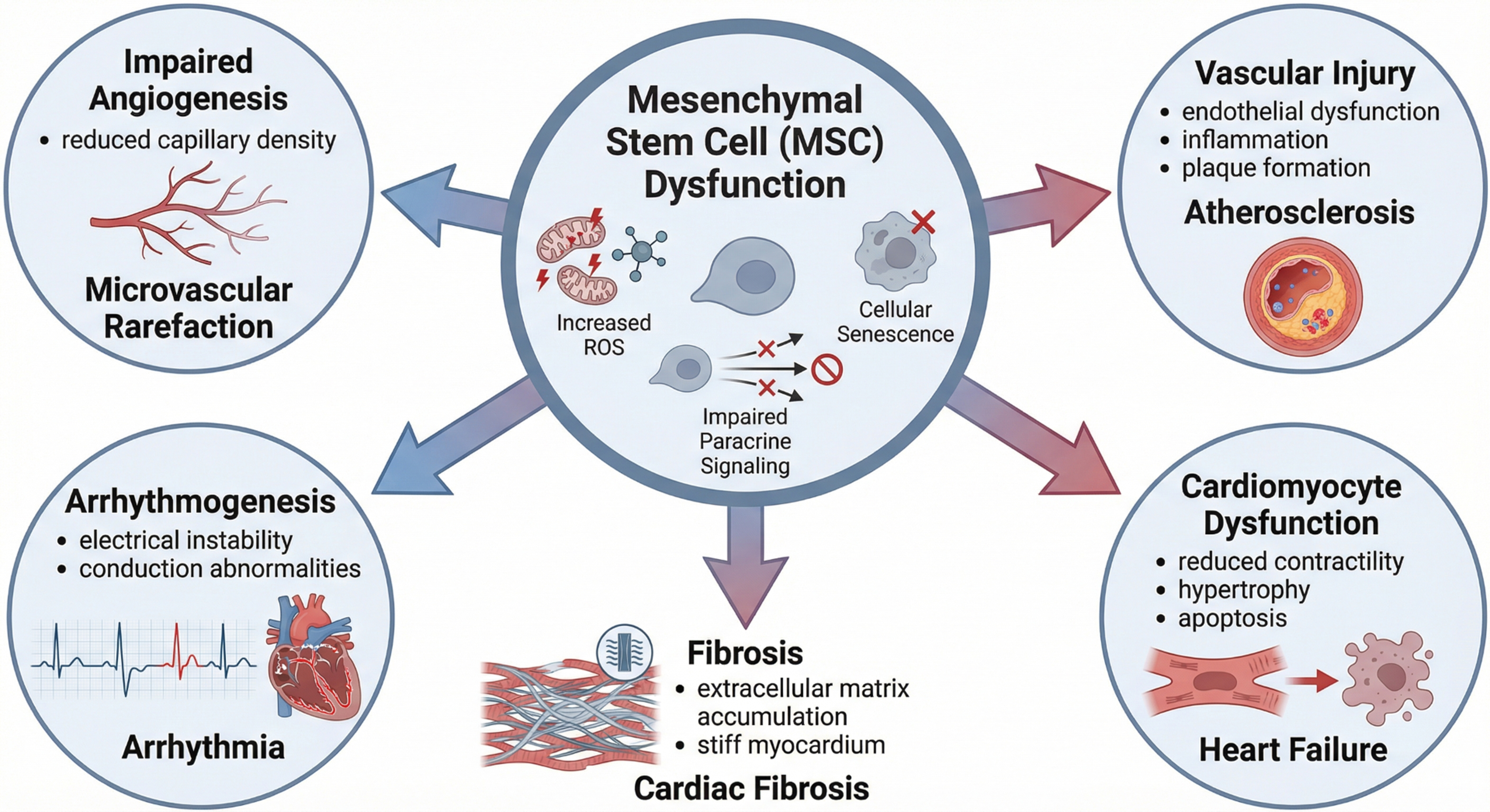
Figure 4. Downstream cardiovascular consequences of MSC dysfunction. Endogenous MSC dysfunction may contribute to multiple adverse cardiovascular outcomes in individuals with obesity. Impaired paracrine signaling and increased ROS production promote endothelial dysfunction, inflammation, and plaque formation, facilitating atherosclerosis. Reduced angiogenic capacity leads to microvascular rarefaction and decreased capillary density. In cardiomyocytes, these changes reduce contractility and promote hypertrophy and apoptosis, culminating in heart failure. Additionally, structural and electrical remodeling contribute to arrhythmogenesis and conduction abnormalities, while increased extracellular matrix deposition promotes cardiac fibrosis, further exacerbating myocardial stiffness and dysfunction. The figure was created in BioRender [Elmaraezy, A. (2026) http://BioRender.com/2mdwbbh] and refined using AI-assisted image-refinement tools (Figurelabs and Nano Banana Pro). MSC: Mesenchymal stem/stromal cell; ROS: reactive oxygen species.
c) Hepatic repair
Metabolic dysfunction-associated steatotic liver disease (MASLD) has become a leading indication for liver transplantation and is now the top indication in the United States among women and patients with hepatocellular carcinoma[75]. MSCs are postulated as central regulators of liver inflammation, fibrosis, and regeneration[76,77]. MSCs decrease insulin resistance, oxidized low-density lipoprotein (LDL), and IL-6 in high-fat-diet-fed obese mice, and injection of their suspension improved glucose tolerance and reduced hepatic steatosis, macrophage infiltration, and pancreatic islet hypertrophy[78]. Dysfunctional endogenous MSCs likely worsen disease progression by failing to control fibrosis and inflammation; however, data directly demonstrating the effect of dysfunctional obese-MSCs on liver disease progression remain limited.
d) Central nervous system repair
Direct evidence that obesity compromises MSC-mediated central nervous system (CNS) repair is sparse. MSCs and their EVs improve functional outcomes in preclinical stroke and traumatic brain injury models[79,80], yet no study has tested cells from donors with obesity in a CNS injury model, and donor adiposity is rarely reported. The most direct evidence is our own: human obese-MSCs displayed altered hydroxymethylation of nuclear-encoded mitochondrial genes governing neuronal development, together with impaired neurogenic differentiation in vitro that improved after epigenetic modulation[18]. This establishes diminished neurogenic capacity rather than failure of CNS repair, which remains untested in vivo. The concern therefore rests on extrapolation from the senescent, pro-inflammatory phenotype that blunts obese-MSC efficacy in renal and cardiac models, supported by evidence that donor characteristics predict MSC potency after brain injury[81]. Separately, obesity suppresses endogenous adult neurogenesis[82,83], though this reflects host rather than graft dysfunction.
VARIABILITY OF THE EVIDENCE BASE
A central challenge in synthesizing existing literature is that the underlying studies differ substantially in species, tissue source, and how obesity is defined, and these differences constrain direct comparison. Mechanistic work on ‘obese’ MSCs spans different species, including human, porcine, and murine MSCs. Human studies most often used AT-MSCs from abdominal subcutaneous fat of bariatric or surgical patients, offering clinical relevance but limited sample sizes, heterogeneous comorbidity and medication profiles, and cross-sectional designs[4,18,67]. Porcine models of diet-induced obesity and metabolic syndrome permit controlled dietary exposure and standardized harvesting and have helped define EV and mitochondrial phenotypes. However, swine metabolic syndrome also encompasses hypertension and dyslipidemia, making obesity-specific effects difficult to isolate[19,25,65,66,72]. Murine high-fat-diet models afford genetic tractability and mechanistic dissection but differ from humans in adipose biology and MSC behavior[23,68,74].
Several additional factors complicate comparisons among research studies. The tissue of origin is an important variable: AT- and BM-MSCs differ in secretome, immunomodulatory potency, and susceptibility to lipotoxic stress, so findings in one compartment do not necessarily generalize to another[7,8,11]. Definitions of obesity are similarly inconsistent. Human studies use varying BMI thresholds, whereas animal studies generally rely on diet duration and weight gain. Moreover, few studies account for coexisting insulin resistance, dyslipidemia, or hypertension. Reporting of MSC characterization and EV isolation also varies, further confounding comparisons. Protocol harmonization does not eliminate inter-donor or inter-laboratory variability. In a multicenter study using a shared workflow, broad source-specific patterns were reproducible, but quantitative readouts still varied by laboratory and operator[9]. Donor metabolic status is increasingly argued to determine adipose-derived MSC potency, which primary reports seldom document[84]. We therefore outline the model underlying key findings throughout this review and specify the source of supporting evidence in the subsequent summary tables. Standardized characterization of MSCs (per ISCT criteria) and EVs (per MISEV2023), together with harmonized definitions of obesity, would substantially strengthen the comparability and translational value of future work[6,62].
INTEGRATED SUMMARY: MECHANISM, FUNCTIONAL CONSEQUENCE, AND THERAPEUTIC TARGET
To connect the mechanistic sections above with the therapeutic strategies that follow, Table 1 maps each principal molecular derangement in obese-MSCs to its downstream functional consequence and the candidate intervention that targets it.
Integrated mapping of molecular mechanisms to functional consequences and candidate therapeutic strategies in obese-MSCs
| Molecular mechanism | Functional consequence in MSCs | Therapeutic target/strategy | Ref. |
| Lipotoxicity and ER stress (palmitate, ceramides; ATF4/CHOP, p38 MAPK) | Mitochondrial depolarization, apoptosis, ROS, inflammatory priming | Lipid buffering/SCD1 induction; AMPK activation; p38 inhibition | [10-16] |
| Mitochondrial dysfunction and altered dynamics (↑DRP1, ↓MFN2; ↓COX-IV) | ↓ATP, ↓respiratory capacity, fragmented mitochondria, ↑superoxide | Mitochondrial-targeted antioxidants (PQQ); restoring mitophagy/transfer | [18-25,37] |
| mitomiR-181a upregulation (TFAM, ETC targets) | Impaired respiration, inflammation, altered MDP output | Anti-mitomiR-181a; MOTS-c supplementation | [18,19,26-29] |
| AMPK-mTORC1 imbalance and autophagy/mitophagy defect | Lipid accumulation, ↓stemness (OCT4/NANOG/SOX2/KLF4), ↓stress resilience | AMPK activators (metformin, AICAR); mTOR modulation | [34-38,85-88] |
| Self-amplifying NAD+ depletion (↑NNMT, miR-34a → SIRT1/NAMPT; PARP) | PGC-1α hyperacetylation, ↓biogenesis, senescence | NAD+ precursors (NMN, NR); SIRT1 support | [39-41,89-91] |
| Epigenetic remodeling (5hmC shifts; miR-34a/miR-155; leptin-histone axis) | Global hypomethylation/locus hypermethylation, metabolic memory, pro-inflammatory bias | Epigenetic modulation; genetic reprogramming (SIRT1, TSG-6) | [4,18,42-51,53-56,92] |
| Altered EV biogenesis and cargo (smaller EVs; loss of pro-angiogenic miRNAs) | Impaired angiogenesis, reduced regenerative signaling | Lean-donor/engineered EVs; cargo enrichment | [63-66,72] |
| Senescence and SASP (↑p16/p21/p53, IL-6, MCP-1, TNF-α) | ↓Engraftment, pro-fibrotic and pro-inflammatory secretome | Senomodulation; preconditioning; allogeneic lean MSCs | [15,67,69,70,93-95] |
THERAPEUTIC TARGETS
Increasing understanding of the mechanisms underlying obese-MSC dysfunction has driven development of therapeutic strategies to blunt it [Table 2]. Metabolic and genetic reprogramming, mitochondrial protection, preconditioning, and allogeneic or EV-based therapies are potentially promising avenues for restoring function to obesity-impaired MSCs. Importantly, evidence for these strategies remains predominantly preclinical, derived from in vitro MSC cultures and animal models. None has been tested in a clinical trial specifically designed to rescue obesity-induced MSC dysfunction. Table 2 therefore identifies the experimental model and evidence level for each approach.
Rescue approaches for dysfunctional MSCs, annotated by experimental model and source of evidence
| Strategy | Main targeted defect | Key mechanisms/outcomes | Experimental model | Evidence source | Ref. |
| AMPK activators (metformin, AICAR) | Senescence, loss of immunosuppression | AMPK activation and metabolic reprogramming; ↑NO, ↓oxidative stress; restored migration and T-cell suppression | Human aging AT-MSCs in vitro; rodent MetS/diabetes models | Preclinical (in vitro + animal) | [85-87] |
| mTOR modulation (DEPTOR axis) | Adipo-osteogenic imbalance | High energy → mTOR-driven adipogenesis; DEPTOR/TAZ axis controls the bone–fat switch | Mouse BM-MSCs; genetic ablation models | Preclinical (animal); indirect for rescue | [88] |
| NAD+ repletion (NMN, NR) | NAD+ loss, mitochondrial dysfunction, loss of stemness | ↑NAD+, SIRT1/SIRT3 activation; improved mitochondrial function; ↑osteogenesis, ↓adipogenesis; rejuvenated stemness | Human and rodent MSCs in vitro; aged bone marrow | Preclinical (in vitro + animal) | [89-91] |
| Mitochondrial-targeted antioxidants (PQQ) | Impaired mitophagy, defective mitochondrial transfer | Restores cardiolipin, mitophagy, mitochondrial health, and intercellular mitochondrial transfer, rescuing efficacy | Obese-mouse MSCs; in vitro transfer assays | Preclinical (in vitro + animal) | [37] |
| Preconditioning (hypoxia, cytokines, adiponectin) | Reduced immunomodulation and metabolism | Hypoxia/leptin and TNF-α/IFN-γ priming; adiponectin-p38-HIF-1α shifts metabolism to glycolysis; ↑immunoregulatory factors and survival | Human and rodent MSCs in vitro; rodent in vivo | Preclinical (in vitro + animal) | [94,95] |
| Genetic reprogramming (SIRT1, DEPTOR, TSG-6) | Skewed differentiation, aging, inflammatory phenotype | SIRT1 overexpression, DEPTOR deletion, TSG-6 modulation redirect fate and enhance therapeutic effects | Mouse/human MSCs; gene-modified cells | Preclinical; early/proof-of-concept | [88,92] |
| Senolytic/senomodulatory approaches | Senescent-cell burden and SASP in the obese niche | Clearing p16-positive senescent cells improves glucose tolerance, insulin sensitivity, and adipogenesis and lowers circulating inflammatory mediators in obese mice; rationale for reducing senescent burden before or during MSC harvest | Obese mouse models (genetic and pharmacologic senolysis) | Preclinical (animal); yet untested for MSC rescue | [93] |
| Allogeneic lean-donor MSCs/EVs | Global dysfunction of autologous obese-MSCs | Allogeneic MSCs and their EVs improve obesity-related metabolic and atrophy phenotypes via paracrine and AMPK/autophagy pathways | Rodent obesity/diabetes models; human MSC-derived EVs | Preclinical (animal); human EV trials in other indications | [96,97] |
AMPK activation acts as a metabolic switch, restoring mitochondrial biogenesis and reducing oxidative stress in MSCs. Metformin reverses senescence-associated dysfunction via AMPK signaling, enhancing MSC migration, immunomodulatory capacity, and therapeutic efficacy in models of metabolic syndrome and diabetes[85,86]. In aging human adipose MSCs, metformin reduces oxidative stress and senescence while restoring adipogenic capacity and insulin sensitivity through AMPK activation[87]. These data support metformin (and other AMPK activators such as AICAR) as candidates to counteract obesity-induced MSC alterations. Modulating mTOR signaling may rebalance MSC lineage commitment and restore stemness: the DEP-domain-containing mTOR-interacting protein (DEPTOR) promotes adipogenesis while suppressing osteogenesis, whereas DEPTOR ablation restores bone mass and reduces marrow fat[88]. Targeted mTOR modulation may counter obesity-induced MSC dysfunction, although direct evidence for functional rescue remains limited and warrants further investigation.
NAD+ precursors such as nicotinamide riboside (NR) or nicotinamide mononucleotide (NMN) enhance SIRT1 and sirtuin-3 (SIRT3) activity, improve mitochondrial function, reduce senescence markers, and restore differentiation potential in MSCs[89,90]. NR also enhances MSC stemness, lowers ROS, and inhibits terminal adipocyte differentiation[91]. Collectively, these findings provide a strong mechanistic rationale for testing NAD+ repletion in obesity-impaired MSCs, although direct rescue studies in obese-donor MSCs remain limited. Several mitochondrial-targeted approaches have shown potential. Pyrroloquinoline quinone (PQQ) rescues cardiolipin content, mitophagy, and mitochondrial donation, improving therapeutic efficacy[37], whereas silencing miR-181a improves mitochondrial gene expression and reduces oxidative stress in models of mitochondrial dysfunction[27]. MOTS-c has broader metabolic and mitochondrial effects in experimental systems, collectively supporting MSC stemness, differentiation, and paracrine function[29]. Yet, direct evidence that MOTS-c restores the function of obesity-impaired MSCs is currently lacking. Genetic modification offers a complementary route: AT-MSCs engineered to modulate regulators such as tumor necrosis factor-stimulated gene-6 (TSG-6) have been proposed to correct inflammatory phenotypes and metabolic complications of obesity, although data remain scarce[92].
Because senescence is a convergent endpoint of the mechanisms described above, reducing senescent-cell burden represents a conceptually distinct strategy. Removal of senescent cells improves metabolic function in obese mice[93]. Genetic or pharmacologic senolysis improves glucose handling and insulin sensitivity, restores adipogenesis, and reduces circulating inflammatory mediators and monocyte trafficking, with parallel gains in renal and cardiac functional measures[93]. The implication is that senescent burden is not merely a marker of the obese niche but a causal contributor to it. Whether senolytic pre-treatment of a prospective donor, or senomodulation of harvested cells ex vivo, can restore the reparative competence of obese-MSCs has not been directly tested and represents a logical next step.
Hypoxic preconditioning enhances MSC cardioprotection by inducing leptin expression, while pro-inflammatory cytokines [tumor necrosis factor-α (TNF-α) and interferon-γ (IFN-γ)] promote an immunosuppressive MSC phenotype[94]. Obesity-related adiponectin deficiency impairs MSC survival and function; priming obese-MSCs with globular adiponectin restores glycolysis via the adiponectin receptor-1 (AdipoR1)/p38/hypoxia-inducible factor-1α (HIF-1α) axis, boosts immunoregulatory gene expression, and improves therapeutic efficacy[95]. These findings support hypoxia-, cytokine-, and adipokine-based metabolic reprogramming strategies. In contrast to autologous obese-MSCs, allogeneic MSCs from lean, healthy donors ameliorate obesity, improve insulin sensitivity, enhance mitochondrial biogenesis and thermogenesis, and reduce inflammation[96,97]. Lean-MSC-EVs further mitigate diabetes- and obesity-induced muscle atrophy via AMPK/Unc-51-like kinase-1 (ULK1)-mediated autophagy[96]. These data support using allogeneic MSCs or engineered EVs when autologous cells are compromised.
CLINICAL TRANSLATION AND CURRENT EVIDENCE IN HUMANS
A glaring weakness of the strategies above is that none has advanced to human testing for the specific goal of restoring the competence of obesity-impaired MSCs. The clinical experience that exists concerns MSC therapy for metabolic disease as an indication, rather than restoration of endogenous MSC function, and was not designed to address the donor-quality problem.
A 2025 systematic review and meta-analysis included 13 randomized trials in type-1 and type-2 diabetes involving 507 patients. At 12 months, MSC therapy was associated with lower glycated hemoglobin (HbA1c) [mean difference -0.72%; 95% confidence interval (CI) -1.11 to -0.33], a reduced daily insulin requirement (-14.50 U/day; 95%CI -19.45 to -9.55), higher fasting C-peptide (+0.24 ng/mL; 95%CI 0.05 to 0.43), and lower postprandial glucose. Fasting glucose, by contrast, did not separate from control, and between-study heterogeneity was high for C-peptide and fasting glucose in particular, so these pooled estimates should be read as encouraging rather than definitive[98]. A separate synthesis of type-2 diabetes trials reported reduced anti-diabetic drug requirements over 12 months with effective doses ranging from approximately 1 × 10⁶ to 3.7 × 10⁶ cells/kg and no severe adverse events[99]. Reported routes are predominantly intravenous or intra-pancreatic, and optimal dose, route, and timing remain unsettled.
Two caveats are essential for interpreting these results. First, both autologous and allogeneic sources have been used across trials, and their relative efficacy remains unresolved; at least one pooled analysis has reported more favorable outcomes with autologous than allogeneic MSCs, whereas subgroup analyses elsewhere have favored other cell types entirely, and certainty of evidence has been graded low to very low in guideline-oriented reviews[98,99]. Second, and more pertinently, these trials were not designed to test whether donor adiposity degrades cell potency: donor BMI and metabolic status are rarely reported, participants are not stratified by donor obesity, and potency assays that might detect the relevant deficits are not routinely applied[84]. The clinical literature therefore neither confirms nor refutes the translational relevance of obesity-induced MSC dysfunction.
Cell-free EV therapeutics are at an earlier clinical stage still. A systematic appraisal of the registered EV trial landscape identified 471 studies spanning more than 200 conditions, the vast majority being diagnostic; among the smaller therapeutic subset, MSC-derived EVs predominated and respiratory indications were the most common[100]. Narrowing to MSC-EV therapeutics registered over the decade to 2024 yields 66 trials, across which neither potency criteria nor dose selection follow any shared framework[101]. No registered trial has evaluated engineered or lean-donor EVs specifically to address obesity-induced MSC or EV dysfunction. Likewise, MSC-directed conditioning with AMPK activators, NAD+ precursors, PQQ, anti-mitomiR-181a, adiponectin, or senolytics has not been clinically tested for this purpose. Several of these agents have independent human safety data, which may lower the barrier to future MSC-focused trials, but efficacy for MSC rescue in humans is unproven. Bridging this gap will require standardized MSC and EV characterization, validated potency assays, uniform definitions of the obese phenotype, routine reporting of donor metabolic status, and trial designs that assess restored reparative function rather than surrogate metabolic endpoints alone.
LIMITATIONS
The conclusions that can be drawn from current evidence remain tentative due to several methodological and empirical limitations within the existing body of evidence. First, reversibility remains unsettled: existing evidence for a durable “obesogenic memory” in adipose tissue and MSCs after weight loss[15,57-59] conflicts with recent single-nucleus evidence that senescence in adipose precursor and vascular cells is substantially reversed by weight loss[60]. Reconciling these findings will require studies specifying the phenotype, cell compartment, and timescale being assessed. Second, much of the mechanistic literature is cross-sectional and associative, derived from small human cohorts or from animal models whose metabolic phenotype does not necessarily map onto human obesity, limiting causal and translational inference. Third, heterogeneity in tissue source, species, obesity definition, and MSC/EV characterization constrains cross-study comparison and formal meta-analysis, and inter-donor variability persists even under harmonized manufacturing[9]. Fourth, most proposed rescue strategies have been validated only on isolated readouts, such as a single mitochondrial or senescence marker, rather than on integrated in vivo restoration of reparative function, and standardized potency assays are lacking. Fifth, the epitranscriptomic layer discussed above is inferred largely from adipose tissue and from osteoporosis and aging models; direct m6A profiling of MSCs from donors with obesity has not been reported. Finally, the near-absence of clinical data for MSC-directed conditioning in obesity means that safety, durability, and efficacy in humans are untested, and existing trials do not report the donor characteristics that would make them informative here.
Addressing these gaps will require prospective, adequately powered studies with uniform definitions of obesity and standardized MSC (ISCT) and EV (MISEV2023) characterization. It will also require longitudinal designs that test reversibility before and after weight loss or pharmacologic conditioning, with pre-specified phenotypes; head-to-head comparison of autologous rescue vs. allogeneic healthy-donor approaches; routine reporting of donor metabolic status; and functional, in vivo endpoints of tissue repair. Defining validated biomarkers of “MSC competence” would additionally allow patient selection and monitoring in future trials[6,62].
CONCLUSIONS
Obesity profoundly alters MSC biology and can compromise their reparative potential. The obese microenvironment, characterized by chronic metabolic stress, lipotoxicity, oxidative stress, and inflammation, induces mitochondrial dysfunction, epigenetic remodeling, and dysregulation of key metabolic pathways including AMPK-mTOR signaling and NAD+ homeostasis. These changes impair MSC bioenergetics, reduce stemness, promote senescence, and shift MSCs toward a pro-inflammatory phenotype. In parallel, obesity alters the molecular cargo and biological activity of MSC-derived EVs, further limiting their capacity to mediate tissue repair.
Consequently, obese-MSCs exhibit reduced angiogenic, immunomodulatory, and regenerative capacity and demonstrate diminished therapeutic efficacy in several preclinical disease models. These findings raise important considerations for the use of autologous MSC therapies in individuals with obesity. Future research should define the reversibility of obesity-induced MSC dysfunction, identify biomarkers of stem cell competence, and optimize therapeutic strategies tailored to metabolically compromised patients. Emerging approaches, including metabolic reprogramming, mitochondrial protection, NAD+ replenishment, and use of allogeneic MSCs or engineered EVs, may help restore stem cell function and improve regenerative outcomes, but translation will depend on standardized characterization and rigorous clinical evaluation.
DECLARATIONS
Acknowledgments
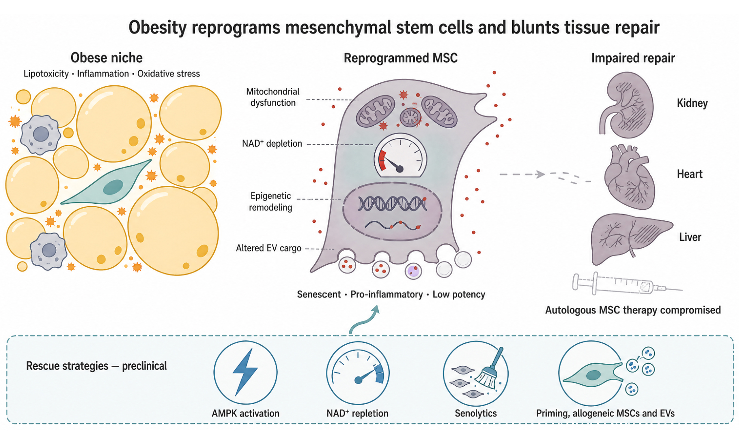
The authors used BioRender (BioRender.com), licensed to Mayo Clinic, to construct all schematic figures and the graphical abstract [Elmaraezy, A. (2026) https://BioRender.com/l737afg].
Authors’ contributions
Conceptualization and design of the review: Elmaraezy A, Lerman LO
Literature search, screening, and data extraction: Elmaraezy A
Drafting of the manuscript: Elmaraezy A
Figure and table preparation: Elmaraezy A, Zhu XY
Critical revision for important intellectual content: Zhu XY, Eirin A, Lerman LO
Supervision and final approval of the submitted version: Lerman LO
All authors have read and approved the final manuscript and agree to be accountable for all aspects of the work.
Availability of data and materials
Not applicable.
AI and AI-assisted tools statement
AI-assisted image-refinement tools, including FigureLabs (web-based application; released August 5, 2025) and Nano Banana Pro (Gemini 3 Pro Image, Google; released November 20, 2025), were used solely to improve visual clarity and layout. All figures are original to this work and were reviewed, verified, and edited by the authors. The authors are responsible for the accuracy and scientific integrity of the content presented in any images generated using AI tools.
Financial support and sponsorship
This work was partly supported by the National Institutes of Health (Nos. DK120292, DK122734, HL158691, and AG062104).
Conflicts of interest
Lerman LO is an advisor to CureSpec, Ribocure Pharmaceuticals, and LiveKidney.bio, outside the submitted work. The other authors declare no conflicts of interest.
Ethical approval and consent to participate
Not applicable.
Consent for publication
Not applicable.
Copyright
© The Author(s) 2026.
REFERENCES
1. Liu J, Fang Z, Lu Q, Wang Y, Zhang L. Projecting global trends and inequalities in adult overweight and obesity, 2023-2040: findings from the NCD-RisC database. Obesity. 2025;33:1955-67.
2. NCD Risk Factor Collaboration (NCD-RisC). Worldwide trends in underweight and obesity from 1990 to 2022: a pooled analysis of 3663 population-representative studies with 222 million children, adolescents, and adults. Lancet. 2024;403:1027-50.
3. Bianchettin RG, Gawey BJ, Acosta AJ, et al. Clinical assessment of people with obesity: focus on adiposity-related multimorbidity. Mayo Clin Proc. 2025;100:1005-29.
4. Xing L, Zhu X, Klomjit N, et al. Human obesity decreases the anti-inflammatory functionality of adipose tissue-derived mesenchymal stem/stomal cells by upregulating IL-1β expression. Stem Cells. 2025;43:sxaf058.
5. Pérez LM, de Lucas B, Gálvez BG. Unhealthy stem cells: when health conditions upset stem cell properties. Cell Physiol Biochem. 2018;46:1999-2016.
6. Dominici M, Le Blanc K, Mueller I, et al. Minimal criteria for defining multipotent mesenchymal stromal cells. The International Society for Cellular Therapy position statement. Cytotherapy. 2006;8:315-7.
7. Strioga M, Viswanathan S, Darinskas A, Slaby O, Michalek J. Same or not the same? Comparison of adipose tissue-derived versus bone marrow-derived mesenchymal stem and stromal cells. Stem Cells Dev. 2012;21:2724-52.
8. Mohamed-Ahmed S, Fristad I, Lie SA, et al. Adipose-derived and bone marrow mesenchymal stem cells: a donor-matched comparison. Stem Cell Res Ther. 2018;9:168.
9. Calcat-I-Cervera S, Rendra E, Scaccia E, et al. Harmonised culture procedures minimise but do not eliminate mesenchymal stromal cell donor and tissue variability in a decentralised multicentre manufacturing approach. Stem Cell Res Ther. 2023;14:120.
10. Engin AB. Mechanism of obesity-related lipotoxicity and clinical perspective. Adv Exp Med Biol. 2024;1460:131-66.
11. Ulum B, Teker HT, Sarikaya A, et al. Bone marrow mesenchymal stem cell donors with a high body mass index display elevated endoplasmic reticulum stress and are functionally impaired. J Cell Physiol. 2018;233:8429-36.
12. Ly LD, Xu S, Choi SK, et al. Oxidative stress and calcium dysregulation by palmitate in type 2 diabetes. Exp Mol Med. 2017;49:e291.
13. Lu J, Wang Q, Huang L, et al. Palmitate causes endoplasmic reticulum stress and apoptosis in human mesenchymal stem cells: prevention by AMPK activator. Endocrinology. 2012;153:5275-84.
14. Dalla Valle A, Vertongen P, Spruyt D, et al. Induction of stearoyl-CoA 9-desaturase 1 protects human mesenchymal stromal cells against palmitic acid-induced lipotoxicity and inflammation. Front Endocrinol. 2019;10:726.
15. Polat EG, Şeker ME, Pervin B, Ulum B, Aerts-Kaya F. Obesity-induced loss of function of bone marrow mesenchymal stromal cells is linked to cellular stress and irreversible at advanced stages. J Cell Mol Med. 2025;29:e70776.
16. Yaghooti H, Mohammadtaghvaei N, Mahboobnia K. Effects of palmitate and astaxanthin on cell viability and proinflammatory characteristics of mesenchymal stem cells. Int Immunopharmacol. 2019;68:164-70.
17. Yan W, Diao S, Fan Z. The role and mechanism of mitochondrial functions and energy metabolism in the function regulation of the mesenchymal stem cells. Stem Cell Res Ther. 2021;12:140.
18. Eirin A, Thaler R, Glasstetter LM, et al. Obesity-driven mitochondrial dysfunction in human adipose tissue-derived mesenchymal stem/stromal cells involves epigenetic changes. Cell Death Dis. 2024;15:387.
19. Meng Y, Eirin A, Zhu XY, et al. Obesity-induced mitochondrial dysfunction in porcine adipose tissue-derived mesenchymal stem cells. J Cell Physiol. 2018;233:5926-36.
20. de Mello AH, Costa AB, Engel JDG, Rezin GT. Mitochondrial dysfunction in obesity. Life Sci. 2018;192:26-32.
21. Wang B, Zhang G, Hu Y, et al. Uncovering impaired mitochondrial and lysosomal function in adipose-derived stem cells from obese individuals with altered biological activity. Stem Cell Res Ther. 2024;15:12.
22. Pérez LM, Bernal A, de Lucas B, et al. Altered metabolic and stemness capacity of adipose tissue-derived stem cells from obese mouse and human. PLoS One. 2015;10:e0123397.
23. Zheng P, Ma W, Gu Y, et al. High-fat diet causes mitochondrial damage and downregulation of mitofusin-2 and optic atrophy-1 in multiple organs. J Clin Biochem Nutr. 2023;73:61-76.
24. Finocchietto P, Perez H, Blanco G, et al. Inhibition of mitochondrial fission by Drp-1 blockade by short-term leptin and Mdivi-1 treatment improves white adipose tissue abnormalities in obesity and diabetes. Pharmacol Res. 2022;178:106028.
25. Farahani RA, Farah MC, Zhu XY, et al. Metabolic syndrome impairs 3D mitochondrial structure, dynamics, and function in swine mesenchymal stem cells. Stem Cell Rev Rep. 2020;16:933-45.
26. Liu L, Wang Y, Fan H, et al. MicroRNA-181a regulates local immune balance by inhibiting proliferation and immunosuppressive properties of mesenchymal stem cells. Stem Cells. 2012;30:1756-70.
27. Indrieri A, Carrella S, Romano A, et al. miR-181a/b downregulation exerts a protective action on mitochondrial disease models. EMBO Mol Med. 2019;11:e8734.
28. Kornicka K, Houston J, Marycz K. Dysfunction of mesenchymal stem cells isolated from metabolic syndrome and type 2 diabetic patients as result of oxidative stress and autophagy may limit their potential therapeutic use. Stem Cell Rev Rep. 2018;14:337-45.
29. Wan W, Zhang L, Lin Y, et al. Mitochondria-derived peptide MOTS-c: effects and mechanisms related to stress, metabolism and aging. J Transl Med. 2023;21:36.
30. Luo YH, Xie L, Li JY, Xie Y, Li MQ, Zhou L. Serum MOTS-C levels are decreased in obese children and associated with vascular endothelial function. Diabetes Metab Syndr Obes. 2023;16:1013-20.
31. Cataldo LR, Fernández-Verdejo R, Santos JL, Galgani JE. Plasma MOTS-c levels are associated with insulin sensitivity in lean but not in obese individuals. J Investig Med. 2018;66:1019-22.
32. Yoon SH, Yuan F, Zhu X, et al. Systemic MOTS-c levels are increased in adults with obesity in association with metabolic dysregulation and remain unchanged after weight loss. J Clin Transl Endocrinol. 2026;43:100429.
33. Sequeira IR, Woodhead JST, Chan A, et al. Plasma mitochondrial derived peptides MOTS-c and SHLP2 positively associate with android and liver fat in people without diabetes. Biochim Biophys Acta Gen Subj. 2021;1865:129991.
34. Inoki K, Kim J, Guan KL. AMPK and mTOR in cellular energy homeostasis and drug targets. Annu Rev Pharmacol Toxicol. 2012;52:381-400.
35. Marcondes-de-Castro IA, Reis-Barbosa PH, Marinho TS, Aguila MB, Mandarim-de-Lacerda CA. AMPK/mTOR pathway significance in healthy liver and non-alcoholic fatty liver disease and its progression. J Gastroenterol Hepatol. 2023;38:1868-76.
36. Tao Z, Aslam H, Parke J, Sanchez M, Cheng Z. Mechanisms of autophagic responses to altered nutritional status. J Nutr Biochem. 2022;103:108955.
37. Sagar S, Faizan MI, Chaudhary N, et al. Obesity impairs cardiolipin-dependent mitophagy and therapeutic intercellular mitochondrial transfer ability of mesenchymal stem cells. Cell Death Dis. 2023;14:324.
38. Al-Sammarraie SHA, Ayaz-Güner Ş, Acar MB, et al. Mesenchymal stem cells from adipose tissue prone to lose their stemness associated markers in obesity related stress conditions. Sci Rep. 2024;14:19702.
39. Kraus D, Yang Q, Kong D, et al. Nicotinamide N-methyltransferase knockdown protects against diet-induced obesity. Nature. 2014;508:258-62.
40. Choi SE, Fu T, Seok S, et al. Elevated microRNA-34a in obesity reduces NAD+ levels and SIRT1 activity by directly targeting NAMPT. Aging Cell. 2013;12:1062-72.
41. Jukarainen S, Heinonen S, Rämö JT, et al. Obesity is associated with low NAD+/SIRT pathway expression in adipose tissue of BMI-discordant monozygotic twins. J Clin Endocrinol Metab. 2016;101:275-83.
42. Afarideh M, Thaler R, Khani F, et al. Global epigenetic alterations of mesenchymal stem cells in obesity: the role of vitamin C reprogramming. Epigenetics. 2021;16:705-17.
43. Xie S, Choudhari S, Wu CL, et al. Aging and obesity prime the methylome and transcriptome of adipose stem cells for disease and dysfunction. FASEB J. 2023;37:e22785.
44. Tryggestad JB, Teague AM, Sparling DP, Jiang S, Chernausek SD. Macrophage-derived microRNA-155 increases in obesity and influences adipocyte metabolism by targeting peroxisome proliferator-activated receptor gamma. Obesity. 2019;27:1856-64.
45. Pan Y, Hui X, Hoo RLC, et al. Adipocyte-secreted exosomal microRNA-34a inhibits M2 macrophage polarization to promote obesity-induced adipose inflammation. J Clin Invest. 2019;129:834-49.
46. Wang L, Sun M, Cao Y, et al. miR-34a regulates lipid metabolism by targeting SIRT1 in non-alcoholic fatty liver disease with iron overload. Arch Biochem Biophys. 2020;695:108642.
47. Briones-Espinoza MJ, Cortés-García JD, Vega-Cárdenas M, et al. Decreased levels and activity of Sirt1 are modulated by increased miR-34a expression in adipose tissue mononuclear cells from subjects with overweight and obesity: a pilot study. Diabetes Metab Syndr. 2020;14:1347-54.
48. Mokhberian N, Bolandi Z, Eftekhary M, et al. Inhibition of miR-34a reduces cellular senescence in human adipose tissue-derived mesenchymal stem cells through the activation of SIRT1. Life Sci. 2020;257:118055.
49. Karkeni E, Astier J, Tourniaire F, et al. Obesity-associated inflammation induces microRNA-155 expression in adipocytes and adipose tissue: outcome on adipocyte function. J Clin Endocrinol Metab. 2016;101:1615-26.
50. Onodera Y, Teramura T, Takehara T, Obora K, Mori T, Fukuda K. miR-155 induces ROS generation through downregulation of antioxidation-related genes in mesenchymal stem cells. Aging Cell. 2017;16:1369-80.
51. Bispo AFS, de Jesus Simao J, Moraes MA, et al. Effects of obesity-associated plasma markers on adipose stem cell function and epigenetic regulation. Obesity. 2025;33:1529-42.
52. Grun LK, Maurmann RM, Scholl JN, et al. Obesity drives adipose-derived stem cells into a senescent and dysfunctional phenotype associated with P38MAPK/NF-KB axis. Immun Ageing. 2023;20:51.
53. Kang Q, Zhu X, Ren D, et al. Adipose METTL14-elicited N6-methyladenosine promotes obesity, insulin resistance, and NAFLD through suppressing β adrenergic signaling and lipolysis. Adv Sci. 2023;10:e2301645.
54. Wang X, Wu R, Liu Y, et al. m6A mRNA methylation controls autophagy and adipogenesis by targeting Atg5 and Atg7. Autophagy. 2020;16:1221-35.
55. Wu Y, Xie L, Wang M, et al. Mettl3-mediated m6A RNA methylation regulates the fate of bone marrow mesenchymal stem cells and osteoporosis. Nat Commun. 2018;9:4772.
56. He M, Lei H, He X, et al. METTL14 regulates osteogenesis of bone marrow mesenchymal stem cells via inducing autophagy through m6A/IGF2BPs/Beclin-1 signal axis. Stem Cells Transl Med. 2022;11:987-1001.
57. Hinte LC, Castellano-Castillo D, Ghosh A, et al. Adipose tissue retains an epigenetic memory of obesity after weight loss. Nature. 2024;636:457-65.
58. Silva KR, Liechocki S, Carneiro JR, et al. Stromal-vascular fraction content and adipose stem cell behavior are altered in morbid obese and post bariatric surgery ex-obese women. Stem Cell Res Ther. 2015;6:72.
59. Adnan A, Juntunen M, Tyrväinen T, et al. Effects of bariatric surgery-related weight loss on the characteristics, metabolism, and immunomodulation of adipose stromal/stem cells in a follow-up study. Stem Cells Int. 2025;2025:1212255.
60. Miranda AMA, McAllan L, Mazzei G, et al. Selective remodelling of the adipose niche in obesity and weight loss. Nature. 2025;644:769-79.
61. Keshtkar S, Azarpira N, Ghahremani MH. Mesenchymal stem cell-derived extracellular vesicles: novel frontiers in regenerative medicine. Stem Cell Res Ther. 2018;9:63.
62. Welsh JA, Goberdhan DCI, O’Driscoll L, et al.; MISEV Consortium. Minimal information for studies of extracellular vesicles (MISEV2023): from basic to advanced approaches. J Extracell Vesicles. 2024;13:e12404.
63. Togliatto G, Dentelli P, Gili M, et al. Obesity reduces the pro-angiogenic potential of adipose tissue stem cell-derived extracellular vesicles (EVs) by impairing miR-126 content: impact on clinical applications. Int J Obes. 2016;40:102-11.
64. Eirin A, Meng Y, Zhu XY, et al. The micro-RNA cargo of extracellular vesicles released by human adipose tissue-derived mesenchymal stem cells is modified by obesity. Front Cell Dev Biol. 2021;9:660851.
65. Conley SM, Shook JE, Zhu XY, et al. Metabolic syndrome induces release of smaller extracellular vesicles from porcine mesenchymal stem cells. Cell Transplant. 2019;28:1271-8.
66. Eirin A, Ferguson CM, Zhu XY, et al. Extracellular vesicles released by adipose tissue-derived mesenchymal stromal/stem cells from obese pigs fail to repair the injured kidney. Stem Cell Res. 2020;47:101877.
67. Conley SM, Hickson LJ, Kellogg TA, et al. Human obesity induces dysfunction and early senescence in adipose tissue-derived mesenchymal stromal/stem cells. Front Cell Dev Biol. 2020;8:197.
68. Zhu Z, Gan X, Yu H. NF‑κB‑miR15a‑bFGF/VEGFA axis contributes to the impaired angiogenic capacity of BM‑MSCs in high fat diet‑fed mice. Mol Med Rep. 2017;16:7609-16.
69. Serena C, Keiran N, Ceperuelo-Mallafre V, et al. Obesity and type 2 diabetes alters the immune properties of human adipose derived stem cells. Stem Cells. 2016;34:2559-73.
70. Acar MB, Ayaz-Güner Ş, Di Bernardo G, et al. Obesity induced by high-fat diet is associated with critical changes in biological and molecular functions of mesenchymal stromal cells present in visceral adipose tissue. Aging. 2020;12:24894-913.
71. Klomjit N, Conley SM, Zhu XY, et al. Effects of obesity on reparative function of human adipose tissue-derived mesenchymal stem cells on ischemic murine kidneys. Int J Obes. 2022;46:1222-33.
72. Farahani RA, Zhu XY, Tang H, et al. Metabolic syndrome alters the cargo of mitochondria-related microRNAs in swine mesenchymal stem cell-derived extracellular vesicles, impairing their capacity to repair the stenotic kidney. Stem Cells Int. 2020;2020:8845635.
73. Yu S, Klomjit N, Jiang K, et al. Human obesity attenuates cardioprotection conferred by adipose tissue-derived mesenchymal stem/stromal cells. J Cardiovasc Transl Res. 2023;16:221-32.
74. Hong S, Huang W, Zhu X, et al. Obesity blunts amelioration of cardiac hypertrophy and fibrosis by human mesenchymal stem/stromal cell-derived extracellular vesicles. Am J Physiol Heart Circ Physiol. 2023;325:H163-71.
75. Younossi ZM, Kalligeros M, Henry L. Epidemiology of metabolic dysfunction-associated steatotic liver disease. Clin Mol Hepatol. 2025;31:S32-50.
76. Yao S, Cen Y, Lou G, Liu Y. Therapeutic potentials of mesenchymal stem cells and their extracellular vesicles on liver diseases by modulating mitochondrial function of macrophages. Int Immunopharmacol. 2025;165:115486.
77. Cen Y, Lou G, Qi J, Zheng M, Liu Y. A new perspective on mesenchymal stem cell-based therapy for liver diseases: restoring mitochondrial function. Cell Commun Signal. 2023;21:214.
78. Shree N, Venkategowda S, Venkatranganna MV, Datta I, Bhonde RR. Human adipose tissue mesenchymal stem cells as a novel treatment modality for correcting obesity induced metabolic dysregulation. Int J Obes. 2019;43:2107-18.
79. Shen Z, Tang X, Zhang Y, et al. Efficacy and safety of mesenchymal stem cell therapies for ischemic stroke: a systematic review and meta-analysis. Stem Cells Transl Med. 2024;13:886-97.
80. Chen C, Peng C, Hu Z, Ge L. Effects of bone marrow mesenchymal stromal cells-derived therapies for experimental traumatic brain injury: a meta-analysis. Heliyon. 2024;10:e25050.
81. Kota DJ, Prabhakara KS, Toledano-Furman N, et al. Prostaglandin E2 indicates therapeutic efficacy of mesenchymal stem cells in experimental traumatic brain injury. Stem Cells. 2017;35:1416-30.
82. Park HR, Park M, Choi J, Park KY, Chung HY, Lee J. A high-fat diet impairs neurogenesis: involvement of lipid peroxidation and brain-derived neurotrophic factor. Neurosci Lett. 2010;482:235-9.
83. Li J, Tang Y, Cai D. IKKβ/NF-κB disrupts adult hypothalamic neural stem cells to mediate a neurodegenerative mechanism of dietary obesity and pre-diabetes. Nat Cell Biol. 2012;14:999-1012.
84. Khalid M, Frommer ML, Abu-Hanna J, et al. Influence of donor obesity on adipose-derived stem cell function and therapeutic efficacy. Cells. 2026;15:946.
85. Liu F, Han R, Nie S, et al. Metformin rejuvenates Nap1l2-impaired immunomodulation of bone marrow mesenchymal stem cells via metabolic reprogramming. Cell Prolif. 2024;57:e13612.
86. Mikłosz A, Nikitiuk BE, Chabowski A. Using adipose-derived mesenchymal stem cells to fight the metabolic complications of obesity: where do we stand? Obes Rev. 2022;23:e13413.
87. Le Pelletier L, Mantecon M, Gorwood J, et al. Metformin alleviates stress-induced cellular senescence of aging human adipose stromal cells and the ensuing adipocyte dysfunction. Elife. 2021;10:e62635.
88. Ouyang Z, Kang D, Li K, et al. DEPTOR exacerbates bone-fat imbalance in osteoporosis by transcriptionally modulating BMSC differentiation. Biomed Pharmacother. 2022;151:113164.
89. Wang H, Sun Y, Pi C, et al. Nicotinamide mononucleotide supplementation improves mitochondrial dysfunction and rescues cellular senescence by NAD+/Sirt3 pathway in mesenchymal stem cells. Int J Mol Sci. 2022;23:14739.
90. Song J, Li J, Yang F, et al. Nicotinamide mononucleotide promotes osteogenesis and reduces adipogenesis by regulating mesenchymal stromal cells via the SIRT1 pathway in aged bone marrow. Cell Death Dis. 2019;10:336.
91. Chinnapaka S, Malekzadeh H, Tirmizi Z, Arellano JA, Ejaz A. Nicotinamide riboside improves stemness of human adipose-derived stem cells and inhibits terminal adipocyte differentiation. Pharmaceuticals. 2023;16:1134.
92. Lopez-Yus M, García-Sobreviela MP, Del Moral-Bergos R, Arbones-Mainar JM. Gene therapy based on mesenchymal stem cells derived from adipose tissue for the treatment of obesity and its metabolic complications. Int J Mol Sci. 2023;24:7468.
93. Palmer AK, Xu M, Zhu Y, et al. Targeting senescent cells alleviates obesity-induced metabolic dysfunction. Aging Cell. 2019;18:e12950.
94. Pham DV, Nguyen TK, Park PH. Adipokines at the crossroads of obesity and mesenchymal stem cell therapy. Exp Mol Med. 2023;55:313-24.
95. Pham DV, Nguyen TK, Nguyen BL, et al. Adiponectin restores the obesity-induced impaired immunomodulatory function of mesenchymal stromal cells via glycolytic reprogramming. Acta Pharm Sin B. 2024;14:273-91.
96. Song J, Liu J, Cui C, et al. Mesenchymal stromal cells ameliorate diabetes-induced muscle atrophy through exosomes by enhancing AMPK/ULK1-mediated autophagy. J Cachexia Sarcopenia Muscle. 2023;14:915-29.
97. Wang Y, Fu J, He W, et al. Bone marrow mesenchymal stem cells ameliorate diet-induced obesity by activating thermogenesis and alleviating inflammation in adipose tissue. Biochem Biophys Res Commun. 2025;747:151172.
98. Kashbour M, Abdelmalik A, Yassin MNA, et al. Mesenchymal stem cell-based therapy for type 1 & 2 diabetes mellitus patients: a systematic review and meta-analysis of randomized controlled trials. Diabetol Metab Syndr. 2025;17:189.
99. Mathur A, Taurin S, Alshammary S. The safety and efficacy of mesenchymal stem cells in the treatment of type 2 diabetes - a literature review. Diabetes Metab Syndr Obes. 2023;16:769-77.
100. Mizenko RR, Feaver M, Bozkurt BT, et al. A critical systematic review of extracellular vesicle clinical trials. J Extracell Vesicles. 2024;13:e12510.
Cite This Article
 Open Access
How to Cite
Download Citation
Export Citation File:
Type of Import
Tips on Downloading Citation
Citation Manager File Format
Type of Import
Direct Import: When the Direct Import option is selected (the default state), a dialogue box will give you the option to Save or Open the downloaded citation data. Choosing Open will either launch your citation manager or give you a choice of applications with which to use the metadata. The Save option saves the file locally for later use.
Indirect Import: When the Indirect Import option is selected, the metadata is displayed and may be copied and pasted as needed.
About This Article
Special Topic
Copyright

Data & Comments
Data
0









Comments
Comments must be written in English. Spam, offensive content, impersonation, and private information will not be permitted. If any comment is reported and identified as inappropriate content by OAE staff, the comment will be removed without notice. If you have any queries or need any help, please contact us at support@oaepublish.com.