



Noncoding RNAs in cardiovascular aging and diseases
 0
0  ,
, Abstract
This review comprehensively explores the roles of noncoding RNAs (ncRNAs) in cardiovascular diseases (CVDs), highlighting their involvement in major pathological programs and cellular functions. The diverse types of ncRNAs, including microRNAs (miRNAs), long noncoding RNAs (lncRNAs), and circular RNAs (circRNAs), and their mechanisms in regulating gene expression and cellular functions are reviewed. We also examine the potential of ncRNAs as diagnostic biomarkers and therapeutic targets, emphasizing recent advancements in delivery systems and clinical trials. Challenges such as achieving tissue-specific delivery, minimizing off-target effects, and ensuring long-term safety are addressed. By synthesizing current research and addressing translational challenges, this review aims to elucidate the transformative potential of ncRNAs in cardiovascular medicine, paving the way for innovative, targeted therapies
Keywords
INTRODUCTION
Cardiovascular diseases (CVDs) remain the leading cause of global morbidity and mortality, imposing an unprecedented burden on healthcare systems and economies worldwide[1]. Despite significant advancements in diagnostic and therapeutic approaches, the complex molecular mechanisms driving CVD progression and pathogenesis remain not fully elucidated. This knowledge gap highlights the urgent need for innovative strategies to improve patient outcomes. In recent years, noncoding RNAs (ncRNAs), a heterogeneous class of RNA molecules that do not encode proteins, have emerged as key players in cardiovascular biology and disease[2]. Once dismissed as mere transcriptional byproducts, ncRNAs are now recognized as critical regulators of gene expression, acting at transcriptional, post-transcriptional, and epigenetic levels to fine-tune cellular processes essential for cardiovascular homeostasis[3].
The essential role of ncRNAs in maintaining cardiovascular homeostasis is robustly demonstrated by loss-of-function genetic models. For example, cardiac-specific deletion of miR-208a disrupts the stress-responsive switch of myosin heavy chain genes, leading to aberrant cardiac remodeling and impaired contractility even in the absence of pathological stress[4]. Similarly, deletion of the endothelial-enriched miR-126 compromises vascular integrity by attenuating angiogenic signaling and promoting capillary leakage, thereby demonstrating its non-redundant function in vascular homeostasis[5]. The lncRNA Myosin Heavy Chain Associated RNA Transcript (MHRT) is essential for repressing the chromatin remodeler Brg1; its loss in the adult heart triggers pathological hypertrophy and fatal heart failure, confirming its critical role in maintaining cardiac gene expression programs and structural integrity[6]. These findings collectively underscore that ncRNAs are not merely stress-responsive elements but are fundamental components essential for the basal stability of the cardiovascular system.
The advent of high-throughput sequencing technologies and bioinformatics tools has revolutionized our understanding of ncRNAs, revealing their extensive involvement in key pathological processes such as inflammatory signaling[7,8], matrix remodeling[9,10], cell death[11,12], angiogenesis[10,13], and vascular remodeling[7,14]. These processes are integral to the development and progression of major CVDs, including atherosclerosis, myocardial infarction, heart failure, arrhythmias, and endothelial dysfunction. Among the diverse subtypes of ncRNAs, microRNAs (miRNAs), long noncoding RNAs (lncRNAs), and circular RNAs (circRNAs) have been extensively studied for their roles in cardiovascular pathophysiology[15]. Additionally, emerging classes of ncRNAs, such as transfer RNA-derived small RNAs (tsRNAs) and piwi-interacting RNAs (piRNAs), are increasingly recognized for their regulatory functions and clinical relevance as disease drivers, biomarkers, and therapeutic targets. Specific tsRNAs (e.g., tRF-21-NB8PLML3E[16]) are dysregulated in pathological cardiac hypertrophy and correlate with clinical parameters such as ventricular wall dimensions, establishing their dual roles as pathogenic drivers and diagnostic biomarkers[17,18]. piRNAs similarly demonstrate significant clinical relevance through multiple mechanisms: their detection in blood extracellular vesicles enables high-accuracy classification of diseases such as Alzheimer’s and cholangiocarcinoma, underscoring their utility as non-invasive biomarkers[19,20]. Furthermore, piRNAs modulate critical pathways, including calcium signaling and epigenetic regulation, highlighting their therapeutic potential for conditions such as cardiac arrhythmias and cancer[21-23].
Beyond their biological roles, ncRNAs demonstrate high stability and specificity in biofluids, positioning them as promising noninvasive biomarkers for the diagnosis, prognosis, and risk stratification of CVDs[24,25]. This stability arises from protection against degradation through encapsulation in extracellular vesicles (EVs) or association with RNA-binding proteins and lipoproteins, which confers a comparative advantage over other cell-free nucleic acids such as mRNA[24,25]. This robustness is evidenced by the consistent detection of specific ncRNAs, including miRNAs and transfer RNA (tRNA) fragments, in biofluids such as plasma and cerebrospinal fluid, where they remain resistant to endogenous nucleases[26,27]. Their specificity stems from distinct, tissue-enriched expression patterns; for instance, certain tissue-specific miRNAs and tRNA-derived fragments (tRFs) can accurately indicate pathological states such as ischaemic heart disease and hypertension[28,29]. Technological advances, particularly in RNA sequencing and metal-organic framework (MOF)-based extraction methods, have further validated these properties by enabling robust quantification of distinct ncRNA signatures in biofluids[30]. Furthermore, the therapeutic potential of ncRNAs is being actively explored, with strategies such as antisense oligonucleotides, RNA mimics, and small molecule inhibitors demonstrating potential in preclinical and clinical studies[31]. However, the translation of ncRNA-based therapies into clinical practice faces significant challenges, including delivery specificity, off-target effects, and scalability. Innovative delivery platforms, such as lipid nanoparticles, exosomes, and viral vectors, are being developed to address these challenges and improve the precision and efficacy of ncRNA therapeutics[32].
This review offers an in-depth examination of the functional diversity and mechanistic roles of ncRNAs in cardiovascular biology, with the discussion organized from ncRNA classification to cardiovascular aging, major disease settings, clinical biomarker applications, and therapeutic strategies. By synthesizing recent mechanistic and translational findings, this review aims to clarify how ncRNAs may reshape cardiovascular medicine and support the development of more precise, targeted interventions. A preliminary version of this study has been published as a preprint[33].
TYPES OF NONCODING RNAs IN CARDIOVASCULAR DISEASES
ncRNAs comprise a diverse group of RNA molecules that do not encode proteins but are essential to regulate gene expression and cellular processes in cardiovascular biology and disease. Recent advances in high-throughput sequencing technologies have uncovered a wide range of roles and mechanisms of ncRNAs in CVDs, including transcriptional regulation, post-transcriptional modification, and epigenetic regulation [Table 1]. Despite significant progress, the complete landscape of ncRNAs’ regulatory networks in CVDs is not fully understood, necessitating further exploration of their interactions with cellular signaling pathways.
Examples of ncRNAs in cardiovascular diseases
| ncRNA type | Example | Function | Detailed signaling targets | Associated disease |
| miRNA | miR-21 | Promotes cardiac fibrosis | Transforming growth factor (TGF)β/Smad7 | Cardiac fibrosis[34] |
| miRNA | miR-155 | Pro-inflammatory, promotes atherosclerosis | B-cell lymphoma 6 (BCL6); signal transducer and activator of transcription 3 (STAT3) | Atherosclerosis[35] |
| miRNA | miR-126 | Protects vascular integrity | Vascular endothelial growth factor (VEGF) | Vascular repair[36] |
| miRNA | miR-133 | Ensures normal cardiac development and function | Serum response factor (SRF)/myocardin/myocyte enhancer factor-2 (MEF2) | Cardiac development[37] |
| lncRNA | Metastasis associated lung adenocarcinoma transcript 1 (MALAT1) | Regulates endothelial cell proliferation and angiogenesis | VEGF | Angiogenesis[38] |
| lncRNA | Myocardial infarction associated transcript (MIAT) | Attenuates H9C2 cell pyroptosis | Splicing factor 1 (SF1)/calcitonin gene-related peptide (CGRP) | Myocardial infarction[39] |
| circRNA | Heart-related circular RNA (HRCR) | Protects against hypertrophy by sequestering miR-223 | HRCR/miR-223/ARC | Cardiac hypertrophy[40] |
| circRNA | (Circular antisense non-coding RNA in the INK4 locus) cANRIL | Regulates cell proliferation and apoptosis in vascular smooth muscle cells | Apoptosis | Atherosclerosis[41] |
| lncRNA | (Wisp2 super-enhancer-associated RNA) wisper | Exacerbates myocardial fibrosis and remodeling | Wisper/T-cell intracellular antigen 1-related (TIAR)/procollagen-lysine, 2-oxoglutarate 5-dioxygenase 2 (PLOD2) | Heart failure[42] |
| lncRNA | LINC00607 | Contributes to endothelial dysfunction under high glucose and TNFα conditions | VEGF; extracellular matrix (ECM) remodling; HIF-1α | Vascular disease[43,44] |
miRNAs
miRNAs are small, single-stranded RNA molecules approximately 22 nucleotides in length that bind to complementary sequences on target messenger RNAs (mRNAs), leading to mRNA degradation or translational repression. In cardiovascular diseases, miRNAs serve as crucial regulators of processes such as inflammation, fibrosis, apoptosis, and angiogenesis. miR-21 is a representative profibrotic miRNA in cardiovascular disease, but its context-specific mechanisms are discussed in later sections to avoid redundancy. Current research identifies miR-21 as a promising therapeutic target, although achieving tissue-specific inhibition without off-target effects in non-cardiac tissues remains a significant challenge[45].
miR-155, a pro-inflammatory miRNA, plays a crucial role in the progression of atherosclerosis[35,46,47]. Modulating miR-155 in animal models has demonstrated promising results in reducing atherosclerotic burden, but translating these findings to human therapies requires a deeper understanding of the pleiotropic effects. Similarly, miR-126, which is predominantly found in endothelial cells, plays a protective role in maintaining vascular integrity and repair[36,48]. Given its protective role in vascular biology, miR-126 holds significant potential as a biomarker for early-stage vascular diseases, although challenges in reliably measuring circulating miR-126 levels in clinical practice remain. Dysregulated miRNAs are often detected in plasma and tissue samples from patients with CVDs, underscoring their potential as biomarkers and therapeutic targets.
lncRNAs
lncRNAs are RNA transcripts exceeding 200 nucleotides in length, exhibiting diverse functions such as chromatin remodeling, transcriptional regulation, and post-transcriptional modification. lncRNAs such as MALAT1 (metastasis-associated lung adenocarcinoma transcript 1) and MIAT (myocardial infarction-associated transcript) have been widely investigated in cardiovascular diseases. lncRNA MALAT1 regulates endothelial cell proliferation and angiogenesis[38]. Emerging evidence indicates that inhibiting MALAT1 may reduce pathological angiogenesis in conditions such as diabetic retinopathy, underscoring its dual roles in health and disease. lncRNA MIAT is involved in myocardial infarction and contributes to cardiac fibrosis and dysfunction[39,49]. Although targeting MIAT therapeutically has potential, its involvement in multiple signaling pathways complicates the development of specific inhibitors, highlighting the need for precision medicine. lncRNAs hold significant therapeutic potential due to their ability to interact with multiple molecular pathways, making them promising targets for the treatment of cardiovascular diseases. However, the low expression levels and nuclear localization of lncRNAs pose challenges for their therapeutic application in vivo.
circRNAs
circRNAs are single-stranded RNA molecules characterized by a covalently closed circular structure formed via back-splicing. Unlike linear RNAs, circRNAs are resistant to exonucleases, thereby conferring enhanced stability. In cardiovascular diseases, circRNAs act as sponges for miRNAs, indirectly regulating mRNA targets. For instance, circular antisense non-coding RNA in the INK4 locus (circANRIL), which is transcribed from the same locus as Antisense non-coding RNA in the INK4 locus (ANRIL), regulates cell proliferation and apoptosis in vascular smooth muscle cells, thereby contributing to atherogenesis[41,50]. The dual roles of circANRIL in promoting inflammation and cell survival render it a complex therapeutic target, necessitating further studies to balance its pro- and anti-atherogenic effects. Another example is circRNA Heart-related Circular RNA (HRCR), which safeguards the heart against hypertrophy and heart failure by sponging miR-223[40]. circRNAs are also emerging as promising diagnostic markers due to their abundance in blood and tissue and their disease-specific expression profiles. While promising, translating circRNA-based therapies, such as HRCR, into clinical practice will require overcoming delivery challenges due to their large size and stable structure.
Other noncoding RNAs
In addition to miRNAs, lncRNAs, and circRNAs, other classes of ncRNAs have recently attracted attention for their roles in cardiovascular diseases. Traditionally known for guiding ribosomal RNA (rRNA) modifications, small nucleolar RNAs (snoRNAs) have been implicated in cardiovascular stress responses. For instance, SNORD113 and SNORD114 play a role in cardiomyocyte survival under ischemic conditions[51,52]. Recent studies highlight the role of snoRNAs in cellular stress responses, suggesting their potential as novel therapeutic targets for ischemic heart diseases. snoRNAs also regulate alternative splicing and gene expression, further broadening their functional repertoire in cardiovascular diseases.
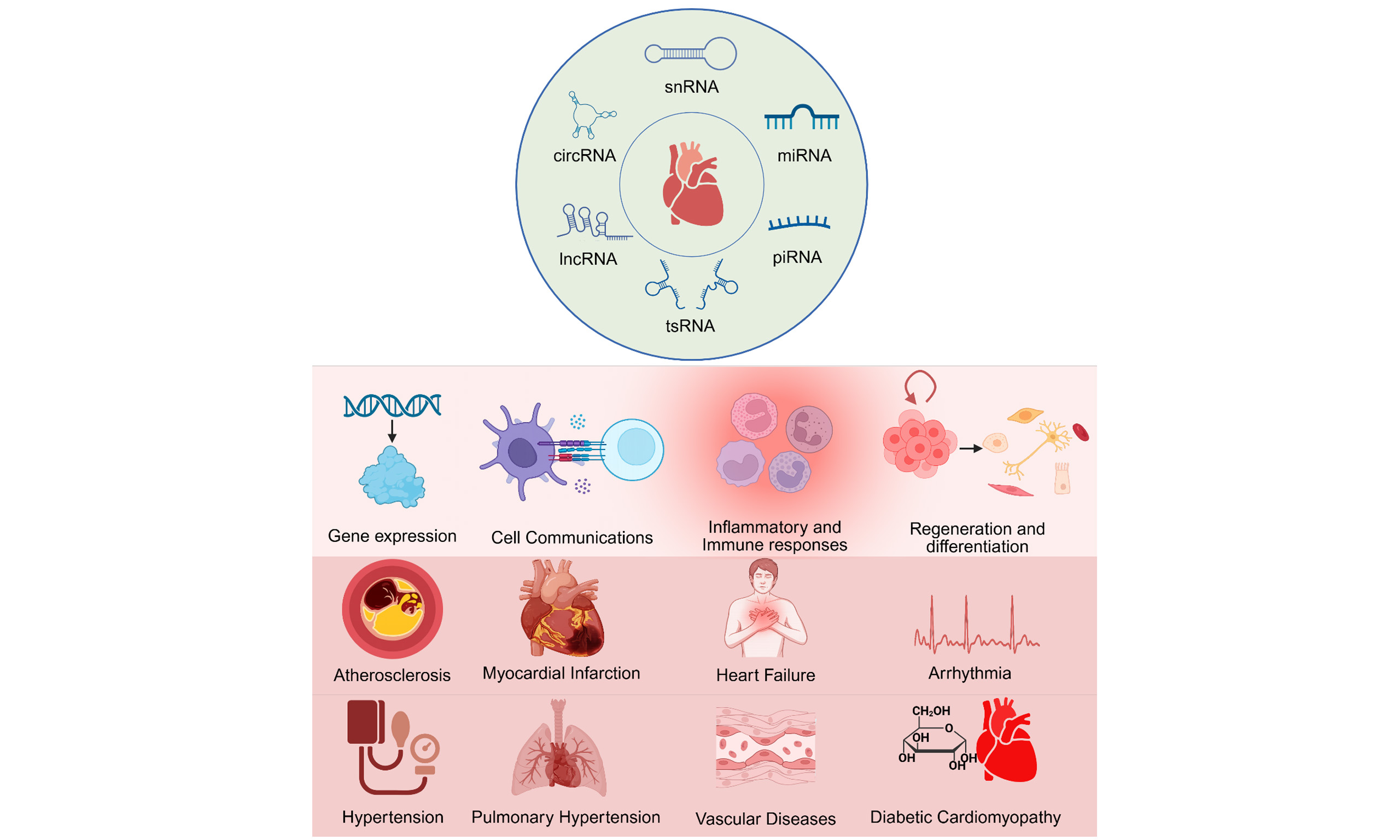
Small nuclear RNAs (snRNAs), core RNA components of spliceosomal small nuclear ribonucleoproteins, also warrant brief consideration because the graphical abstract includes this class of ncRNAs. U1, U2, U4, U5, and U6 snRNAs participate in pre-mRNA splice-site recognition and spliceosome catalysis; therefore, snRNA-related dysregulation is most relevant to cardiovascular disease through altered RNA splicing programs rather than through canonical miRNA-like targeting mechanisms. Consistent with this concept, dysregulated alternative splicing and spliceosome-associated pathways have been implicated in cardiac remodeling, cardiomyopathy, and heart failure, although direct cardiovascular studies focused on individual snRNAs remain limited[53,54].
piRNAs, initially identified in germline cells, are small RNAs that associate with PIWI proteins and participate in epigenetic regulation and genome stability. Emerging evidence indicates that piRNAs may regulate cardiovascular pathologies by modulating transposable elements and influencing inflammatory and oxidative stress pathways[55-57]. Although their role in cardiovascular diseases is still in its early stages, piRNAs represent a promising frontier for epigenetic therapeutics.
tsRNAs are small RNA molecules generated by specific cleavage of tRNAs. tsRNAs are implicated in stress responses, regulation of protein synthesis, and the formation of stress granules. In cardiovascular diseases, tsRNAs are thought to influence cardiac remodeling and endothelial function[58-60]. The biogenesis of tsRNAs involves specific cleavage events within the nucleus and mitochondria. In the nucleus, tsRNAs are generated by enzymes such as angiogenin and RNase Z, which cleave precursor tRNAs at specific sites to produce mature tsRNAs[61,62]. These nuclear tsRNAs can then be exported to the cytoplasm to exert their regulatory functions. The biogenesis of tsRNAs in the mitochondria is a complex process involving the cleavage of mitochondrial tRNAs by specific enzymes. These mitochondrial tsRNAs have been noted to play crucial roles in regulating mitochondrial function and dynamics. For instance, some mitochondrial tsRNAs modulate the activity of mitochondrial respiratory chain complexes, thereby influencing cellular energy metabolism[63,64]. Additionally, mitochondrial tsRNAs have been implicated in regulating mitochondrial fission and fusion, critical processes for maintaining mitochondrial homeostasis. Mitochondrial tsRNAs may also be involved in the response to oxidative stress, potentially contributing to the protection of cells against oxidative damage[65-67]. The emerging role of tsRNAs in cardiovascular diseases underscores the need for high-throughput studies to elucidate their specific targets and functions.
NONCODING RNAS IN CARDIOVASCULAR AGING
Cardiovascular aging arises from linked disturbances in endothelial redox balance, mitochondrial function, cellular senescence, and extracellular matrix turnover [Figure 1A]. In this context, ncRNAs are better understood as mechanistic regulators than as passive markers, because they link molecular stress signals to endothelial dysfunction, vascular stiffening, and age-associated cardiovascular decline[68-70].
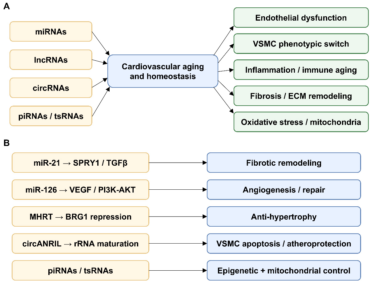
Figure 1. Mechanistic overview of ncRNAs in cardiovascular aging and disease. (A) Summarizes how miRNAs, lncRNAs, circRNAs, piRNAs, and tsRNAs converge on endothelial dysfunction, vascular smooth muscle phenotypic switching, inflammation and immune aging, fibrosis and extracellular matrix remodeling, and oxidative stress or mitochondrial dysfunction; (B) Highlights representative ncRNA mechanisms, including miR-21-SPRY1/TGFβ-driven fibrotic remodeling, miR-126-VEGF/PI3K-AKT-mediated angiogenesis and repair, MHRT-mediated BRG1 repression, circANRIL-associated rRNA maturation, and piRNA/tsRNA-mediated epigenetic and mitochondrial control. lncRNAs: Long noncoding RNAs; circRNAs: circular RNAs; piRNAs: piwi-interacting RNAs; tsRNAs: transfer RNA-derived small RNAs; rRNA: ribosomal RNA; miRNAs: microRNAs; ECM: extracellular matrix; VSMC: vascular smooth muscle cell; MHRT: myosin heavy chain associated RNA transcript; BRG1: brahma-related gene 1; VEGF: vascular endothelial growth factor; circANRIL: circular antisense non-coding RNA in the INK4 locus; SPRY1: sprouty RTK signaling antagonist; TGFβ: transforming growth factor-beta; PI3K: phosphoinositide 3-kinase; AKT: protein kinase B.
Oxidative stress and endothelial dysfunction
Oxidative stress is a major driver of vascular aging because it reduces nitric oxide bioavailability and weakens endothelial function. Several ncRNAs sit directly in this pathway. miR-34a is increased in senescent endothelial cells and represses sirtuin 1 (SIRT1), thereby favoring reactive oxygen species accumulation and endothelial senescence[71]. miR-217 acts on the same SIRT1-centered axis and likewise accelerates endothelial aging while impairing angiogenic capacity[72]. In parallel, the lncRNAs H19 and maternally expressed 3 (MEG3) modulate endothelial stress responses through STAT3- and p53-linked pathways, indicating that both small and long ncRNAs participate in the control of endothelial homeostasis during aging[73,74].
Mitochondrial dysfunction
Mitochondrial dysfunction is another defining feature of cardiovascular aging because it reduces energy efficiency and amplifies oxidative injury. miR-181c is a representative mitochondria-associated miRNA that can translocate to mitochondria and alter mitochondrial gene expression, electron transport chain activity, and cardiac bioenergetics[75]. These observations suggest that age-related mitochondrial decline is shaped, at least in part, by ncRNA-dependent control of organellar programs rather than by secondary damage alone.
Cellular senescence and senescence-associated secretory phenotype
Cellular senescence contributes to cardiovascular aging through durable growth arrest and the development of a senescence-associated secretory phenotype. The shared SIRT1-centered actions of miR-34a and miR-217 place these miRNAs near the core of endothelial senescence control[71,72]. H19 depletion induces a premature senescent endothelial phenotype, whereas MEG3 has been linked to impaired endothelial regeneration and heightened senescence-related signaling[73,74]. Taken together, these findings indicate that ncRNAs influence both the onset of senescence and the pro-inflammatory microenvironment that sustains vascular aging.
Extracellular matrix remodeling and fibrosis
Age-associated cardiovascular remodeling is marked by extracellular matrix accumulation, vascular stiffening, and fibrosis. In this setting, miR-21 is best viewed as a convergence node between stress signaling and fibroblast activation rather than as an isolated marker. Its profibrotic actions link ncRNA dysregulation to matrix deposition, myofibroblast persistence, and the structural deterioration of the aging heart and vasculature[34,76].
Overall, the available evidence argues against viewing cardiovascular aging as a collection of isolated ncRNA changes. Instead, oxidative stress, mitochondrial dysfunction, cellular senescence, and matrix remodeling appear to be connected outputs of overlapping ncRNA-target-pathway networks.
Noncoding RNAs in key cardiovascular pathologies
ncRNAs play a pivotal role in regulating key cardiovascular pathologies. Their ability to modulate gene expression and cellular functions has been extensively studied in various cardiovascular conditions, including atherosclerosis, myocardial infarction, heart failure, arrhythmias, and vascular diseases.
Atherosclerosis
In atherosclerosis, ncRNAs regulate lipid metabolism, endothelial function, and inflammatory responses. For example, miR-33 regulates cholesterol homeostasis by targeting genes involved in high-density lipoprotein (HDL) formation and cholesterol efflux. Inhibition of miR-33 increases plasma HDL levels and enhances reverse cholesterol transport[77-79]. Although inhibiting miR-33 has demonstrated promising results in preclinical models, its clinical application remains challenging due to potential off-target effects and unintended disruptions of lipid metabolism. Additionally, lncRNA ANRIL is linked to atherosclerosis susceptibility, with its expression correlating to coronary artery disease risk by regulating vascular smooth muscle cell proliferation and apoptosis[41,80]. Future studies should aim to clarify the tissue-specific roles of ANRIL isoforms, as their functional diversity complicates therapeutic development. Similarly, circRNA cANRIL, transcribed from the same locus, regulates INK4/ARF (a family of cyclin-dependent kinase inhibitors/ADP-ribosylation factor) expression and influences atherosclerosis risk[81,82]. The stability and abundance of circRNAs, such as cANRIL, make them promising candidates for biomarker development, but further studies are required to validate their diagnostic utility in large cohorts.
Myocardial infarction and cardiac injury
Following myocardial infarction (MI), ncRNAs play critical roles in regulating cardiac remodeling, fibrosis, and angiogenesis. In the post-infarction heart, miR-21 is consistently upregulated and promotes fibroblast survival and fibrotic remodeling, in part through SPRY1 (Sprouty RTK Signaling Antagonist 1)-dependent signaling[76,83,84]. Therapeutic strategies targeting miR-21 therefore remain of interest after MI, but precise delivery methods are essential to ensure specificity and minimize systemic effects. In contrast, miR-1 and miR-133 are downregulated following MI, contributing to arrhythmogenesis and adverse remodeling. Restoring miR-1 levels enhances cardiac function and reduces arrhythmias[85,86]. Despite promising results, the clinical translation of miR-1 therapy is constrained by challenges in achieving stable and controlled expression levels in vivo. Additionally, lncRNA MIAT contributes to cardiac fibrosis and dysfunction by acting as a competing endogenous RNA (ceRNA) for miR-24, thereby regulating fibrosis-related gene expression[87]. Targeting MIAT provides dual benefits by modulating both fibrosis and inflammation, but the challenge lies in effectively delivering lncRNA inhibitors to fibrotic regions of the heart.
Heart failure
In heart failure (HF), ncRNAs regulate hypertrophy, apoptosis, and contractility. miR-208, a cardiac-specific miRNA, regulates β-myosing heavy chain (MHC) expression, and its upregulation is linked to pathological hypertrophy and HF[88-90]. Silencing miR-208 prevents cardiac remodeling and enhances cardiac function in hypertensive rats[91]. Although miR-208 inhibition demonstrates therapeutic potential, its role in maintaining cardiac homeostasis raises concerns regarding potential side effects, such as impaired adaptive hypertrophy. Similarly, lncRNA MHRT prevents hypertrophy by sequestering (Brahma-related gene 1) BRG1, a chromatin remodeling factor that activates stress-response genes. Overexpression of MHRT attenuates hypertrophy and improves cardiac function[6,92]. Further research on MHRT could offer valuable insights into the role of chromatin remodeling in cardiac diseases and facilitate the development of novel epigenetic therapies.
Arrhythmias and conduction disorders
ncRNAs play a critical role in maintaining cardiac electrophysiological stability. miR-1 and miR-133 regulate ion channel expression, and their dysregulation is associated with arrhythmias. Overexpression of miR-1 contributes to atrial fibrillation by targeting potassium channels[93,94]. Given its dual role in arrhythmias and cardiac remodeling, miR-1 is both a therapeutic target and a biomarker, but its clinical application requires precision to avoid off-target effects on normal cardiac electrical activity. Additionally, lncRNA KCNQ1OT1 regulates potassium channel expression; its dysregulation disrupts cardiac repolarization, contributing to long QT syndrome and arrhythmias[95,96]. Further research into lncRNA-based modulation of ion channels could facilitate the development of targeted therapies for inherited arrhythmia syndromes, including long QT syndrome.
Vascular diseases and endothelial dysfunction
ncRNAs regulate endothelial cell function and maintain vascular integrity. miR-126, highly expressed in endothelial cells, promotes angiogenesis by targeting negative regulators of the VEGF pathway. Decreased miR-126 levels are linked to impaired endothelial function and atherosclerosis[36,97,98]. Therapeutic delivery of miR-126 mimics has promise to enhance angiogenesis in ischemic tissues, but achieving efficient and sustained delivery in vivo remains challenging. Similarly, lncRNA MALAT1 regulates endothelial cell proliferation and vessel growth, with its knockdown causing endothelial dysfunction and reduced capillary density[99,100]. However, targeting MALAT1 requires balancing its pro-angiogenic roles in vascular diseases against its potential pro-tumorigenic effects, given its involvement in cancer. CircRNA cZNF292 promotes angiogenesis, highlighting its role in vascular diseases[101,102]. The stability and functional specificity of cZNF292 underscore its potential as a therapeutic target, though further validation in human studies is required to fully elucidate its role in vascular repair.
Hypertension
In hypertension, specific ncRNAs directly regulate vascular tone and remodeling through defined molecular targets. miR-155 suppresses human angiotensin II type 1 receptor expression, thereby modulating angiotensin II signaling and endothelial responses relevant to blood pressure control[103]. In parallel, the miR-143/145 cluster preserves the contractile phenotype of vascular smooth muscle cells, whereas loss of miR-143/145 promotes phenotypic switching and vascular remodeling[104]. These findings indicate that hypertension-related ncRNAs are not merely circulating markers but active regulators of vascular homeostasis.
Pulmonary arterial hypertension
In pulmonary arterial hypertension, ncRNAs have been causally linked to pulmonary vascular remodeling through defined proliferative and antiapoptotic pathways. miR-204 is markedly downregulated in pulmonary artery smooth muscle cells from patients with pulmonary arterial hypertension, and its loss activates the Src-STAT3-NFAT pathway to promote proliferation and apoptosis resistance[105]. By contrast, hypoxia-induced miR-210 exerts an antiapoptotic effect in pulmonary artery smooth muscle cells and contributes to the persistence of hypoxia-driven remodeling[106]. Together, these studies establish a direct mechanistic link between ncRNA dysregulation and the progressive vascular occlusion that characterizes pulmonary arterial hypertension.
Diabetic cardiomyopathy
In diabetic cardiomyopathy, ncRNAs contribute to myocardial injury through coordinated control of senescence, apoptosis, and fibrosis. miR-34a is increased in the diabetic heart and activates a pro-aging program associated with SIRT1 repression and cardiomyocyte injury[107]. In parallel, cardiac miR-133a is reduced in diabetes, and restoration of miR-133a attenuates fibrosis and structural remodeling[108]. Supporting this axis, the lncRNA HOX transcript antisense RNA (HOTAIR) functions as a competing endogenous RNA for miR-34a to preserve SIRT1 signaling and reduce oxidative stress, inflammation, and myocyte death in diabetic hearts[109]. In addition to these fibrosis- and senescence-related pathways, recent studies have highlighted the importance of ncRNA-mediated metabolic and mitochondrial regulation in diabetic cardiomyopathy. The miR-320/CD36 positive feedback loop promotes lipotoxicity, reactive oxygen species production, and diabetic diastolic dysfunction under hyperglycemic conditions[110]. Furthermore, mitochondrial localization of Argonaute RISC Catalytic Component 2 (AGO2), a core component of the miRNA machinery, is reduced in diabetic hearts, leading to impaired mitochondrial gene translation, electron transport chain imbalance, and oxidative stress, whereas restoration of mitochondrial AGO2 ameliorates cardiac dysfunction in diabetic models[111]. Collectively, these findings support diabetic cardiomyopathy as a mechanistically defined model of ncRNA-driven metabolic cardiac remodeling.
Mechanisms of ncRNAs in cardiovascular disease pathophysiology
ncRNAs play critical roles in the pathophysiology of CVDs, regulating gene expression, cell communication, inflammatory responses, and cellular differentiation [Figure 1B]. Their intricate molecular mechanisms underscore their potential as therapeutic targets.
Regulation of gene expression
ncRNAs regulate gene expression at the transcriptional, post-transcriptional, and epigenetic levels. miRNAs bind to the 3’ untranslated region (3’UTR) of target mRNAs, repressing translation or promoting mRNA degradation. For example, miR-155 promotes inflammatory responses by targeting suppressors of cytokine signaling, thereby enhancing pro-inflammatory pathways[112,113]. Conversely, miR-126, highly expressed in endothelial cells, regulates vascular integrity and angiogenesis by modulating VEGF signaling[114,115]. lncRNAs regulate gene expression by interacting with chromatin-modifying complexes. For instance, MALAT1 interacts with chromatin-modifying complexes to regulate endothelial function and vascular repair[116,117]. Additionally, circRNAs function as miRNA sponges, indirectly regulating gene expression. circRNA HRCR, for example, sequesters miR-223, thereby protecting the heart from hypertrophy and failure by preserving target mRNAs essential for cardiac function[118-120]. While these findings underscore the regulatory potential of ncRNAs, future research should focus on their dynamic regulation across different stages of cardiovascular diseases to enable the development of precise therapeutic interventions.
ncRNAs in cell communication
ncRNAs mediate intercellular communication via extracellular vesicles, such as exosomes, that transport ncRNAs to recipient cells. Exosomal miRNAs, including miR-143 and miR-145, are transferred between endothelial cells and vascular smooth muscle cells (VSMCs), where they regulate vascular remodeling and stability-processes essential for maintaining vascular homeostasis and repair[121,122]. Similarly, lncRNAs such as H19 are carried by exosomes derived from cardiac fibroblasts and regulate endothelial cell migration and angiogenesis, highlighting their role in cell-cell signaling during cardiovascular repair[123-125]. Furthermore, circRNAs, such as circRNA cZNF609, regulate angiogenesis by sponging miR-145 in endothelial cells, thereby promoting vascular repair[126-128]. Mechanistically, cZNF609 acts as a molecular sponge for miR-145, leading to the de-repression of key pro-angiogenic targets such as the transcription factor FLI1 and the cell-cycle regulator CDKN1B/p27. The consequent upregulation of these factors enhances endothelial cell proliferation and cell-cycle progression[101,128]. Furthermore, emerging evidence indicates that cZNF609 can be translated into a functional protein via a cap-independent mechanism under specific conditions, revealing an additional layer of functional complexity in vascular biology[129]. The evolutionary conservation and inherent stability of cZNF609 underscore its fundamental role in maintaining vascular homeostasis, highlighting its dual significance as a key regulatory molecule and a promising therapeutic candidate for ischemic vascular diseases. Emerging evidence indicates that the exosome-mediated transport of ncRNAs is modulated by cellular stress, emphasizing the need for further research to elucidate how pathological conditions, such as ischemia or hypoxia, affect this process.
ncRNAs in inflammation and immune response
ncRNAs are key regulators of inflammatory pathways and immune system activation in CVDs. Pro-inflammatory miRNAs, such as miR-155, promote the release of cytokines like Tumor Necrosis Factor alpha (TNF-α) and Interleukin-6 (IL-6) by targeting SH2-domain-containing inositol 5-phosphatase 1 (SHIP1) and Suppressor of Cytokine Signaling 1 (SOCS1), thereby contributing to vascular inflammation and atherogenesis[130-132]. In comparison with its more central profibrotic role, miR-21 contributes here mainly as an amplifier of injury-associated inflammatory signaling through SPRY1/Extracellular signal-Regulated Kinase (ERK)/Nuclear factor kappa-light-chain-enhancer of activated B cells (NF-kappaB)-linked pathways rather than as the dominant inflammatory ncRNA[76,133]. In contrast, some lncRNAs demonstrate anti-inflammatory effects. For example, the lncRNA MANTIS suppresses inflammation in endothelial cells by enhancing chromatin accessibility to angiogenesis-related genes[134,135]. circRNAs also play key roles in immune modulation; for instance, circANRIL inhibits inflammatory pathways in vascular cells by regulating rRNA maturation, thus influencing macrophage survival and apoptosis[41,136]. Despite the therapeutic potential of targeting ncRNAs in inflammatory pathways, their pleiotropic effects pose challenges in achieving specificity without disrupting other essential cellular functions.
ncRNAs in cell differentiation and regeneration
ncRNAs play essential roles in cell fate determination and tissue regeneration, processes that are critical for cardiac repair following injury. miRNAs, including miR-143 and miR-145, regulate the differentiation of VSMCs from progenitor cells, thereby promoting vascular stability during development and injury repair[137,138]. Similarly, miR-1 and miR-133 are critical for cardiac progenitor cell differentiation into cardiomyocytes, a process essential for cardiac regeneration[86,139]. lncRNAs also contribute to tissue regeneration; for example, lncRNA FOXF1 Adjacent Non-Coding Developmental Regulatory RNA (FENDRR) regulates mesenchymal progenitor cell differentiation into cardiomyocytes by interacting with chromatin modifiers[140]. Additionally, circRNAs, such as circRNA CIRB-PRKCB, regulate stem cell differentiation and cardiac repair by sponging miR-9, which modulates key regenerative pathways[141,142]. While ncRNAs hold great promise to enhance cardiac regeneration, variability in ncRNA expression across different patient populations underscores the need for personalized approaches to optimize therapeutic efficacy.
ncRNA-encoded peptides
An increasing body of evidence shows that some lncRNAs and circRNAs are not purely noncoding but can also encode short functional peptides. These peptides can modulate transcriptional and signaling pathways in the heart and vasculature, affecting myocardial contraction, pressure-overload remodeling, and valve calcification. Examples such as the circSLC8a1-derived isoform and the circZBTB44-encoded peptide suggest that this emerging field warrants brief attention in cardiovascular ncRNA reviews[143-145].
Clinical applications of ncRNAs in cardiovascular diseases
ncRNAs are now being considered not only as mechanistic contributors to cardiovascular disease but also as clinically useful molecules for diagnosis, prognosis, and intervention. For clarity, biomarker applications and therapeutic strategies [Table 2] are discussed separately below.
ncRNA-based therapeutic strategies
| Strategy | Example | Function | Detailed signaling targets | Disease application |
| Antisense oligonucleotides (ASOs) | ASOs targeting miR-155 | Inhibit pro-inflammatory miRNA | BCL6; C-C motif chemokine ligand 2 (CCL2); Intercellular Adhesion Molecule 1 (ICAM-1) | Atherosclerosis[146] |
| miRNA mimics | miR-126 mimics | Enhance endothelial repair and angiogenesis | VEGF; ECM remodling | Ischemic conditions[147] |
| Exosome-based delivery | Engineered exosomes carrying miR-126 | Improve angiogenesis in ischemic tissues | HIF-1α | Ischemic heart disease[148] |
| Antisense oligonucleotide | Silencing wisper | Attenuate pathological development of MI-induced fibrosis | Wisper/TIAR/PLOD2 | Heart failure[42] |
| siRNA | Inclisiran | lower LDL cholesterol | PCSK9 | Hyperlipidemia[149] |
ncRNAs as biomarkers for diagnosis and prognosis
The stability and tissue enrichment of ncRNAs in circulating biofluids make them attractive biomarker candidates in cardiovascular disease. Because many ncRNAs are packaged in exosomes, lipoproteins, or RNA-binding protein complexes, they can remain detectable in blood and plasma under routine sampling conditions. Circulating miR-1, miR-133, and miR-208 have been associated with myocardial infarction severity and prognosis, reflecting cardiomyocyte injury and remodeling[90,150]. miR-499 has likewise been linked to cardiac injury and disease progression[151,152]. Beyond miRNAs, lncRNA MIAT has been reported to be upregulated in myocardial infarction and to correlate with adverse cardiovascular status[153,154], whereas circANRIL has been connected to atherosclerosis and coronary artery disease through effects on inflammatory and apoptotic pathways[155-157]. The main challenge now is less the discovery of candidates than the standardization of measurement, cohort validation, and benchmarking against established markers such as troponins and natriuretic peptides.
Therapeutic delivery of ncRNAs
Therapeutic delivery remains a central bottleneck for ncRNA-based treatment. Any useful platform must protect the RNA cargo, favor delivery to the intended tissue, and limit off-target exposure. Current delivery strategies therefore need to be judged not only by proof-of-concept efficacy, but also by how well they solve these practical constraints.
Lipid nanoparticles
Lipid Nanoparticles (LNPs) are among the most mature delivery systems for synthetic ncRNAs, including small interfering RNAs (siRNAs) and antisense oligonucleotides (ASOs). By encapsulating RNA cargo in ionizable lipid-based particles, LNPs improve nuclease protection, circulation stability, and cellular uptake. The approval and clinical use of the LNP-formulated siRNA patisiran demonstrate that systemic RNA delivery can be achieved when the formulation, target organ, and disease biology are appropriately matched[158]. Broader reviews of nucleic acid therapeutics further support LNPs as a clinically validated but still tissue-selectivity-limited platform[32]. For cardiovascular use, however, the key question is whether comparable delivery efficiency and reproducibility can be achieved for targets beyond the current liver-centered and lipid-lowering settings.
GalNAc conjugation
GalNAc conjugation is highly effective for hepatocyte-directed delivery because multivalent N-acetylgalactosamine ligands bind the asialoglycoprotein receptor on hepatocytes and support efficient uptake of siRNA or ASO cargo[159,160]. In cardiovascular medicine, its relevance is currently most plausible where systemic lipid metabolism or liver-derived factors are involved, rather than for direct delivery to the heart or vessel wall. The broader value of this platform will therefore depend on whether future target selection can align cardiovascular benefit with its liver-centered pharmacology.
Exosome-based delivery
Exosomes are attractive delivery vehicles because they are endogenous carriers of ncRNAs and, in principle, can be engineered for cell-selective transport. Experimental studies using exosomes loaded with miR-126 mimics have shown improved angiogenesis in ischemic models[98,148]. Even so, the translational question is not whether exosomes can carry ncRNAs, but whether they can be produced, purified, and characterized at a scale and consistency compatible with clinical use.
Modulating ncRNAs for therapeutic gain
Therapeutic modulation of ncRNAs aims either to suppress maladaptive signals or to restore protective ones. The most credible strategies are those in which the ncRNA, the disease mechanism, and the delivery route can be aligned in a biologically coherent way.
Inhibiting pathogenic ncRNAs
ASOs and miRNA sponges are commonly used to inhibit pathogenic ncRNAs. miR-155 is often discussed in this context because of its link to vascular inflammation and because targeted miR-155 silencing has shown therapeutic potential in experimental atherosclerosis[146]. For miR-21, the translational rationale is strongest in fibrosis-dominant settings, where inhibitory strategies have shown antifibrotic effects after myocardial injury[34,161]. The practical limitation is not simply whether these molecules work in vitro or in animal models, but whether sufficient tissue selectivity can be achieved without perturbing physiologic ncRNA functions elsewhere.
Using ncRNA mimics
miRNA mimics are designed to restore beneficial ncRNAs that are reduced in disease. Delivery of miR-126 mimics, for example, has been used to enhance endothelial repair and angiogenesis in ischemic settings[147,162]. In parallel, CDR132L, which targets miR-132, has advanced as a clinically relevant example of therapeutic ncRNA modulation in heart failure[163-165]. For this class of therapy, long-term safety, dosing durability, and tissue specificity will likely determine clinical usefulness as much as initial efficacy.
Targeting lncRNAs and circRNAs
Disease-associated lncRNAs and circRNAs are also being explored as therapeutic targets. Silencing MALAT1, for example, has been associated with improved endothelial function and reduced vascular inflammation[99,116]. circRNA-directed strategies remain earlier in development, but they are of interest because circular transcripts often show strong stability and context-specific regulation. The field still needs clearer rules for target selection and mechanism validation before these approaches can move confidently toward translation.
Clinical trials and future perspectives
A small but important group of ncRNA-directed therapies has already reached the clinic or advanced clinical testing. Inclisiran, an siRNA targeting proprotein convertase subtilisin/kexin type 9 (PCSK9), provides a clear example of successful cardiovascular translation through durable low-density lipoprotein (LDL) cholesterol lowering[149]. Vutrisiran, although developed for transthyretin amyloidosis rather than common atherosclerotic disease, further shows that RNA interference therapeutics can achieve clinically meaningful cardiac benefit in a defined patient population[166-168]. Together, these programs suggest that translation is feasible, but also that success depends heavily on disease context, target choice, and deliverability.
The next stage of the field will depend on solving a smaller set of concrete problems: off-target activity, long-term safety, scalable manufacturing, and reproducible delivery to the relevant tissue. Precision medicine approaches may help by matching ncRNA profiles to narrower therapeutic indications, but cost and production complexity remain real barriers. Progress is therefore likely to come from incremental, indication-specific advances rather than from a single broadly applicable ncRNA platform.
Public databases and annotation resources
As shown in Figure 2, public resources such as miRBase, LNCipedia, and circBase provide standardized annotation, sequence retrieval, and cross-study comparisons that facilitate biomarker discovery and functional validation[169-171].
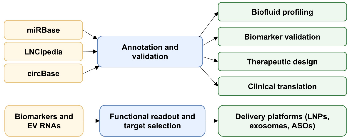
Figure 2. Translational workflow from ncRNA discovery to clinical use. Public resources such as miRBase, LNCipedia, and circBase support annotation and discovery; biofluid profiling and biomarker validation refine candidate selection; and delivery platforms such as lipid nanoparticles, exosomes, and antisense oligonucleotides enable therapeutic translation. LNPs: Lipid nanoparticles; ASOs: antisense oligonucleotides.
CONCLUSION
ncRNAs have redefined our understanding of CVDs by serving as pivotal regulators of gene expression and cellular processes. Their involvement in key pathological mechanisms, including inflammation, fibrosis, apoptosis, and angiogenesis, underscores their potential as biomarkers and therapeutic targets. Recent advancements in sequencing technologies, delivery platforms, and functional studies have paved the way for ncRNA-based diagnostics and therapies, exemplified by ongoing clinical trials for specific ncRNA modulators. However, significant challenges remain, including achieving tissue-specific delivery, minimizing off-target effects, ensuring long-term safety, and overcoming the complexity of large-scale production. Addressing these barriers requires interdisciplinary collaboration and innovative approaches to fully realize the translational potential of ncRNAs. As research continues to unravel the intricacies of ncRNA biology, their integration into clinical practice promises to revolutionize the diagnosis, prognosis, and treatment of CVDs, offering a new frontier in precision medicine.
DECLARATIONS
Acknowledgement
Graphic Abstract is created in BioRender. Chen P (2026) https://BioRender.com/645jhop.
Authors’ contributions
Designed the framework of the article and wrote the first draft of the manuscript: Geng B, Zhu H.
Provided suggestions and edited the manuscript: Chen P, Li D, Xie R, Pawlik TM
Availability of data and materials
Not applicable.
AI and AI-assisted tools statement
During the preparation of this manuscript, the AI tool Gemini (version 2.5, released 2025-03-25) was used solely for language editing. The tool did not influence the study design, data collection, analysis, interpretation, or the scientific content of the work. All authors take full responsibility for the accuracy, integrity, and final content of the manuscript.
Financial support and sponsorship
Zhu H laboratory is supported by the NIH grants: HL153876, EY030621, EY032583 and the American Heart Association grant: 23TPA1142638.
Conflicts of interest
Zhu H is an Editorial Board Member of The Journal of Cardiovascular Aging. Zhu H was not involved in any steps of editorial processing, notably including reviewers’ selection, manuscript handling and decision making. The other authors declare that there are no conflicts of interest.
Ethical approval and consent to participate
Not applicable.
Consent for publication
Not applicable.
Copyright
© The Author(s) 2026.
REFERENCES
1. Vaduganathan M, Mensah GA, Turco JV, Fuster V, Roth GA. The global burden of cardiovascular diseases and risk: a compass for future health. J Am Coll Cardiol. 2022;80:2361-71.
2. Nemeth K, Bayraktar R, Ferracin M, Calin GA. Non-coding RNAs in disease: from mechanisms to therapeutics. Nat Rev Genet. 2023;25:211-32.
3. Beermann J, Piccoli M, Viereck J, Thum T. Non-coding RNAs in development and disease: background, mechanisms, and therapeutic approaches. Physiol Rev. 2016;96:1297-325.
4. Van Rooij E, Sutherland LB, Qi X, Richardson JA, Hill J, Olson EN. Control of stress-dependent cardiac growth and gene expression by a microRNA. Science. 2007;316:575-9.
5. Wang S, Aurora AB, Johnson BA, et al. The endothelial-specific microRNA miR-126 governs vascular integrity and angiogenesis. Dev Cell. 2008;15:261-71.
6. Han P, Li W, Lin C, et al. A long noncoding RNA protects the heart from pathological hypertrophy. Nature. 2014;514:102-6.
7. Sun J, Chen G, Jing Y, et al. LncRNA expression profile of human thoracic aortic dissection by high-throughput sequencing. Cell Physiol Biochem. 2018;46:1027-41.
8. Traxler D, Spannbauer A, Einzinger P, et al. Early elevation of systemic plasma clusterin after reperfused acute myocardial infarction in a preclinical porcine model of ischemic heart disease. Int J Mol Sci. 2020;21:4591.
9. Yang M, Zhang M, Li Z, et al. A landscape of long non-coding RNAs reveals the leading transcriptome alterations in murine aorta during aging. Genomics. 2023;115:110573.
10. Baulina N, Osmak G, Kiselev I, et al. NGS-identified circulating miR-375 as a potential regulating component of myocardial infarction associated network. J Mol Cell Cardiol. 2018;121:173-9.
11. Kim S, Lee S, Park J, Lee S, Kim H. Postmortem-derived exosomal microRNA 486-5p as potential biomarkers for ischemic heart disease diagnosis. Int J Mol Sci. 2024;25:9619.
12. Liao J, Wang J, Liu Y, Li J, Duan L. Transcriptome sequencing of lncRNA, miRNA, mRNA and interaction network constructing in coronary heart disease. BMC Med Genomics. 2019;12:124.
13. Ramachandran V, Bhagavatheeswaran S, Shanmugam S, et al. Deep sequencing unveils altered cardiac miRNome in congenital heart disease. Mol Genet Genomics. 2022;297:1123-39.
14. Niu X, Liang G, Chen S, et al. MicroRNA profiling reveals novel biomarkers for cardiovascular and psychological health in plateau psycho CVD. Sci Rep. 2025;15:12488.
15. Van Rooij E, Kauppinen S. Development of microRNA therapeutics is coming of age. EMBO Mol Med. 2014;6:851-64.
16. Xu J, Qian B, Wang F, et al. Global profile of tRNA-derived small rnas in pathological cardiac hypertrophy plasma and identification of tRF-21-NB8PLML3E as a new hypertrophy marker. Diagnostics. 2023;13:2065.
17. Zhao L, Peng Y, Su P. Expression profiles and functional analysis of tRNA-derived small RNAs in epicardial adipose tissue of patients with heart failure. Ann Med. 2023;55:2267981.
18. Rajagopalan V, Chakraborty S, Lin R. Novel transcriptomic interactomes of noncoding RNAs in the heart under altered thyroid hormonal states. Int J Mol Sci. 2023;24:6560.
19. Jain G, Stuendl A, Rao P, et al. A combined miRNA-piRNA signature to detect Alzheimer’s disease. Transl Psychiatry. 2019;9:250.
20. Gu X, Wang C, Deng H, et al. Exosomal piRNA profiling revealed unique circulating piRNA signatures of cholangiocarcinoma and gallbladder carcinoma. Acta Biochim Biophys Sin. 2020;52:475-84.
21. Huang X, Yuan T, Tschannen M, et al. Characterization of human plasma-derived exosomal RNAs by deep sequencing. BMC Genomics. 2013;14:319.
22. Ogawa Y, Taketomi Y, Murakami M, Tsujimoto M, Yanoshita R. Small RNA Transcriptomes of two types of exosomes in human whole saliva determined by next generation sequencing. Biol Pharm Bull. 2013;36:66-75.
23. Yuan T, Huang X, Woodcock M, et al. Plasma extracellular RNA profiles in healthy and cancer patients. Sci Rep. 2016;6:19413.
24. Jouravleva K, Zamore PD. A guide to the biogenesis and functions of endogenous small non-coding RNAs in animals. Nat Rev Mol Cell Biol. 2025;26:347-70.
25. Gao F, Wang F, Cao H, Chen Y, Diao Y, Kapranov P. Evidence for existence of multiple functional human small RNAs derived from transcripts of protein-coding genes. Int J Mol Sci. 2023;24:4163.
26. Wong L, Tsang YS, Kenny R, Lyburn M, Mcmahon LP. MiR-423-5p as Optimal Endogenous Control for Quantification of Circulating MicroRNAs in Patients With CKD. Kidney Int Rep. 2023;8:2150-2.
27. Chen L, Kim VN. Small and long non-coding RNAs: past, present, and future. Cell. 2024;187:6451-85.
28. Caporali A, Anwar M, Devaux Y, et al. Non-coding RNAs as therapeutic targets and biomarkers in ischaemic heart disease. Nat Rev Cardiol. 2024;21:556-73.
29. Riffo-campos AL, Perez-hernandez J, Martinez-arroyo O, et al. Biofluid specificity of long non-coding RNA profile in hypertension: relevance of exosomal fraction. Int J Mol Sci. 2022;23:5199.
30. Wang Y, Trinh MP, Zheng Y, Guo K, Jimenez LA, Zhong W. Analysis of circulating non-coding RNAs in a non-invasive and cost-effective manner. Trends Analyt Chem. 2019;117:242-62.
31. Rupaimoole R, Slack FJ. MicroRNA therapeutics: towards a new era for the management of cancer and other diseases. Nat Rev Drug Discov. 2017;16:203-22.
32. Kulkarni JA, Witzigmann D, Thomson SB, et al. The current landscape of nucleic acid therapeutics. Nat Nanotechnol. 2021;16:630-43.
33. Geng B, Zhang Z, Xu Y, et al. The landscape of noncoding RNAs in cardiovascularaging and disease. Scierxiv 2025;2025060231.
34. Yuan J, Chen H, Ge D, et al. Mir-21 promotes cardiac fibrosis after myocardial infarction via targeting smad7. Cell Physiol Biochem. 2017;42:2207-19.
35. Fitzsimons S, Oggero S, Bruen R, et al. microRNA-155 is decreased during atherosclerosis regression and is increased in urinary extracellular vesicles during atherosclerosis progression. Front. Immunol. 2020;11:576516.
36. Ohta M, Kihara T, Toriuchi K, et al. IL-6 promotes cell adhesion in human endothelial cells via microRNA-126-3p suppression. Exp Cell Res. 2020;393:112094.
37. Liu N, Bezprozvannaya S, Williams AH, et al. microRNA-133a regulates cardiomyocyte proliferation and suppresses smooth muscle gene expression in the heart. Genes Dev. 2008;22:3242-54.
38. Michalik KM, You X, Manavski Y, et al. Long noncoding RNA MALAT1 regulates endothelial cell function and vessel growth. Circ Res. 2014;114:1389-97.
39. Zhou J. LncRNA MIAT promotes hypoxia‐induced H9C2 cell pyroptosis via binding to SF1 to inhibit CGRP transcription. Exp Physiol. 2021;107:58-67.
40. Wang K, Long B, Liu F, et al. A circular RNA protects the heart from pathological hypertrophy and heart failure by targeting miR-223. Eur Heart J. 2016;37:2602-11.
41. Fang J, Pan Z, Guo X. Research advance of ANRIL on atherosclerosis by regulating cell proliferation and apoptosis. J. Zhejiang Univ. (Med Sci.). 2020;49:113-7.
42. Micheletti R, Plaisance I, Abraham BJ, et al. The long noncoding RNA wisper controls cardiac fibrosis and remodeling. Sci Transl Med. 2017;9:eaai9118.
43. Boos F, Oo JA, Warwick T, et al. The endothelial-enriched lncRNA LINC00607 mediates angiogenic function. Basic Res Cardiol. 2023;118:5.
44. Sriram K, Luo Y, Yuan D, et al. Vascular regulation by super enhancer-derived LINC00607. Front. Cardiovasc. Med. 2022;9:881916.
45. Surina, Fontanella RA, Scisciola L, Marfella R, Paolisso G, Barbieri M. miR-21 in human cardiomyopathies. Front. Cardiovasc. Med. 2021;8:767064.
46. Ma YL, Ma ZJ, Wang M, Liao MY, Yao R, Liao YH. MicroRNA-155 induces differentiation of RAW264.7 cells into dendritic-like cells. Int J Clin Exp Pathol. 2015;8:14050-62.
47. Wu XY, Fan WD, Fang R, Wu GF. Regulation of microRNA-155 in endothelial inflammation by targeting nuclear factor (NF)-κB P65. J Cell Biochem. 2014;115:1928-36.
48. Xiao Q, Wang D, Sheng Y, Huang J, Ha X. MicroRNA-126 regulates thrombosis through endothelial progenitor cells. DNA Cell Biol. 2023;42:315-21.
49. Toraih EA, El-wazir A, Alghamdi SA, et al. Association of long non-coding RNA MIAT and MALAT1 expression profiles in peripheral blood of coronary artery disease patients with previous cardiac events. Genet Mol Biol. 2019;42:509-18.
50. Akan G, Nyawawa E, Nyangasa B, et al. Severity of coronary artery disease is associated with diminished circANRIL expression: A possible blood based transcriptional biomarker in East Africa. J Cell Mol Med. 2023;28:e18093.
51. Van Ingen E, Engbers PA, Woudenberg T, et al. C/D box snoRNA SNORD113-6 guides 2’-O-methylation and protects against site-specific fragmentation of tRNALeu(TAA) in vascular remodeling. Mol Ther Nucleic Acids. 2022;30:162-72.
52. van Ingen E, van den Homberg DAL, van der Bent ML, et al. C/D box snoRNA SNORD113-6/AF357425 plays a dual role in integrin signalling and arterial fibroblast function via pre-mRNA processing and 2’O-ribose methylation. Hum Mol Genet. 2022;31:1051-66.
53. Will CL, Luhrmann R. Spliceosome structure and function. Cold Spring Harb Perspect Biol. 2010;3:a003707.
54. Cao J, Wei Z, Nie Y, Chen H. Therapeutic potential of alternative splicing in cardiovascular diseases. eBioMedicine. 2024;101:104995.
55. Yang F, Xi R. Silencing transposable elements in the drosophila germline. Cell Mol Life Sci. 2016;74:435-48.
56. Sokolova OA, Iakushev E, Stoliarenko AD, Mikhaleva EA, Gvozdev VA, Klenov MS. The interplay of transposon silencing genes in the drosophila melanogaster germline. Mol Biol. (Mosk.). 2011;45:633-41.
57. Anzelon TA, Chowdhury S, Hughes SM, Xiao Y, Lander GC, Macrae IJ. Structural basis for piRNA targeting. Nature. 2021;597:285-9.
58. Wang J, Dong P, Xu X, et al. Identification of tRNA-derived fragments and their potential roles in atherosclerosis. Curr Med Sci. 2021;41:712-21.
59. Soares AR, Santos M. Discovery and function of transfer RNA‐derived fragments and their role in disease. Wiley Interdiscip Rev RNA. 2017;8:e1423.
60. Fu X, He X, Yang Y, et al. Identification of transfer RNA-derived fragments and their potential roles in aortic dissection. Genomics. 2021;113:3039-49.
61. Kim HK, Yeom J, Kay MA. Transfer RNA-derived small RNAs: another layer of gene regulation and novel targets for disease therapeutics. Mol Ther. 2020;28:2340-57.
62. Zhou M, He X, Zhang J, Mei C, Zhong B, Ou C. tRNA-derived small RNAs in human cancers: roles, mechanisms, and clinical application. Mol Cancer. 2024;23:76.
63. Rashad S, Al-mesitef S, Mousa A, et al. Translational response to mitochondrial stresses is orchestrated by tRNA modifications. bioRxiv 2024.
64. Brogli R, Cristodero M, Schneider A, Polacek N. A ribosome-bound tRNA half stimulates mitochondrial translation during stress recovery in trypanosoma brucei. Cell Rep. 2023;42:113112.
65. Zhang L, Liu J, Hou Y. Classification, function, and advances in tsRNA in non-neoplastic diseases. Cell Death Dis. 2023;14:748.
66. Cao J, Cowan DB, Wang D. tRNA-derived small RNAs and their potential roles in cardiac hypertrophy. Front. Pharmacol. 2020;11:572941.
67. Zuo J, Zhang Z, Li M, et al. The crosstalk between reactive oxygen species and noncoding RNAs: from cancer code to drug role. Mol Cancer. 2022;21:30.
68. He S, Huang B, Xu F, et al. Functions and application of circRNAs in vascular aging and aging-related vascular diseases. J Nanobiotechnol. 2025;23:216.
69. Wang Z, Zhang X, Li W, Yang B. Decoding noncoding RNAs in regulating cardiovascular aging and age-related conditions: the emerging landscape. Sci. China Life Sci. 2025;69:353-83.
70. Jusic A, Thomas PB, Wettinger SB, et al. Noncoding RNAs in age-related cardiovascular diseases. Ageing Res Rev. 2022;77:1016.
71. Ito T, Yagi S, Yamakuchi M. MicroRNA-34a regulation of endothelial senescence. Biochem Biophys Res Commun. 2010;398:735-40.
72. Menghini R, Casagrande V, Cardellini M, et al. MicroRNA 217 modulates endothelial cell senescence via silent information regulator 1. Circulation. 2009;120:1524-32.
73. Hofmann P, Sommer J, Theodorou K, et al. Long non-coding RNA H19 regulates endothelial cell aging via inhibition of STAT3 signalling. Cardiovasc Res. 2019;115:230-42.
74. Boon RA, Hofmann P, Michalik KM, et al. Long noncoding RNA Meg3 controls endothelial cell aging and function: implications for regenerative angiogenesis. J Am Coll Cardiol. 2016;68:2589-91.
75. Das S, Bedja D, Campbell N, et al. miR-181c regulates the mitochondrial genome, bioenergetics, and propensity for heart failure in vivo. PLoS ONE. 2014;9:e96820.
76. Li D, Mao C, Zhou E, et al. MicroRNA-21 Mediates a positive feedback on angiotensin II-induced myofibroblast transformation. J Inflamm Res. 2020;13:1007-20.
77. Rayner KJ, Suárez Y, Dávalos A, et al. MiR-33 contributes to the regulation of cholesterol homeostasis. Science. 2010;328:1570-3.
78. Najafi-shoushtari SH, Kristo F, Li Y, et al. MicroRNA-33 and the SREBP host genes cooperate to control cholesterol homeostasis. Science. 2010;328:1566-9.
79. Dávalos A, Fernández-hernando C. From evolution to revolution: miRNAs as pharmacological targets for modulating cholesterol efflux and reverse cholesterol transport. Pharmacol Res. 2013;75:60-72.
80. Correction to: LincRNA-p21 Regulates neointima formation, vascular smooth muscle cell proliferation, apoptosis, and atherosclerosis by enhancing p53 activity. Circulation. 2019;139.
81. Razeghian-jahromi I, Karimi Akhormeh A, Zibaeenezhad MJ. The role of ANRIL in atherosclerosis. Dis Markers. 2022;2022:1-10.
82. Chi J, Li J, Jia J, Zhang T, Liu X, Yi L. Long non-coding RNA ANRIL in gene regulation and its duality in atherosclerosis. CURR MED SCI. 2017;37:816-22.
83. Sothivelr V, Hasan MY, Mohd Saffian S, Zainalabidin S, Ugusman A, Mahadi MK. Revisiting miRNA-21 as a therapeutic strategy for myocardial infarction: a systematic review. J Cardiovasc Pharmacol. 2022;80:393-406.
84. Wu Y, Zhang K, Liu R, et al. MicroRNA-21-3p accelerates diabetic wound healing in mice by downregulating SPRY1. Aging. 2020;12:15436-45.
85. Gui Y, Li D, Chen J, et al. Soluble epoxide hydrolase inhibitors, t-AUCB, downregulated miR-133 in a mouse model of myocardial infarction. Lipids Health Dis. 2018;17:129.
86. Al-muhtaresh HA, Salem AH, Al-kafaji G. Upregulation of circulating cardiomyocyte-enriched miR-1 and miR-133 associate with the risk of coronary artery disease in type 2 diabetes patients and serve as potential biomarkers. J Cardiovasc Transl Res. 2019;12:347-57.
87. Qu X, Du Y, Shu Y, et al. MIAT Is a Pro-fibrotic long non-coding RNA governing cardiac fibrosis in post-infarct myocardium. Sci Rep. 2017;7:42657.
88. Mohammadi A, Balizadeh Karami AR, Dehghan Mashtani V, et al. Evaluation of oxidative stress, apoptosis, and expression of microRNA-208a and microRNA-1 in cardiovascular patients. Rep Biochem Mol Biol. 2021;10:183-96.
89. Chistiakov DA, Orekhov AN, Bobryshev YV. Cardiac-specific miRNA in cardiogenesis, heart function, and cardiac pathology (with focus on myocardial infarction). J Mol Cell Cardiol. 2016;94:107-21.
90. Boštjančič E, Zidar N, Štajer D, Glavač D. MicroRNAs miR-1, miR-133a, miR-133b and miR-208 are dysregulated in human myocardial infarction. Cardiology. 2010;115:163-9.
91. Montgomery RL, Hullinger TG, Semus HM, et al. Therapeutic inhibition of miR-208a improves cardiac function and survival during heart failure. Circulation. 2011;124:1537-47.
92. Kontaraki JE, Marketou ME, Kochiadakis GE, et al. The long non-coding RNAs MHRT,FENDRR and CARMEN, their expression levels in peripheral blood mononuclear cells in patients with essential hypertension and their relation to heart hypertrophy. Clin Exp Pharmacol Physiol. 2018;45:1213-7.
93. Liao C, Gui Y, Guo Y, Xu D. The regulatory function of microRNA-1 in arrhythmias. Mol Biosyst. 2016;12:328-33.
94. Sun L, Sun S, Zeng S, Li Y, Pan W, Zhang Z. Expression of circulating microRNA-1 and microRNA-133 in pediatric patients with tachycardia. Mol Med Rep. 2015;11:4039-46.
95. Chen L, Sampson KJ, Kass RS. Cardiac delayed rectifier potassium channels in health and disease. Card Electrophysiol Clin. 2016;8:307-22.
96. Shimizu W, Horie M. Phenotypic manifestations of mutations in genes encoding subunits of cardiac potassium channels. Circ Res. 2011;109:97-109.
97. Cao WJ, Rosenblat JD, Roth NC, et al. Therapeutic angiogenesis by ultrasound-mediated microRNA-126-3p delivery. Arterioscler Thromb Vasc Biol. 2015;35:2401-11.
98. Zhang Y, Xu Y, Zhou K, Kao G, Xiao J. MicroRNA‑126 and VEGF enhance the function of endothelial progenitor cells in acute myocardial infarction. Exp Ther Med. 2021;23:142.
99. Li D, Zhang C, Li J, et al. Long non-coding RNA MALAT1 promotes cardiac remodeling in hypertensive rats by inhibiting the transcription of MyoD. Aging. 2019;11:8792-809.
100. Liu H, Zhang Z, Xiong W, et al. Long non‐coding RNA MALAT1 mediates hypoxia‐induced pro‐survival autophagy of endometrial stromal cells in endometriosis. J Cell Mol Med. 2018;23:439-52.
101. Zhou Q, Boeckel JN, Yao J, et al. Diagnosis of acute myocardial infarction using a combination of circulating circular RNA cZNF292 and clinical information based on machine learning. MedComm. 2023;4:e299.
102. Zhang T, Huang W. Angiogenic circular RNAs: A new landscape in cardiovascular diseases. Microvasc Res. 2020;129:103983.
103. Martin MM, Lee EJ, Buckenberger JA, Schmittgen TD, Elton TS. MicroRNA-155 regulates human angiotensin II type 1 receptor expression in fibroblasts. J Biol Chem. 2006;281:18277-84.
104. Elia L, Quintavalle M, Zhang J, et al. The knockout of miR-143 and -145 alters smooth muscle cell maintenance and vascular homeostasis in mice: correlates with human disease. Cell Death Differ. 2009;16:1590-8.
105. Courboulin A, Paulin R, Giguère NJ, et al. Role for miR-204 in human pulmonary arterial hypertension. J Exp Med. 2011;208:535-48.
106. Gou D, Ramchandran R, Peng X, et al. miR-210 has an antiapoptotic effect in pulmonary artery smooth muscle cells during hypoxia. Am J Physiol Lung Cell Mol Physiol. 2012;303:L682-91.
107. Fomison-nurse I, Saw EEL, Gandhi S, et al. Diabetes induces the activation of pro-ageing miR-34a in the heart, but has differential effects on cardiomyocytes and cardiac progenitor cells. Cell Death Differ. 2018;25:1336-49.
108. Chen S, Puthanveetil P, Feng B, Matkovich SJ, Dorn GW, Chakrabarti S. Cardiac miR‐133a overexpression prevents early cardiac fibrosis in diabetes. J Cell Mol Med. 2014;18:415-21.
109. Gao L, Wang X, Guo S, et al. LncRNA HOTAIR functions as a competing endogenous RNA to upregulate SIRT1 by sponging miR‐34a in diabetic cardiomyopathy. J Cell Physiol. 2018;234:4944-58.
110. Kankuri E. Positive feedback loop of miR-320 and CD36 regulates the hyperglycemic memory-induced diabetic diastolic cardiac dysfunction. Mol Ther Nucleic Acids. 2023;32:318-21.
111. Zhan J, Jin K, Xie R, et al. AGO2 protects against diabetic cardiomyopathy by activating mitochondrial gene translation. Circulation. 2024;149:1102-20.
112. Gedikbasi A, Adas G, Isiksacan N, et al. The effect of host miRNAs on prognosis in COVID-19: miRNA-155 may promote severity via targeting suppressor of cytokine signaling 1 (SOCS1) gene. Genes. 2022;13:1146.
113. Adel RM, Helal H, Ahmed Fouad M, Sobhy Abd-elhalem S. Regulation of miRNA-155-5p ameliorates NETosis in pulmonary fibrosis rat model via inhibiting its target cytokines IL-1β, TNF-α and TGF-β1. Int Immunopharmacol. 2024;127:111456.
114. Zhang L, Ouyang P, He G, et al. Exosomes from microRNA‐126 overexpressing mesenchymal stem cells promote angiogenesis by targeting the PIK3R2‐mediated PI3K/Akt signalling pathway. J Cell Mol Med. 2020;25:2148-62.
115. Ge Y, Wang Q, Qin X, et al. Tetrahedral framework nucleic acids connected with microRNA-126 mimics for applications in vascular inflammation, remodeling, and homeostasis. ACS Appl Mater Interfaces. 2022;14:19091-103.
116. Xue Y, Li Z, Liu W, Shan J, Wang L, Su Q. Down-regulation of lncRNA MALAT1 alleviates vascular lesion and vascular remodeling of rats with hypertension. Aging. 2019;11:5192-205.
117. Zhang Y, Guan X, Wang H, Wang Y, Yue D, Chen R. Long non‑coding RNA metastasis‑associated lung adenocarcinoma transcript 1 regulates renal cancer cell migration via cofilin‑1. Oncol Lett. 2020.
118. Chen Y, Zhou J, Wei Z, et al. Identification of circular RNAs in cardiac hypertrophy and cardiac fibrosis. Front Pharmacol. 2022;13:940768.
119. Hu Y, Cao H, Sheng J, et al. Functional role of circRNA CHRC through miR-431-5p/KLF15 signaling axis in the progression of heart failure. J Genet Genomics. 2024;51:844-54.
120. Yang M, Wang H, Han S, et al. Circular RNA expression in isoproterenol hydrochloride-induced cardiac hypertrophy. Aging. 2020;12:2530-44.
121. Pan S, Zhao X, Shao C, et al. STIM1 promotes angiogenesis by reducing exosomal miR-145 in breast cancer MDA-MB-231 cells. Cell Death Dis. 2021;12:38.
122. Zhou H, Zhou J, Teng H, Yang H, Qiu J, Li X. MiR-145 enriched exosomes derived from bone marrow-derived mesenchymal stem cells protects against cerebral ischemia-reperfusion injury through downregulation of FOXO1. Biochem Biophys Res Commun. 2022;632:92-9.
123. Gao W, Zhang Y, Yuan L, Huang F, Wang YS. Long non‐coding RNA H19‐overexpressing exosomes ameliorate UVB‐induced photoaging by upregulating SIRT1 via sponging miR‐138. Photochem Photobiol. 2023;99:1456-67.
124. Wang J, Cao B, Sun R, Chen Y, Feng J. RETRACTED: Exosome-transported long non-coding ribonucleic acid H19 induces blood-brain barrier disruption in cerebral ischemic stroke via the H19/micro ribonucleic acid-18a/vascular endothelial growth factor axis. Neuroscience. 2022;500:41-51.
125. Hashemi M, Moosavi MS, Abed HM, et al. Long non-coding RNA (lncRNA) H19 in human cancer: from proliferation and metastasis to therapy. Pharmacol Res. 2022;184:106418.
126. Arderiu G, Peña E, Aledo R, et al. MicroRNA-145 regulates the differentiation of adipose stem cells toward microvascular endothelial cells and promotes angiogenesis. Circ Res. 2019;125:74-89.
127. Liu X, Zhong L, Jiang W, Wen D. Repression of circRNA_000684 inhibits malignant phenotypes of pancreatic ductal adenocarcinoma cells via miR-145-mediated KLF5. Pancreatology. 2021;21:406-17.
128. Wang J, Lin Y, Jiang DH, Yang X, He XG. CircRNA ZNF609 promotes angiogenesis in nasopharyngeal carcinoma by regulating miR‐145/STMN1 axis. Kaohsiung J Med Sci. 2021;37:686-98.
129. Legnini I, Di Timoteo G, Rossi F, et al. Circ-ZNF609 is a circular RNA that can be translated and functions in myogenesis. Mol Cell. 2017;66:22-37.e9.
130. Ye J, Guo R, Shi Y, Qi F, Guo C, Yang L. miR-155 regulated inflammation response by the SOCS1-STAT3-PDCD4 axis in atherogenesis. Mediators Inflamm. 2016;2016:1-14.
131. Billeter AT, Hellmann J, Roberts H, et al. MicroRNA‐155 potentiates the inflammatory response in hypothermia by suppressing IL‐10 production. FASEB J. 2014;28:5322-36.
132. Pathak S, Grillo AR, Scarpa M, et al. MiR-155 modulates the inflammatory phenotype of intestinal myofibroblasts by targeting SOCS1 in ulcerative colitis. Exp Mol Med. 2015;47:e164.
133. Liu E, Lv L, Zhan Y, et al. METTL3/N6‐methyladenosine/ miR‐21‐5p promotes obstructive renal fibrosis by regulating inflammation through SPRY1/ERK/NF‐κB pathway activation. J Cell Mol Med. 2021;25:7660-74.
134. Kong C, Lyu D, He C, Li R, Lu Q. Dioscin elevates lncRNA MANTIS in therapeutic angiogenesis for heart diseases. Aging Cell. 2021;20:e13392.
135. Leisegang MS, Fork C, Josipovic I, et al. Long noncoding RNA MANTIS facilitates endothelial angiogenic function. Circulation. 2017;136:65-79.
136. Gareev I, Kudriashov V, Sufianov A, et al. The role of long non-coding RNA ANRIL in the development of atherosclerosis. Noncoding RNA Res. 2022;7:212-6.
137. Chandy M, Ishida M, Shikatani EA, et al. c-Myb regulates transcriptional activation of miR-143/145 in vascular smooth muscle cells. PLoS One. 2018;13:e0202778.
138. Lan S, Albinsson S. Regulation of IRS-1, insulin signaling and glucose uptake by miR-143/145 in vascular smooth muscle cells. Biochem Biophys Res Commun. 2020;529:119-25.
139. Werner JH, Rosenberg JH, Um JY, Moulton MJ, Agrawal DK. Molecular discoveries and treatment strategies by direct reprogramming in cardiac regeneration. Transl Res. 2019;203:73-87.
140. Li X, Ni L, Wang W, Zong L, Yao B. LncRNA fendrr inhibits hypoxia/reoxygenation-induced cardiomyocyte apoptosis by downregulating p53 expression. J Pharm Pharmacol. 2020;72:1211-20.
141. Zhou J, Qiu C, Fan Z, Liu T, Liu T. Circular RNAs in stem cell differentiation: a sponge-like role for miRNAs. Int J Med Sci. 2021;18:2438-48.
142. Wang F, Li X, Li Z, Wang S, Fan J. Functions of circular RNAs in regulating adipogenesis of mesenchymal stem cells. Stem Cells International. 2020;2020:1-12.
143. Zhang L, Tang M, Diao H, Xiong L, Yang X, Xing S. LncRNA-encoded peptides: unveiling their significance in cardiovascular physiology and pathology-current research insights. Cardiovasc Res. 2023;119:2165-78.
144. Li F, Du WW, Li X, et al. A new protein isoform encoded by human circular RNA circSLC8a1 contributes to cardiac remodelling. Cardiovasc Res. 2025;121:882-99.
145. Hu D, Lin Y, Huang H, et al. CircZBTB44-encoded peptide ZBTB44-342aa alleviates aortic valve calcification via cGAS-STING inhibition. Circ Res. 2026;138.
146. Liu Y, He M, Yuan Y, et al. Neutrophil-membrane-coated biomineralized metal-organic framework nanoparticles for atherosclerosis treatment by targeting gene silencing. ACS Nano. 2023;17:7721-32.
147. Kmiotek‐wasylewska K, Łabędź‐masłowska A, Bobis‐wozowicz S, et al. Induced pluripotent stem cell‐derived extracellular vesicles enriched withmiR‐126 induce proangiogenic properties and promote repair of ischemic tissue. FASEB J. 2024;38:e23415.
148. Gao S, Gao H, Dai L, et al. miR-126 regulates angiogenesis in myocardial ischemia by targeting HIF-1α. Exp Cell Res. 2021;409:112925.
149. Ray KK, Landmesser U, Leiter LA, et al. Inclisiran in patients at high cardiovascular risk with elevated LDL cholesterol. N Engl J Med. 2017;376:1430-40.
150. Boštjančič E, Brandner T, Zidar N, Glavač D, Štajer D. Down-regulation of miR-133a/b in patients with myocardial infarction correlates with the presence of ventricular fibrillation. Biomed Pharmacother. 2018;99:65-71.
151. Xin Y, Yang C, Han Z. Circulating miR-499 as a potential biomarker for acute myocardial infarction. Ann. Transl. Med. 2016;4:135.
152. Adachi T, Nakanishi M, Otsuka Y, et al. Plasma microRNA 499 as a biomarker of acute myocardial infarction. Clin Chem. 2010;56:1183-5.
153. Tan J, Ma X, Wang G, Jiang C, Gong H, Liu H. LncRNA MIAT knockdown alleviates oxygen-glucose deprivation‑induced cardiomyocyte injury by regulating JAK2/STAT3 pathway via miR-181a-5p. J Cardiol. 2021;78:586-97.
154. Hu K, Yan T, Cao K, et al. A tRNA-derived fragment of ginseng protects heart against ischemia/reperfusion injury via targeting the lncRNA MIAT/VEGFA pathway. Mol Ther Nucleic Acids. 2022;29:672-88.
155. Holdt LM, Stahringer A, Sass K, et al. Circular non-coding RNA ANRIL modulates ribosomal RNA maturation and atherosclerosis in humans. Nat Commun. 2016;7:12429.
156. Shi P, Ji H, Zhang H, Yang J, Guo R, Wang J. circANRIL reduces vascular endothelial injury, oxidative stress and inflammation in rats with coronary atherosclerosis. Exp Ther Med. 2020;20:2245-51.
157. Qin Q, Zheng P, Tu R, Huang J, Cao X. Integrated bioinformatics analysis for the identification of hub genes and signaling pathways related to circANRIL. PeerJ. 2022;10:e13135.
158. Adams D, Gonzalez-duarte A, O’riordan WD, et al. Patisiran, an RNAi therapeutic, for hereditary transthyretin amyloidosis. N Engl J Med. 2018;379:11-21.
159. Nair JK, Willoughby JLS, Chan A, et al. Multivalent N-acetylgalactosamine-conjugated siRNA localizes in hepatocytes and elicits robust RNAi-mediated gene silencing. J Am Chem Soc. 2014;136:16958-61.
160. Springer AD, Dowdy SF. GalNAc-siRNA conjugates: leading the way for delivery of RNAi therapeutics. Nucleic Acid Ther. 2018;28:109-18.
161. Zhou XL, Xu H, Liu ZB, Wu QC, Zhu RR, Liu JC. miR‐21 promotes cardiac fibroblast‐to‐myofibroblast transformation and myocardial fibrosis by targeting Jagged1. J Cell Mol Med. 2018;22:3816-24.
162. Zhou Z, Schober A, Nazari-jahantigh M. Dicer promotes endothelial recovery and limits lesion formation after vascular injury through miR-126-5p. Int J Cardiol. 2018;273:199-202.
163. Batkai S, Genschel C, Viereck J, et al. CDR132L improves systolic and diastolic function in a large animal model of chronic heart failure. Eur Heart J. 2021;42:192-201.
164. Täubel J, Hauke W, Rump S, et al. Novel antisense therapy targeting microRNA-132 in patients with heart failure: results of a first-in-human phase 1b randomized, double-blind, placebo-controlled study. Eur Heart J. 2021;42:178-88.
165. Bauersachs J, Solomon SD, Anker SD, et al. Efficacy and safety of CDR132L in patients with reduced left ventricular ejection fraction after myocardial infarction: rationale and design of the HF-REVERT trial. Eur J Heart Fail. 2024;26:674-82.
166. Nie T, Heo Y, Shirley M. Vutrisiran: a review in polyneuropathy of hereditary transthyretin-mediated amyloidosis. Drugs. 2023;83:1425-32.
167. Garcia-Pavia P, Grogan M, Kale P, et al. Impact of vutrisiran on exploratory cardiac parameters in hereditary transthyretin-mediated amyloidosis with polyneuropathy. Eur J Heart Fail. 2024;26:397-4.
168. Fontana M, Berk JL, Gillmore JD, et al. Vutrisiran in patients with transthyretin amyloidosis with cardiomyopathy. N Engl J Med. 2025;392:33-44.
169. Griffiths-jones S. miRBase: microRNA sequences, targets and gene nomenclature. Nucleic Acids Res. 2006;34:D140-4.
170. Volders P, Anckaert J, Verheggen K, et al. LNCipedia 5: towards a reference set of human long non-coding RNAs. Nucleic Acids Res. 2019;47:D135-9.
Cite This Article
 Open Access
How to Cite
Download Citation
Export Citation File:
Type of Import
Tips on Downloading Citation
Citation Manager File Format
Type of Import
Direct Import: When the Direct Import option is selected (the default state), a dialogue box will give you the option to Save or Open the downloaded citation data. Choosing Open will either launch your citation manager or give you a choice of applications with which to use the metadata. The Save option saves the file locally for later use.
Indirect Import: When the Indirect Import option is selected, the metadata is displayed and may be copied and pasted as needed.
About This Article
Copyright

Data & Comments
Data
0










Comments
Comments must be written in English. Spam, offensive content, impersonation, and private information will not be permitted. If any comment is reported and identified as inappropriate content by OAE staff, the comment will be removed without notice. If you have any queries or need any help, please contact us at support@oaepublish.com.