





Platelet-derived extracellular vesicles and post-ablation survival in hepatocellular carcinoma: a prospective cohort study
 0

Abstract
Aim: Extracellular vesicles (EVs) mediate intercellular communication and modulate antitumor immunity, but their utility as peri-interventional immune biomarkers for locoregional ablation in hepatocellular carcinoma (HCC) is poorly characterized. We asked whether peri-interventional EV phenotypes, combined with systemic cytokine and T-cell profiles, capture microwave ablation (MWA)-induced immune remodeling and inform post-ablation outcomes.
Methods: In a prospective single-center cohort study, 53 patients (38 HCC, 15 non-HCC liver metastases) underwent MWA with plasma sampling pre- and 1 h post-MWA and longitudinal follow-up. EVs were enriched by tetraspanin-directed immunomagnetic isolation and profiled for 37 surface epitopes by bead-based flow cytometry. T helper cell type 1/2/17 (Th1/Th2/Th17) cytokines and circulating T-cell subsets were quantified in parallel.
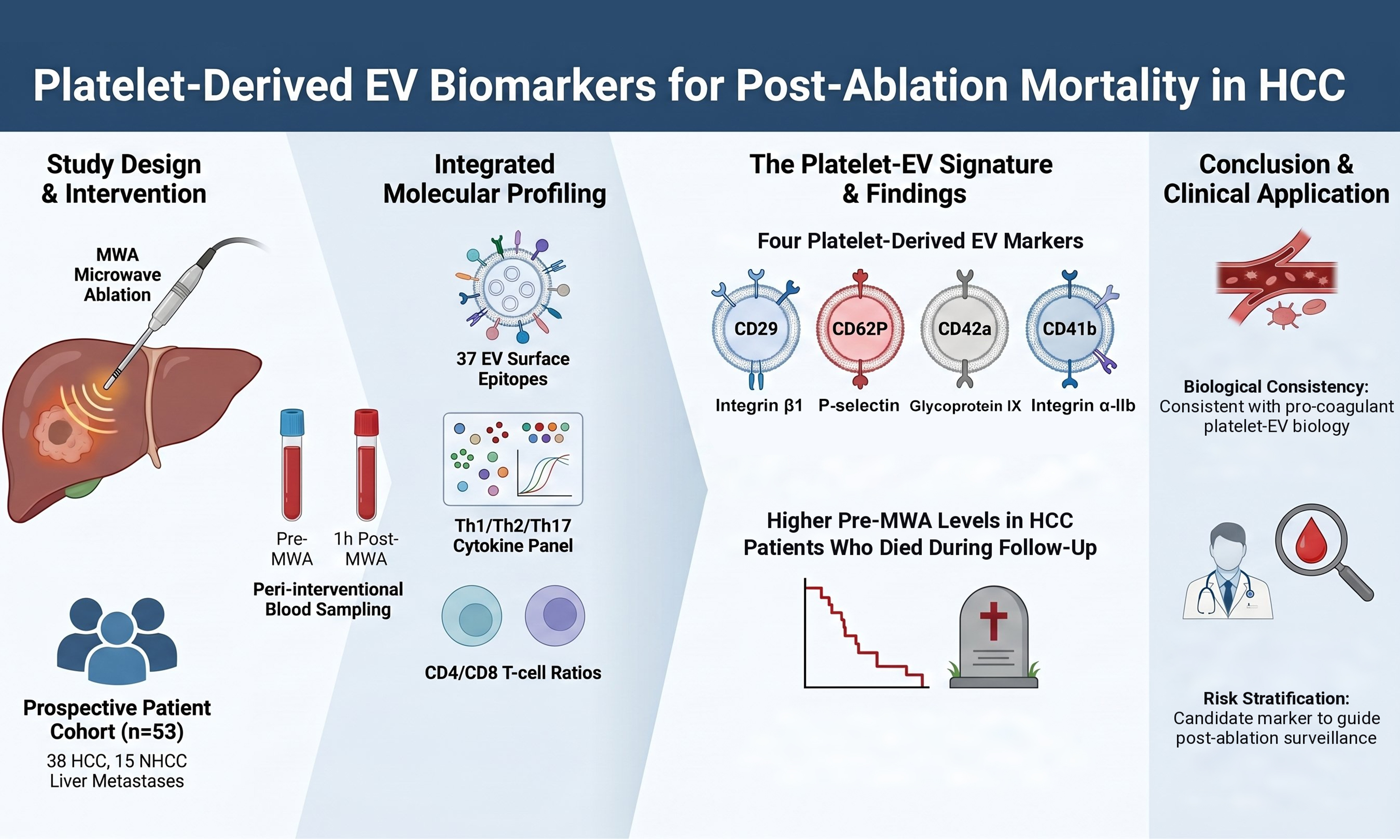
Results: At baseline, HCC patients showed an EV signature enriched for liver cancer stem cell-associated (CD133.1, CD44, CD24) and immune cell-derived markers (CD209, CD86, CD40, CD20, CD8), with higher interleukin-6 (IL-6) and lower interleukin-4 (IL-4) than non-HCC patients. MWA induced entity-specific EV remodeling; within HCC, cirrhosis and recurrence were associated with divergent post-MWA trajectories of CD4-, CD9-, and CD44-positive EVs, and non-recurrent patients showed higher pre-interventional CD45. Pre-interventional platelet-adhesion EV markers (CD29, CD41b, CD42a, CD62P) were concordantly higher in HCC patients who died than in survivors (Cohen’s d 0.44-0.74).
Conclusion: Peri-interventional multi-analyte profiling links defined EV subsets to systemic immune status and oncological outcome in HCC. The directional platelet-EV signal is consistent with the pro-coagulant, tumor-promoting role of platelet-derived microvesicles in advanced hepatic malignancy, nominating these and immune-modulatory EV populations as exploratory biomarkers to guide post-MWA surveillance, pending validation in adequately powered cohorts.
Keywords
INTRODUCTION
The liver occupies a distinct position in human physiology and plays key roles in digestion, metabolism, and detoxification. Receiving dual blood supply from both the portal vein and the hepatic artery, the liver is the most frequent site of metastatic disease, particularly from colorectal malignancies[1]. Furthermore, as the sixth most common site of primary malignancy, hepatocellular carcinoma (HCC) accounts for 80%-90% of primary liver cancers and ranks as the third leading cause of cancer-related mortality worldwide[2]. Hepatic metastases substantially diminish overall survival in multiple cancer types, including breast, colorectal, esophageal, gastric, and pancreatic cancers[1]. Consequently, innovative diagnostic and therapeutic approaches are urgently required.
Although hepatic resection remains the gold standard therapeutic option for hepatic malignancies, anatomical constraints, extrahepatic metastatic disease, comorbidities, and inadequate hepatic functional reserve frequently preclude surgical intervention. Treatment selection is guided by the Barcelona Clinic Liver Cancer (BCLC) staging system, which integrates tumor burden, disease stage, and hepatic synthetic function as assessed by the Child-Pugh score[3]. Alternative therapeutic modalities include liver transplantation, systemic chemotherapy, and transarterial chemoembolization (TACE). Among locoregional ablative techniques, microwave ablation (MWA), which induces thermal coagulation necrosis using microwave energy, represents a valuable alternative. MWA can be delivered percutaneously, laparoscopically, or intraoperatively and is particularly suitable for patients who are ineligible for resection or as part of multimodal treatment strategies[4]. Compared with other thermal ablation modalities, MWA provides superior advantages, including higher temperatures, accelerated heating kinetics, larger ablation zones, and reduced heat-sink effects[4].
Given the substantial recurrence rates of HCC and hepatic metastases, contemporary oncological strategies increasingly incorporate individualized immunotherapies[5]. Liquid biopsies, particularly extracellular vesicles (EVs), offer novel opportunities for characterizing circulating tumor-derived components and enabling precision oncologic diagnostics[6,7]. EVs are lipid bilayer-enclosed particles ranging from approximately 30 nm to 10 μm in diameter, detectable in virtually all body fluids, that carry proteins, lipids, and nucleic acids and mediate short- and long-distance cellular communication, enabling tumor cells to interact with stromal components and promote tumor progression, dissemination, and therapeutic resistance[8]. Small EVs are typically enriched for tetraspanins CD9, CD63, and CD81, the canonical surface markers used for their identification. EV release is markedly amplified by cell activation and tissue stress - conditions highly relevant to MWA-induced thermal injury[9]. EV-associated proteins such as exosomal programmed death-ligand 1 (PD-L1) have emerged as predictive biomarkers of therapy response[6,10], and EV profiling has demonstrated utility in HCC progression, metastasis, angiogenesis and recurrence[6]. EVs additionally carry a rich cargo of cytokines and chemokines, with vesicle encapsulation protecting these mediators from degradation and enabling targeted delivery to distant immune cells, thereby shaping cytokine signaling in the tumor microenvironment.
The intersection of oncology and immunology is fundamental for personalized cancer therapy. Proinflammatory cytokines such as interleukin-2 (IL-2) and tumor necrosis factor-alpha (TNF-α) are important immunotherapeutic agents. Characterization of circulating cytokine profiles holds promise for optimizing tumor treatment, particularly in hepatic malignancies, given the liver’s resident immune cell populations and its intrinsic role in immune homeostasis. Notably, locoregional therapies, including MWA, have demonstrated immunomodulatory effects on T-lymphocyte populations in HCC, with evidence suggesting that thermal ablation alters the CD4+ and CD8+ T-cell ratios. Beyond simple cytoreduction, MWA may exert dual and potentially opposing immunologic effects[9,11]. Thermal destruction of tumor tissue can induce immunogenic cell death with the release of tumor antigens and damage‑associated molecular patterns, which are taken up by antigen presenting cells and promote the cross priming of tumor specific T cells[9,11]. Conversely, insufficient or incomplete ablation may foster an immunosuppressive milieu and facilitate the outgrowth of residual tumor clones, highlighting the need for biomarkers that capture these divergent treatment‑induced immune trajectories[12,13].
In this study, we aimed to investigate the potential of a multi-analyte approach, including measurement of cytokine levels, lymphocyte subpopulations, and EVs immunophenotyping, to comprehensively characterize the immunomodulatory effects of MWA in hepatic malignancies. By simultaneously analyzing different blood compartments, we sought to highlight the added value of this integrated approach and identify potential biomarkers predictive of therapeutic response.
METHODS
Patients and study design
Blood samples of 53 patients were collected within this prospective single-center cohort study after approval by the ethics committee of Johann Wolfgang Goethe University, Frankfurt (19-443, 10 September 2020). All research was conducted in accordance with the Declarations of Helsinki (2013) and Istanbul (2018). Written informed consent was obtained from all participants prior to inclusion. The participants were included between 2021-2022. The inclusion criteria were age ≥ 18 years, absence of active viral hepatitis infections B and C, and negative human immunodeficiency virus status.
This study was reported in accordance with the Strengthening the Reporting of Observational Studies in Epidemiology (STROBE) statement for cohort studies. The completed checklist is provided as Supplementary Materials.
Study characteristics
Overall, 15 patients with hepatic metastasis herein referred to as non-HCC (NHCC, 28.30%) and 38 HCC patients (71.70%) were included in the underlying study. Patient characteristics are summarized in Table 1 and the overall study workflow from screening through peri-interventional sampling and imaging follow-up is shown in Supplementary Figure 1. Blood samples were collected in ethylenediaminetetraacetic acid (EDTA) tubes immediately within 15 min before and 1 h after MWA intervention. Following collection, plasma was separated by sequential centrifugation at 4 °C (see EV isolation below), aliquoted, and stored at -80 °C until batch analysis in the first quarter of 2023, corresponding to a maximum storage duration of 24 months with a single freeze-thaw cycle prior to EV isolation. All pre-analytical handling followed the recommendations of the International Society on Thrombosis and Haemostasis SSC Collaborative Workshop on plasma microvesicle determination[14]. The 1-h post-interventional time point was selected to capture the acute peri-interventional release phase of platelet- and tissue-injury-derived EVs, which occurs within minutes to hours of thermal and coagulative injury, while preceding the delayed systemic cytokine surge and adaptive T-cell response that evolve over days to weeks after ablation[11]. A single early time point therefore isolates the immediate innate and pro-coagulant vesicular response but cannot resolve the full temporal trajectory of EV release and immune remodeling.
Baseline clinical, demographic, histological and prognostic characteristics of the study cohort at enrolment, comparing patients with HCC and NHCC
| Characteristic | Category | HCC | NHCC | P-Value |
| Cohort size, n | 38 | 15 | ||
| Sex | Female | 7 (18.4%) | 8 (53.3%) | 0.028 |
| Male | 31 (81.6%) | 7 (46.7%) | ||
| Age (years) | Mean ± SD | 67.5 ± 8.9 | 54.5 ± 12.9 | < 0.001 |
| Diagnosis | HCC | 38 (100%) | 0 (0%) | |
| Anal carcinoma | 0 (0%) | 1 (6.7%) | ||
| Breast cancer | 0 (0%) | 4 (26.7%) | ||
| Choroidal melanoma | 0 (0%) | 1 (6.7%) | ||
| Colon carcinoma | 0 (0%) | 1 (6.7%) | ||
| Esophagus carcinoma | 0 (0%) | 1 (6.7%) | ||
| Hepatic NET | 0 (0%) | 1 (6.7%) | ||
| Non-small cell lung cancer | 0 (0%) | 1 (6.7%) | ||
| Pancreas NET | 0 (0%) | 3 (20.0%) | ||
| Pancreas MCN | 0 (0%) | 1 (6.7%) | ||
| Thymus carcinoma | 0 (0%) | 1 (6.7%) | ||
| BCLC stage | 0 | 4 (10.8%) | N/A | |
| A | 21 (56.8%) | |||
| B | 6 (16.2%) | |||
| C | 5 (13.5%) | |||
| D | 1 (2.7%) | |||
| Child-Pugh | A | 24 (64.9%) | N/A | |
| B | 8 (21.6%) | |||
| C | 1 (2.7%) | |||
| No cirrhosis | 4 (10.8%) | |||
| MELD score | Mean ± SD | 9.2 ± 2.8 | N/A | |
| Median (IQR) | 8 (7-10) | |||
| Liver fibrosis | No | 1 (2.6%) | N/A | |
| Yes | 37 (97.4%) | |||
| Liver cirrhosis | No | 5 (13.2%) | N/A | |
| Yes | 33 (86.8%) | |||
| Alcohol abuse | No | 22 (59.5%) | N/A | |
| Yes | 15 (40.5%) | |||
| Hepatitis virus infection | No | 20 (54.1%) | N/A | |
| Yes | 17 (45.9%) | |||
| MASH | No | 30 (81.1%) | N/A | |
| Yes | 7 (18.9%) | |||
| Diabetes mellitus | No | 15 (40.5%) | 14 (93.3%) | 0.002 |
| Yes | 22 (59.5%) | 1 (6.7%) | ||
| AFP before ablation (ng/mL) | Median (IQR) | 7.2 (3.5-17.8) | 3.5 (2.5-4.1) | 0.005 |
| Mean ± SD | 109.6 ± 442.9 | 3.4 ± 1.7 | ||
| Tumor volume (cm3) | Mean ± SD | 4.0 ± 5.4 | 3.5 ± 4.9 | 0.758 |
| Tumor histologic grading | 1 | 7 (22.6%) | 0 (0%) | 0.027 |
| 2 | 21 (67.7%) | 5 (55.6%) | ||
| 2 to 3 | 1 (3.2%) | 0 (0%) | ||
| 3 | 2 (6.5%) | 4 (44.4%) | ||
| Recurrence | No | 33 (86.8%) | 12 (85.7%) | 1.000 |
| Yes | 5 (13.2%) | 2 (14.3%) | ||
| Mortality | No | 28 (73.7%) | 9 (60.0%) | 0.320 |
| Yes | 10 (26.3%) | 6 (40.0%) |
Follow Up investigation
All patients underwent magnetic resonance imaging (MRI) follow-up every three months (median 1.9 years). Recurrence was classified as either local (tumor persistence or regrowth at the ablation zone, defined by arterial-phase hyperenhancement with portal-venous washout) or intrahepatic (a new lesion in a separate hepatic segment, representing de novo tumor development).
MWA
MWA was performed using a Covidien Evident generator (Covidien, Boulder, CO, USA; maximum output 100 W) with a single water-cooled, mini-choked coaxial antenna operating at continuous power. The procedure was delivered in two steps (80 W for 1 min, then 100 W for 3-10 min). Smaller lesions were treated in a single ablation zone; larger lesions required repositioning of the applicator. The system has been shown to be safe and effective for hepatic malignancies[4].
Isolation of EVs
EVs were enriched from pre-cleared plasma using the Exosome Isolation Kit Pan Human (Miltenyi Biotec, Bergisch Gladbach, Germany). Isolation was achieved through positive selection using MicroBeads, which simultaneously targeted all three tetraspanins. Plasma samples (2 mL each) were first pre-cleared by sequential centrifugation at 300 × g for 10 min, 2,000 × g for 30 min, and 10,000 × g for 45 min, all at 4 °C, to remove cellular debris and larger vesicles. After each step, the supernatant was carefully transferred into a fresh tube. Subsequently, 50 µL of EV Isolation MicroBeads was added to the pre-cleared plasma and incubated at room temperature for 1 h. The bead-bound EVs were then loaded onto a μ-column placed in the μMACSTM separator. The columns were washed four times with 200 µL of isolation buffer, and magnetically labeled EVs were eluted using 100 µL of isolation buffer.
Characterization of EVs by nanoparticle tracking analysis (NTA)
EVs were characterized using a NanoSight NS500 instrument (Malvern Panalytical, Malvern, UK) equipped with a 642 nm red laser. The capture parameters were set as follows: camera level 16, shutter slider 1300, and gain slider 512. The size distribution profiles generated by NTA software (v3.2) were averaged across six videos for each sample and subsequently normalized to the total nanoparticle concentration. All experiments were performed using a 1:100 dilution of EVs in phosphate-buffered saline (PBS). Mean particle diameters of 200-226 nm confirmed enrichment for small EVs, consistent with tetraspanin-positive vesicle size ranges[8].
EV identification by Western blot
EVs were lysed in M-Per Mammalian Extraction Reagent with 1× Halt protease inhibitor (Thermo Fisher Scientific, Waltham, MA, USA) using Precellys® Evolution with 1.4 mm ceramic beads (10 s, 6,000 rpm). Lysates were clarified (13,000 × g, 15 min, 4 °C); protein concentrations were measured with Pierce Bradford Plus (Thermo Fisher Scientific, Waltham, MA, USA). Equal protein amounts (40 µg) were resolved by 10% sodium dodecyl sulfate-polyacrylamide gel electrophoresis (SDS-PAGE), transferred onto polyvinylidene difluoride (PVDF) membranes (Millipore, Burlington, MA, USA), blocked in 10% non-fat dry milk for 1 h, and incubated overnight at 4 °C with rabbit monoclonal antibodies against CD9 (D3H4P) and CD63 (E1W3T; both 1:500, Cell Signaling Technology, Danvers, MA, USA). Detection used horseradish peroxidase (HRP)-anti-rabbit IgG (1:5,000; Abcam, Cambridge, UK) with enhanced chemiluminescence (ECL) substrate (Amersham/Cytiva, Marlborough, MA, USA) on a Fusion FX imager (Vilber, Collégien, France).
Multiplex bead-based EV surface analysis
Plasma-derived EV surface expression was analyzed using the MACSPlex Exosome Kit, human (Miltenyi Biotec, Bergisch Gladbach, Germany), targeting 37 EV surface epitopes (CD1c, CD2, CD3, CD4, CD8, CD9, CD11c, CD14, CD19, CD20, CD24, CD25, CD29, CD31, CD40, CD41b, CD42a, CD44, CD45, CD49e, CD56, CD62P, CD63, CD69, CD81, CD86, CD105, CD133.1, CD142, CD146, CD209, CD326, HLA-ABC, HLA-DR DP DQ, MCSP, ROR1, and SSEA-4) along with two matching isotype controls. Briefly, EVs (120 µL per sample) were incubated with 15 µL capture beads (containing all 39 antibody-coated bead populations) overnight at room temperature on a tube rotator (12 rpm). Following washing and centrifugation at 3000 × g, 5 µL of allophycocyanin (APC)-conjugated detection reagents (CD9, CD63, and CD81) were added and incubated for 1 h. After incubation, the capture bead-EV complexes were centrifuged, resuspended in 150 µL MACSPLEX buffer, and analyzed using a FACSCantoTM II flow cytometer (BD Bioscience, San Jose, CA, USA). In this bead-based assay (MACSPlex Exosome Kit, human, Cat. No. 130-108-813), EVs are first captured onto antibody-coated capture beads of approximately 4.8 µm diameter and are then stained with fluorescent anti-CD9, anti-CD63, and anti-CD81 detection antibodies. The FACSCanto II flow cytometer therefore detects and analyzes the micrometre-sized capture bead-EV complexes rather than free exosomes or individual sub-micron EVs, so the lower particle-size detection limit of the instrument does not affect this analysis[15]. The CD133.1 bead population denotes CD133 (prominin-1) detected with the CD133/1 (AC133) antibody clone.
Protein expression was quantified as median fluorescence intensity (MFI). For each bead population, both the mean and the MFI of the captured events were exported. Unless stated otherwise, all analyses used the median values, whereas Table 2 and Supplementary Figure 9 are based on the mean values. To obtain the relative EV surface signal levels, the median signal intensity from each bead population of the patient sample was subtracted from the MFI of the respective beads of samples from the control group.
Pre-MWA levels of platelet-adhesion-associated EV markers in the HCC group: alive (n = 28) vs. deceased (n = 10) at the end of follow-up (median follow-up 1.9 years)
| Alive (n = 28) Median (IQR) | Deceased (n = 10) Median (IQR) | P-value | Cohen’s d | |
| CD29 | 613 (198-1,367) | 1,835 (658-2,740) | 0.18 | 0.74 |
| CD41b | 193 (69-344) | 308 (99-533) | 0.50 | 0.44 |
| CD42a | 639 (299-808) | 1,114 (375-1,751) | 0.18 | 0.50 |
| CD62P | 54 (11-276) | 206 (17-548) | 0.41 | 0.55 |
Cytokine determination
Plasma IL-2, IL-4, IL-6, IL-10, TNF-α, interferon-gamma (IFN-γ), and IL-17A were quantified with the Cytometric Bead Array Human T helper cell type 1/2/17 (Th1/Th2/Th17) Cytokine Kit (BD Biosciences, San Jose, CA, USA) per manufacturer’s instructions. Samples were incubated with capture beads and phycoerythrin (PE)-conjugated detection antibodies for 3 h at room temperature, then acquired on a FACSCalibur flow cytometer and analyzed with Flow Cytometric Analysis Program (FCAP) Array v3.0 software (BD Biosciences, San Jose, CA, USA).
CD4 and CD8 ratio determination
CD4+ and CD8+ cells were quantified using the CD3/CD8/CD45/CD4 Multitest (BD Biosciences, San Jose, CA, USA). Fluorochrome-labeled antibodies were incubated with whole blood for 1 h, washed, and acquired on a FACSCanto II flow cytometer with FACSDiva v6.1.3 software (BD Biosciences, San Jose, CA, USA).
Data and methodology reporting
We have submitted all relevant experimental parameters of our EV workflow to the EV-TRACK (Transparent Reporting and Centralizing Knowledge in Extracellular Vesicle Research) knowledgebase (EV-TRACK ID: EV260032)[16], in line with current MISEV2023 (Minimal Information for Studies of Extracellular Vesicles 2023) recommendations for transparent and reproducible reporting of EV studies[17].
Statistical analysis
Statistical analyses were performed using IBM SPSS Statistics version 27 (IBM Corp., Armonk, NY, USA). Graphs were generated using GraphPad Prism version 9 (GraphPad Software, San Diego, CA, USA). The distribution of continuous variables was assessed using the Shapiro-Wilk test and visual inspection of histograms and quantile-quantile (Q-Q) plots; the majority of variables deviated from a normal distribution. Continuous data are reported as median (interquartile range, IQR), whereas conventional data are reported as mean ± standard deviation (SD); categorical data are presented as absolute and relative frequencies, n (%).
Between-group comparisons of HCC and NHCC patients were performed using the Mann-Whitney U test for continuous variables and the χ² test or Fisher’s exact test for categorical variables, as appropriate. Paired pre- and post-MWA comparisons within each cohort were assessed using the Wilcoxon signed-rank test. Adjustments for multiple testing across the panel of EV markers and cytokines were performed using the Bonferroni method. Between-group effect sizes were additionally quantified as Cohen’s d and interpreted using conventional thresholds of small (≥ 0.2), medium (≥ 0.5), and large (≥ 0.8) effects. Bivariate associations between cytokine concentrations and EV-marker levels were assessed using Pearson’s correlation coefficient for normally distributed variable pairs and Spearman’s rank correlation coefficient for non-normally distributed pairs, and normality was verified using the Shapiro-Wilk test. Statistical significance was defined as a two-sided P-value < 0.05. Analyses were performed on complete cases for each variable without imputation, so the effective sample size varied where individual clinical or histological data were missing, for example n = 37 where one value was missing and n = 31 for tumor grading. Nominal unadjusted P-values are reported throughout the main text, and Bonferroni correction across the 37-marker EV panel and the seven-cytokine panel was applied as a sensitivity analysis. Given the exploratory design, marker-level and subgroup findings are interpreted as hypothesis-generating irrespective of multiplicity correction. Accordingly, throughout the Results, the terms “significant” and “significantly”, as well as asterisks in the tables and figures, indicate nominal statistical significance based on an unadjusted two-sided P < 0.05 for the prespecified group and within-subject comparisons. After Bonferroni correction for multiple comparisons across the marker-level and cytokine analyses, none of the observed differences remained statistically significant.
ChatGPT (GPT-4o, OpenAI, San Francisco, CA, USA) and Claude (Claude Opus 4, Anthropic, San Francisco, CA, USA) were used during manuscript preparation for language editing and editorial review. All scientific content, data interpretation, statistical analyses, and conclusions are the sole responsibility of the authors, who reviewed and approved all output.
RESULTS
Patient characteristics
The demographic and clinical characteristics of the 38 HCC and 15 NHCC patients are summarized in Table 1, and patient flow from screening through follow-up is shown in Supplementary Figure 1. The NHCC group included significantly more female patients (8/15, 53.3%) than the HCC group (7/38, 18.4%; P = 0.028). HCC patients were significantly older (mean 67.5 ± 8.9 years) than NHCC patients (mean 54.5 ± 12.9 years; P < 0.001), and diabetes mellitus was substantially more prevalent in the HCC than in the NHCC cohort (22/37, 59.5% vs. 1/15, 6.7%; P = 0.002).
NHCC primary malignancies were predominantly breast cancer (n = 4, 26.7%) and pancreatic neuroendocrine tumors (n = 3, 20.0%). The full list of entities is provided in Table 1. Within the HCC cohort, most patients were classified as BCLC stage A (21/37, 56.8%) or stage 0 (4/37, 10.8%), with the remainder distributed across stages B (6/37, 16.2%), C (5/37, 13.5%) and D (1/37, 2.7%). Hepatic synthetic function was preserved in the majority of patients, with Child-Pugh class A in 64.9%, B in 21.6%, and C in 2.7%; 10.8% had no underlying cirrhosis. The median pre-interventional MELD score was 8 (IQR: 7-10). Liver fibrosis was present in 37 of 38 HCC patients (97.4%) and liver cirrhosis in 33 (86.8%). Etiologically, 17 HCC patients (45.9%) presented with positive hepatitis B or C virus serology, 15 (40.5%) had a history of alcohol abuse, and seven (18.9%) had metabolic dysfunction-associated steatohepatitis (MASH).
Pre-interventional alpha-fetoprotein concentrations were significantly higher in the HCC than in the NHCC cohort [median 7.2 (IQR 3.5-17.8) ng/mL vs. 3.5 (2.5-4.1) ng/mL; P = 0.005]. Tumor volume was numerically larger in the HCC cohort (mean 4.0 ± 5.4 cm3 vs. 3.5 ± 4.9 cm3), but the difference was not statistically significant (P = 0.758). Histological grading differed significantly between the two groups, with a higher proportion of well-differentiated G1 tumors in the HCC cohort (7/31; 22.6%) than in the NHCC cohort (0/9; 0%; P = 0.027); G2 was the most frequent grade in both groups (HCC 21/31, 67.7%; NHCC 5/9, 55.6%).
During the observation period (median follow-up 1.9 years), recurrence was documented in 5/38 HCC patients (13.2%) and 2/14 NHCC patients (14.3%; P = 1.0). Overall mortality was 10/38 (26.3%) in the HCC cohort and 6/15 (40.0%) in the NHCC cohort.
EV characteristics
NTA confirmed comparable EV size distributions across cohorts, with mean particle diameters of 200-226 nm consistent with small EVs [Figure 1A and Supplementary Figure 2]. Particle concentrations declined approximately 4-fold after MWA in both groups (detailed values in Supplementary Figure 2). EV identity was further confirmed by CD9 and CD63 expression on Western blot [Figure 1B and Supplementary Figure 3]. The immunoblot serves as a qualitative confirmation of EV identity rather than a quantitative assay, and the fainter post-MWA CD9 band is consistent with the approximately four-fold post-ablation reduction in total particle concentration under equal-protein loading, since tetraspanin abundance varies across EV subpopulations and does not provide a per-vesicle normalizer[8].
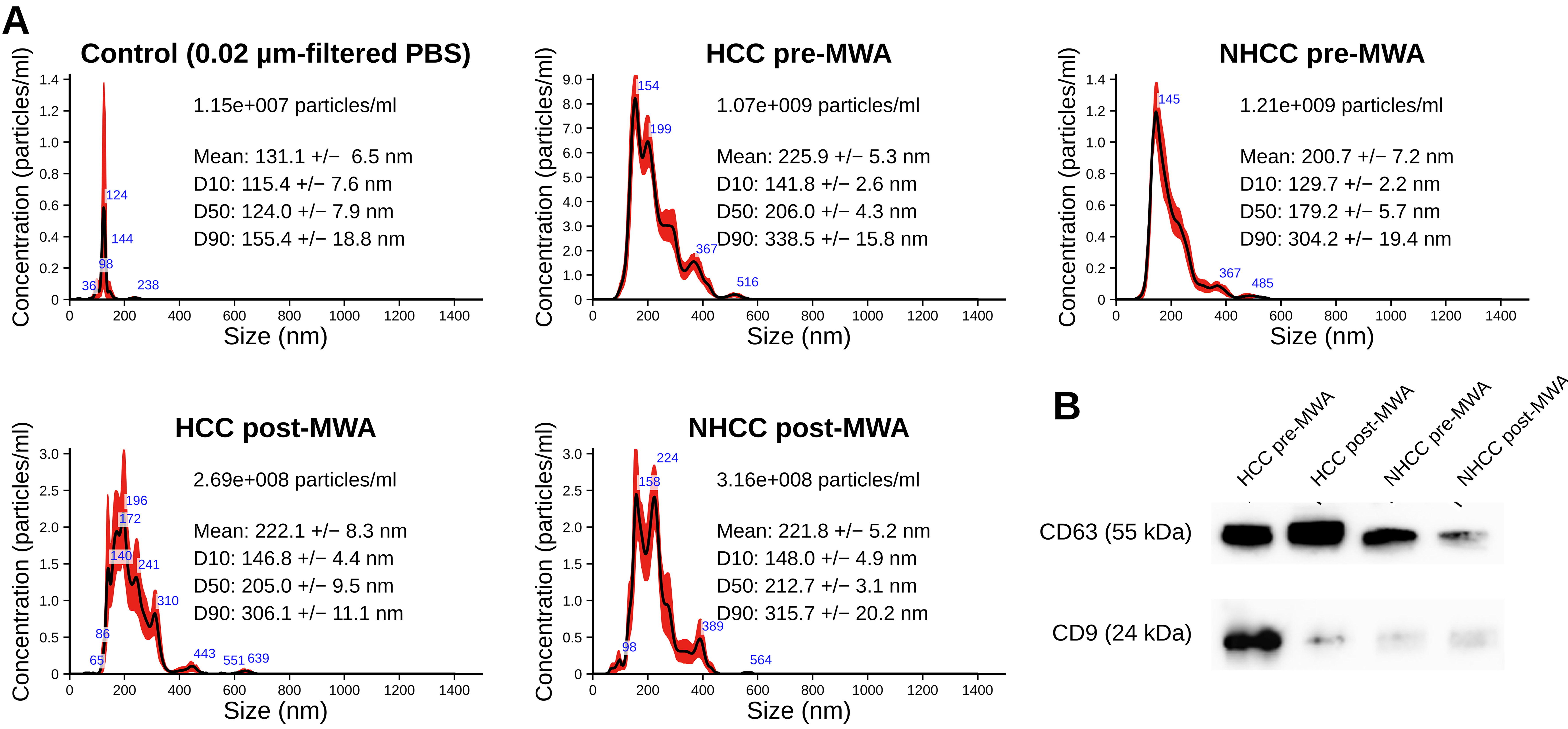
Figure 1. Characterization of EVs. (A) Representative NTA showing particle concentration and size distribution of EVs isolated from plasma of HCC and NHCC patients pre- and post-MWA, compared with resuspension buffer (PBS filtered through a 0.02 µm filter) used for EV isolation; (B) Representative western blot of EV markers CD63 and CD9 in HCC and NHCC patients pre- and post-MWA. EV: Extracellular vesicle; HCC: hepatocellular carcinoma; MWA: microwave ablation; NHCC: non-HCC group (hepatic metastases of extrahepatic primary tumors); NTA: nanoparticle tracking analysis; PBS: phosphate-buffered saline.
Pre- and post-MWA markers
Comparative analysis between the HCC and NHCC groups revealed significant differences in pre-interventional EV surface protein expression, with increased expression of T- and B-cell-associated markers in the HCC group. Overall, EV surface protein levels were elevated in the HCC group both before and after MWA compared with the NHCC group at the corresponding time points [Figure 2]. While most EV surface proteins displayed positive median expression values in both the pre- and post-interventional phases, CD4 and CD1c exhibited median values close to the detection limit in both the HCC and NHCC groups.
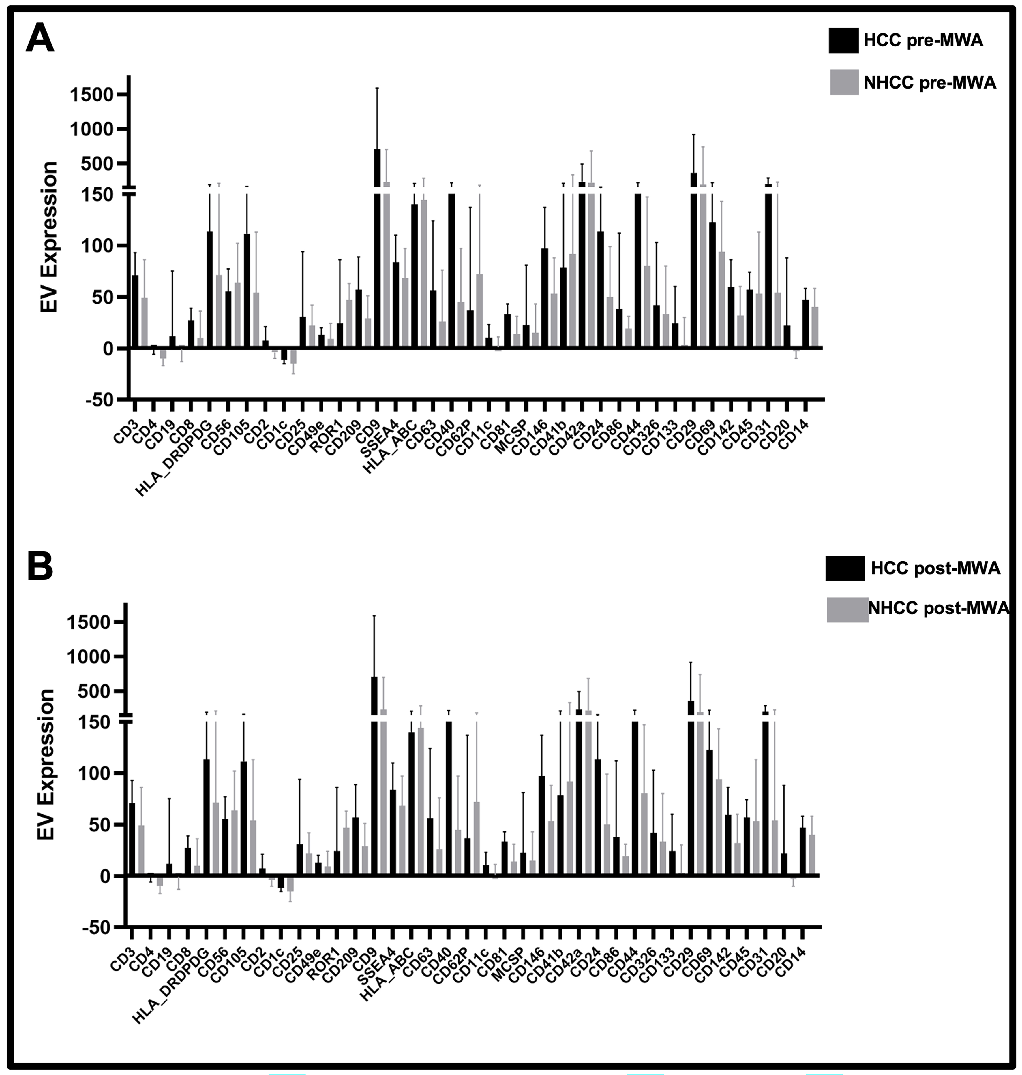
Figure 2. Comparison of EV surface marker levels between HCC and NHCC cohorts: (A) pre-MWA; (B) post-MWA. EV expression is shown as median fluorescence intensity for 37 EV-associated surface proteins in plasma-derived EVs. CD1c: Cluster of differentiation 1c; HLA-ABC: human leukocyte antigen A, B, and C; HLA-DR/DP/DQ: human leukocyte antigen DR, DP, and DQ; MCSP: melanoma-associated chondroitin sulfate proteoglycan; ROR1: receptor tyrosine kinase-like orphan receptor 1; SSEA-4: stage-specific embryonic antigen-4; EV: extracellular vesicle; HCC: hepatocellular carcinoma; MWA: microwave ablation; NHCC: non-HCC group (hepatic metastases of extrahepatic primary tumors).
B-cell-derived CD19- and CD20-positive EVs were detectable exclusively in HCC patients and did not show measurable changes following MWA. For the majority of EV surface proteins, higher median expression values were observed in the HCC group, both before and after MWA. In contrast, ROR1, CD62P, CD56, HLA-ABC, CD41b, and CD42a showed higher median values in the pre-interventional NHCC group. Post-intervention, CD56, ROR1, HLA-ABC, CD62P, CD42a, and CD41b were highly expressed in the NHCC group compared to the HCC group [Figure 2].
Across both cohorts, CD9 showed the highest median expression (approximately 700 to 750 MFI in HCC and 230 to 280 MFI in NHCC), followed by CD29 and CD42a (approximately 230 to 360 MFI in HCC and 190 to 500 MFI in NHCC). The remaining markers showed median values below approximately 200 MFI, the highest among them being CD31, CD40, CD44, CD69, CD105, and HLA-ABC in HCC and CD41b, CD62P, CD69, and HLA-ABC in NHCC, whereas CD2, CD4, CD19, CD1c, CD49e, and CD11c remained at consistently low levels (median below 20 MFI) in both cohorts pre- and post-intervention [Figure 3].
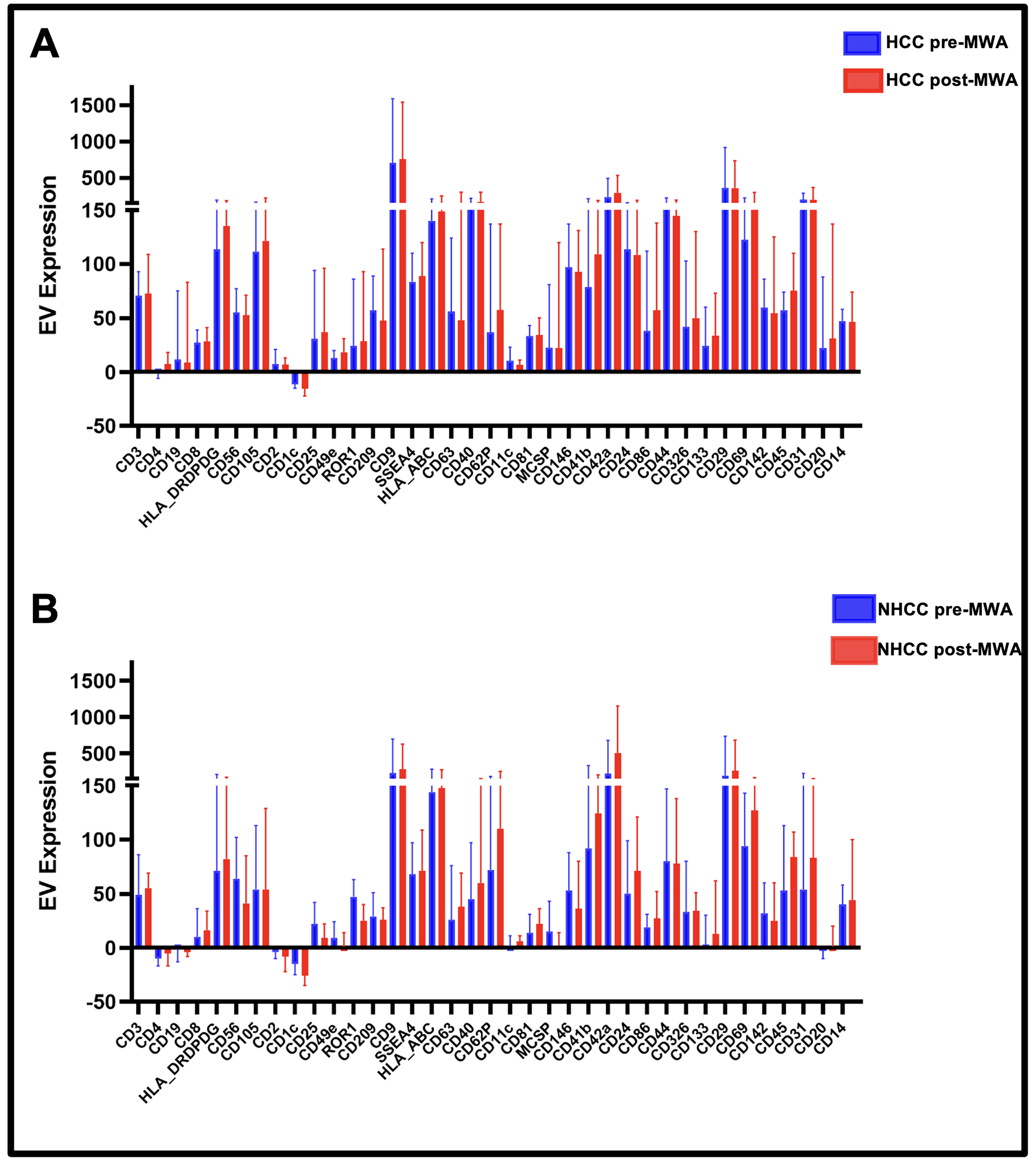
Figure 3. Pre- and post-MWA EV surface marker levels: (A) HCC cohort; (B) NHCC cohort, showing MFI of 37 EV-associated surface proteins in plasma-derived EVs. CD1c: Cluster of differentiation 1c; HLA-ABC: human leukocyte antigen A, B, and C; HLA-DR/DP/DQ: human leukocyte antigen DR, DP, and DQ; MCSP: melanoma-associated chondroitin sulfate proteoglycan; ROR1: receptor tyrosine kinase-like orphan receptor 1; SSEA-4: stage-specific embryonic antigen-4; EV: extracellular vesicle; HCC: hepatocellular carcinoma; MFI: median fluorescence intensity; MWA: microwave ablation; NHCC: non-HCC group (hepatic metastases of extrahepatic primary tumors).
Comparison of pre-treatment EV profiles in HCC vs NHCC
Supplementary Figures 4-6 summarize the pre-interventional EV profiles stratified by exosomal markers [Supplementary Figure 4], tumor tissue-derived EVs [Supplementary Figure 5], and immune cell-derived EVs [Supplementary Figure 6]. The canonical exosomal marker, CD63, was significantly elevated in the HCC cohort [Supplementary Figure 4]. Tumor tissue-derived EVs expressing CD133.1, CD44, and CD24 were significantly higher in the HCC cohort than in the NHCC cohort (P = 0.009, P = 0.029, and P = 0.004, respectively; Supplementary Figure 5). Additional tumor-associated markers, including melanoma-associated chondroitin sulfate proteoglycan (MCSP), CD146, and CD29, showed higher median expression in HCC, without reaching statistical significance. Immune cell-derived EVs expressing CD209 (P = 0.02), CD86 (P = 0.003), CD40 (P = 0.022), CD20 (P = 0.005), CD9 (P = 0.033), and CD8 (P = 0.024) were significantly elevated in the HCC cohort [Supplementary Figure 6]. These findings indicate increased levels of EVs derived from cytotoxic T cell-associated CD8, T helper cell-associated CD40 and CD86, and the B cell marker CD20 in pre-interventional HCC samples compared with NHCC.
Additional pre-MWA elevations in HCC included CD105 (angiogenesis-associated), CD209 (macrophage/dendritic-cell-derived), and CD9. Elevated CD24, CD44, and CD133.1 expression is consistent with their established association with liver-cancer stemness[18]. The distinct HCC marker pattern persisted post-intervention (CD9, CD86, CD81, CD20, CD209; Supplementary Table 1), supporting a tumor-type-specific signature rather than a nonspecific malignancy profile.
Comparison of EV profiles pre- and post-MWA
The median EV surface marker levels in the HCC and NHCC metastasis cohorts before and after MWA were compared using paired Wilcoxon test. The changes in CD44 and MCSP expression are shown in Figure 4. In the HCC cohort, CD44 levels decreased significantly after MWA (P = 0.044), whereas no significant change was observed in the NHCC cohort. Conversely, MCSP levels decreased significantly after MWA in the NHCC cohort (P = 0.016) but remained unchanged in the HCC cohort. Supplementary Figure 7 depicts the immune cell-associated markers CD4, CD9, and CD86. CD4 expression increased significantly after MWA in the HCC cohort (P = 0.021), with no significant changes observed in the NHCC cohort. CD9 levels decreased after MWA in both cohorts, reaching nominal significance in HCC (P = 0.0076), in line with the approximately four-fold reduction in total particle concentration by NTA. Because the bead-based assay reports the relative CD9 surface signal of the captured EV population rather than the total EV output, this change should not be equated with a change in overall EV secretion. CD86 expression also increased post-interventionally in both groups, with a significant increase observed in the NHCC cohort (P = 0.031).
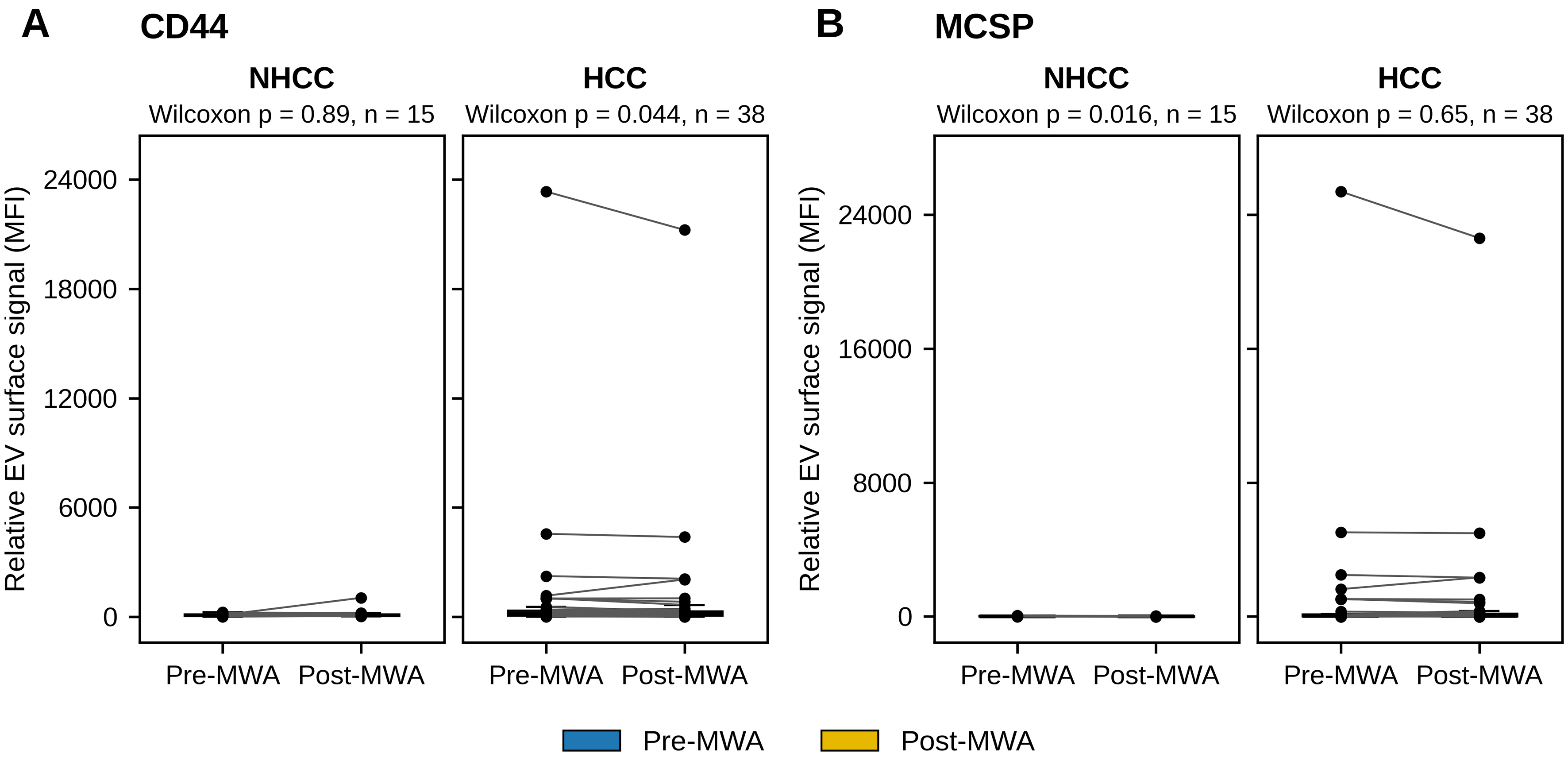
Figure 4. Levels of CD44 and MCSP on plasma-derived EVs in the NHCC (n = 15) and HCC (n = 38) cohorts, shown pre- and post-MWA as box plots (median and IQR) with paired individual values (paired Wilcoxon signed-rank test, nominal P-values): (A) CD44; (B) MCSP. EV: Extracellular vesicle; HCC: hepatocellular carcinoma; IQR: interquartile range; MCSP: melanoma-associated chondroitin sulfate proteoglycan; MFI: median fluorescence intensity; MWA: microwave ablation; NHCC: non-HCC group (hepatic metastases of extrahepatic primary tumors).
Overall, a greater number of EV surface markers exhibited post-interventional changes in the HCC cohort compared with the NHCC cohort, particularly among immune-associated markers. Differential blood cell counts were not available for all patients and were therefore not included in this analysis.
Subgroup analyses based on clinical parameters
The HCC cohort was stratified by cirrhosis status and recurrence, both established modulators of the hepatic immune microenvironment and EV biology[18,19].
EV marker levels were compared between cirrhotic and non-cirrhotic patients with HCC using the Wilcoxon signed-rank test. In the cirrhotic subgroup, all three surface markers changed significantly following MWA: CD4 increased (P = 0.028), whereas CD9 (P = 0.015) and CD44 (P = 0.029) decreased. The non-cirrhotic cohort showed no significant changes in these markers [Supplementary Figure 8]. Because liver fibrosis was near-universal and only five HCC patients were non-cirrhotic, this non-cirrhotic subgroup was too small to support firm conclusions, and the absence of significant post-MWA changes should be interpreted as underpowered rather than as evidence of no effect.
Two distinct stratification approaches have been employed for tumor recurrence. First, all patients were dichotomized into recurrence and non-recurrence cohorts, regardless of tumor histology (HCC or non-HCC metastases).
Between-cohort comparisons of CD3 and CD45 expression yielded nominally lower expression in patients with recurrence at the conventional significance threshold (P = 0.05 and P = 0.024, respectively), driven by isolated high-expression observations in the non-recurrence cohort. These differences did not persist in non-parametric analyses using the Mann-Whitney U test, which is consistent with the influence of distributional outliers and supports the use of rank-based methods as the primary analytical approach across the EV panel.
Marker dynamics following MWA were evaluated in both cohorts using the Wilcoxon signed-rank test [Figure 5]. In patients without documented recurrence (n = 46), CD4 levels increased (P = 0.019) and CD9 levels decreased (P = 0.012) significantly after ablation, whereas neither marker changed significantly in patients with documented recurrence (n = 7, P = 0.13 and P = 0.61). CD62P increased significantly after MWA in the non-recurrence group (P = 0.027), whereas MCSP decreased (P = 0.046), with no significant changes in the recurrence group (P = 0.94 and P = 0.41).
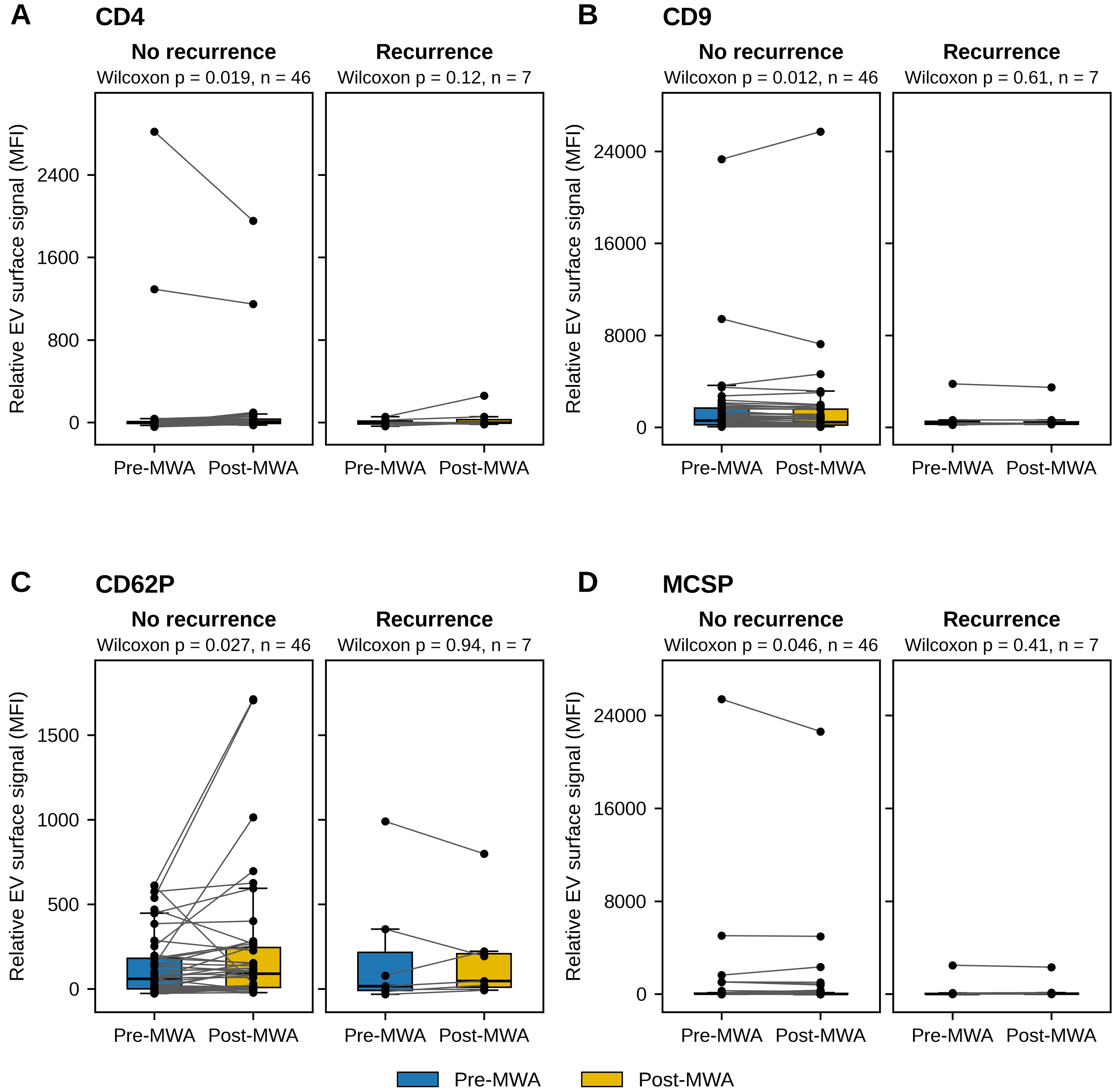
Figure 5. Plasma-derived EV marker levels pre- and post-MWA in the overall patient cohort, stratified by recurrence status (patients without documented recurrence, n = 46, and patients with documented recurrence, n = 7): (A) CD4; (B) CD9; (C) CD62P; (D) MCSP. Data are shown as box plots (median and IQR) with paired individual values. Paired Wilcoxon signed-rank test, nominal P-values. EV: Extracellular vesicle; IQR: interquartile range; MCSP: melanoma-associated chondroitin sulfate proteoglycan; MFI: median fluorescence intensity; MWA: microwave ablation.
Second, a separate analysis was conducted exclusively within the HCC subpopulation.
Baseline analysis revealed significantly elevated CD45 levels in patients with HCC without recurrence (P = 0.048). Post-MWA marker dynamics within each cohort were assessed using the paired t-test on mean fluorescence values [Supplementary Figure 9] and the Wilcoxon signed-rank test on median fluorescence values [Supplementary Figure 10]. CD20 and CD209 increased significantly following ablation in the non-recurrence cohort (P = 0.031 and P = 0.013), whereas CD56 decreased significantly (P = 0.040) [Supplementary Figure 9]. CD9 decreased significantly after MWA in the non-recurrence HCC cohort (P = 0.015), with a non-significant decrease in the recurrence cohort (P = 0.44) [Supplementary Figure 10], consistent with the overall HCC cohort [Supplementary Figure 7] and the cirrhotic subgroup [Supplementary Figure 8].
Cytokine analysis
Baseline cytokine profiles: HCC vs. non-HCC metastases
Cytokines associated with Th1, Th2, and regulatory T cell (Treg) responses were quantified in both cohorts before and after MWA. Cytokine measurements were available for 15 HCC and 15 NHCC patients before MWA and for 14 HCC and 15 NHCC patients after MWA [Supplementary Table 2 and Supplementary Figure 11]. Interleukin-4 (IL-4) was significantly higher in the NHCC cohort (median 6.4 pg/mL vs.
Post-MWA cytokine dynamics
In the HCC cohort, IL-4 (P = 0.037) and IL-10 (P = 0.028) increased nominally after ablation, whereas IL-2 and IL-17A showed non-significant increases and IL-6 a modest, non-significant decrease [Supplementary Figure 12]. In the NHCC cohort, IL-17A decreased significantly following MWA (P = 0.003) [Supplementary Figure 13], whereas the other measured cytokines showed no significant changes.
CD4/CD8 ratio analysis
A total of 37 HCC patients and 13 NHCC patients were evaluated for the levels of CD4+ and CD8+ cells in the blood, enabling CD4/CD8 ratio calculation as a surrogate for T-cell response [Supplementary Table 3]. Baseline analysis revealed a significantly elevated CD4/CD8 ratio in the HCC cohort compared with that in NHCC cohort (P = 0.043). Following MWA, both cohorts demonstrated a significant decrease in the CD4/CD8 ratio [Supplementary Figure 14], indicating a relative increase in CD8+ cell concentration. The NHCC cohort showed a significant decrease (P = 0.013), whereas the HCC cohort demonstrated a highly significant reduction (P = 6.3 × 10⁻¹⁰). These decreased ratios, which are associated with impaired survival outcomes, suggest that the baseline CD4/CD8 ratios may have prognostic value in these patient populations.
Correlation analysis: EV surface proteins and Th1 cytokines
An overall heatmap of pre- and post-interventional EV surface expression across the HCC and NHCC groups is shown in Figure 6A. Cytokine-EV correlation patterns differed by Th subset [Figure 6B]. Th1-associated cytokines (IL-2, TNF-α, IFN-γ) showed moderate-to-high positive correlations with HLA-DR/DP/DQ, CD56, CD9, CD44, CD326, MCSP, CD19, and CD14-positive EVs. IL-2 was significantly correlated with CD56, CD44, CD326, CD69, and CD14 (P < 0.05). Th2/Th17 responses were more heterogeneous: IL-6 was significantly correlated with CD86, CD19, and CD49e; IL-17A with CD44, CD326, CD69, and MCSP. Pre-interventional CD4/CD8 ratios were positively correlated with CD19, CD146, CD44, CD133, CD69, and CD20, whereas post-MWA CD4/CD8 changes showed inverse correlations with these same markers [Figure 6C].
Figure 6. (A) Heatmap of overall fluorescence expression in the pre- and post-interventional HCC and NHCC groups; (B) Correlation matrix of cytokines (IL-2, IL-4, IL-6, IL-10, TNF-α, IFN-γ, and IL-17A) with 37 EV surface markers in the pre-MWA HCC group; (C) Correlations of the 37 EV surface markers with pre-MWA and post-MWA CD4/CD8 ratios, including changes in ratio. Correlation coefficients were calculated using Pearson’s r for normally distributed variables and Spearman’s ρ otherwise, with normality assessed by the Shapiro-Wilk test. Asterisks indicate nominal significance (unadjusted P < 0.05). The reported P-values are nominal and did not remain significant after Bonferroni correction across the marker panel, and the correlations are exploratory. CD1c: Cluster of differentiation 1c; MCSP: melanoma-associated chondroitin sulfate proteoglycan; ROR1: receptor tyrosine kinase-like orphan receptor 1; SSEA-4: stage-specific embryonic antigen-4; EV: extracellular vesicle; HCC: hepatocellular carcinoma; NHCC: non-HCC group (hepatic metastases of extrahepatic primary tumors); MWA: microwave ablation; HLA_ABC: human leukocyte antigen A, B, and C; HLA_DRDPDQ: human leukocyte antigen DR, DP, and DQ; TNF_alpha: tumor necrosis factor-alpha; INF_gamma: interferon-gamma; IL: interleukin.
EV markers and survival outcomes in HCC
Pre-interventional levels of four platelet-adhesion-related EV markers (CD29, CD41b, CD42a, and CD62P) were compared between HCC patients alive (n = 28) and deceased (n = 10) HCC patients at the end of the follow-up [Table 2]. All four markers showed numerically higher median expression in the deceased group. The corresponding standardized effect sizes (Cohen’s d) were 0.74 for CD29, 0.55 for CD62P, 0.50 for CD42a, and 0.44 for CD41b. The direction of effect was concordant across all four platelet-derived markers, placing CD29 in the medium-to-large range and CD41b/CD42a/CD62P in the small-to-medium range. No comparable directional trend was observed in the NHCC cohort. The survival analysis was exploratory and limited by the small number of evaluable patients and mortality events (38 patients and 10 events), resulting in limited statistical power and imprecision in prognostic estimates. Therefore, the observed association between platelet-derived EV markers and post-ablation survival should be considered preliminary and requires validation in larger, independent cohorts with sufficient clinical events.
DISCUSSION
Minimally invasive biomarkers for HCC remain an unmet clinical need, particularly in locoregional therapies such as MWA[1,4,5]. This prospective exploratory study demonstrated that EVs captured by multiplex immunophenotyping provide integrated information on tumor biology, stemness, inflammation, and hemostatic activation in patients undergoing MWA[8]. Distinct EV signatures distinguished HCC from NHCC patients, characterized by enrichment of liver cancer stem cell (LCSC)-associated markers (CD24, CD44, and CD133) and platelet/endothelial markers, reflecting contributions from both tumor cells and cirrhotic vasculature[6]. The emphasis on IL-17A-associated EV signatures is particularly relevant, as intratumoral IL-17-producing cells have been linked to microvessel density and poor survival in HCC[20]. Platelet-enriched EV signatures were more pronounced in advanced disease and macrovascular involvement, consistent with the recognized role of platelet-derived microvesicles in coagulation and cancer biology[18,19,21].
Recent multicenter studies using MACSPlex profiling have reported that EVs from patients with HCC display characteristic signatures enabling discrimination from cirrhotic controls and supporting recurrence detection after curative treatment[6]. The present study extends these observations by demonstrating that EV signatures remain informative in the context of percutaneous thermal ablation and by explicitly incorporating LCSC-associated markers, linking circulating vesicle phenotypes to the well-established concept of liver cancer stem/progenitor cells as highly tumorigenic and chemoresistant subpopulations. Studies investigating radioembolization have similarly shown that locoregional therapies reshape EV profiles, with modulation of T-cell, NK-cell and myeloid-derived EV markers after treatment[22,23]. The present MWA-based study is consistent with this concept but differs by focusing on an earlier disease stage and a purely thermal modality. To our knowledge, this is the first prospective study to combine peri-interventional bead-based EV immunophenotyping with parallel systemic cytokine and circulating T-cell subset profiling around MWA, and to link a concordant four-marker platelet-adhesion EV signature to post-ablation mortality within HCC.
Thus, MWA may exert dual and potentially opposing effects. Thermal destruction of tumor tissue releases neoantigens and damage-associated molecular patterns, facilitating the cross-priming of tumor-specific T cells. Conversely, incomplete ablation or persistence of LCSC-rich clones may result in a pro-tumor EV milieu characterized by sustained LCSC markers, IL-17A, and platelet EVs, promoting residual disease outgrowth. The association between unfavorable EV signatures and early recurrence supports the latter scenario in a subset of patients. These mechanistic scenarios are presented as hypotheses for future functional validation rather than as established causal pathways. The 1-h post-MWA sampling time point was selected to capture early biological changes following ablation, including acute alterations in EV release and the initial immune response to tissue injury. This time point was also operationally robust, as patients remained under standardized post-procedural monitoring, allowing a single controlled venipuncture without requiring a second visit or introducing inter-day variability. We acknowledge that EV release and immune responses are dynamic processes. Therefore, the 1-h sample represents an early post-ablation snapshot rather than the full temporal evolution of these responses.
EV profiling could be integrated into post-MWA risk stratification[6,18]. Patients with unfavorable EV signatures (persistent or rising LCSC-associated and IL-17A+ EVs, high platelet-EV signals) may benefit from intensified surveillance or adjuvant therapies, whereas rapid EV normalization may identify candidates for standard surveillance intervals. EV-based biomarkers may also inform timing and selection of immune checkpoint inhibitors and combination regimens[5]. Platelet-dominated EV signatures additionally raise the possibility that EV profiling could contribute to thrombotic risk assessment in advanced HCC[19,21].
The platelet-EV findings are best interpreted through effect-size magnitude rather than P-values alone. In small or moderate cohorts, standardized effect sizes are more informative than significance testing, since P-values depend strongly on sample size whereas Cohen’s d describes between-group separation independently of n[24]. All four platelet-adhesion-associated EV markers (CD29, CD41b, CD42a, CD62P) showed medium to medium-to-large effect sizes (Cohen’s d 0.44-0.74) in the same direction, with higher expression in patients who subsequently died. This pattern is mechanistically coherent: integrin β1 (CD29) drives HCC progression via the PXN/YWHAZ/AKT axis and is an established prognostic biomarker across solid tumors[6]. P-selectin (CD62P) is up-regulated in advanced cirrhosis and validated as a plasma biomarker of cancer-associated venous thromboembolism[25], and circulating platelet-derived microvesicles carry the bulk of platelet pro-coagulant activity and are causally implicated in the hypercoagulable, pro-metastatic state of advanced HCC[18,19,21,25]. The convergence of four independent platelet-membrane epitopes on the same prognostic direction supports a coherent biological hypothesis that should be tested in adequately powered cohorts. Contemporary HCC literature reinforces this directionality, with platelet-related parameters consistently associated with shorter overall survival[26,27]. Because platelet-derived EVs were quantified in peripheral plasma, their precise anatomical origin cannot be determined in the present study. The observed platelet-EV signal may reflect both local platelet activation associated with tumor ablation and tissue injury and systemic platelet activation induced by the inflammatory and coagulation responses to MWA. Future studies incorporating paired tumor/ablated-tissue and peripheral blood specimens will be needed to determine the relative contribution of local vs. systemic sources.
Several limitations should be acknowledged. The modest sample size and limited number of events (n = 10 deaths in the HCC cohort) render the survival comparison underpowered to detect medium-to-large between-group differences at conventional significance thresholds; post-hoc power is approximately 50% for Cohen’s d = 0.74 and falls below 30% for d ≤ 0.55. The nominal Mann-Whitney P-values in Table 2 (0.18-0.50) therefore reflect this limited power rather than the absence of a real signal, and the effect-size-based interpretation should be regarded as exploratory and hypothesis-generating; validation requires independent cohorts of approximately 75-210 HCC patients with 20-55 events[24]. Clinical heterogeneity with respect to underlying liver disease, tumor burden, and prior treatments may have introduced residual confounding. The NHCC cohort was included as a clinically relevant non-HCC comparator comprising hepatic metastases of extrahepatic primary tumors, but its heterogeneous disease composition and differences in baseline characteristics from the HCC cohort may introduce biological and clinical confounding. Factors such as age, sex, diabetes, and underlying liver disease can independently influence systemic inflammation, immune-cell populations, cytokine profiles, and EV characteristics [Table 1]. Therefore, observed differences between HCC and NHCC should not be interpreted as being solely attributable to tumor type, and larger studies with prospectively matched and more homogeneous comparator cohorts will be needed to distinguish tumor-specific effects from those related to underlying liver disease and other clinical characteristics. The platelet-EV survival signal was, however, derived within the HCC cohort and is therefore independent of this between-group heterogeneity, and the discriminating stem-cell and platelet signatures are concordant with established HCC-specific biology[6,18]. Given the number of biomarkers, variables, and subgroup analyses evaluated relative to the cohort size, the findings should be interpreted as exploratory, despite adjustment for multiple comparisons where applicable. The potential for false-positive associations cannot be excluded, and the observed biomarker associations require validation in larger, independent cohorts before definitive conclusions regarding their clinical relevance can be drawn.
From a methodological standpoint, bead-based EV immunophenotyping is semi-quantitative and cannot resolve vesicle subpopulations by size or unequivocally attribute surface signatures to specific cellular sources[28], and commercial isolation kits may co-isolate plasma lipoproteins and proteins. Cohort-level EV identity was established by tetraspanin-positive bead capture, NTA-derived size distributions consistent with small EVs (mean diameter 200-226 nm), and CD9/CD63 immunoblotting; transmission electron microscopy and explicit non-EV contamination markers (e.g., calnexin, GM130, albumin) were not included on a per-sample basis, a pragmatic compromise for clinical cohorts with more than 100 paired EV preparations. Finally, we did not perform in vitro or in vivo experiments to mechanistically validate the identified EV patterns; their role in tumor progression therefore remains inferential. The platelet-adhesion markers CD29, CD41b, CD42a, and CD62P are not exclusively expressed by platelet-derived vesicles, and peripheral-plasma sampling reflects systemic platelet activation without localizing the vesicle source to tumoral or non-tumoral compartments. The concordant behavior of four platelet-membrane epitopes nonetheless supports a platelet-derived contribution as a hypothesis that requires orthogonal single-vesicle confirmation and, ideally, compartmental gradient sampling[19,21]. Moreover, the paired baseline and 1-h sampling design captures the acute peri-interventional phase but not the later immune-remodeling trajectory, which serial multi-time-point sampling should address in future studies. Although EV phenotyping, cytokine profiling, and immune cell analyses provide complementary biological information, these datasets were not combined into an integrated predictive model because the present study was designed primarily as an exploratory investigation and the cohort size was insufficient to support robust multivariable modeling without a substantial risk of overfitting. Future studies involving larger and independent cohorts, preferably with longitudinal sampling, will be necessary to determine whether integrated multimodal signatures provide incremental clinical value over individual biomarkers and to develop and independently validate predictive models. EV phenotyping, cytokine profiling, and T-cell subset analysis capture distinct features of HCC biology, namely tumor- and platelet-derived vesicle signatures, systemic inflammation, and adaptive immunity, and the multi-analyte design of this exploratory study was chosen to establish the individual behavior and effect size of each domain as the basis for testing, in an adequately powered cohort, whether multidimensional signatures are more predictive than single parameters.
Conclusions
This prospective exploratory cohort study demonstrated that peri-interventional multi-analyte profiling, combining bead-based EV immunophenotyping with systemic cytokine quantification and T-cell subset analysis, captures distinct aspects of tumor biology, LCSC activity, inflammation, and hemostatic activation in patients undergoing MWA for hepatic malignancy. Specific EV signatures, particularly those associated with LCSCs, IL-17A, and platelet-derived microvesicles, have emerged as candidate biomarkers for risk stratification following locoregional ablative therapy[6,18,20,21]. The platelet-EV survival signal, observed concordantly across four functionally related markers (CD29, CD41b, CD42a, and CD62P) in a direction consistent with the established pro-coagulant and tumor-promoting biology of platelet-derived microvesicles in advanced HCC[18,19,21,25] should be regarded as exploratory and hypothesis-generating, warranting validation in adequately powered prospective cohorts[24].
Routine clinical translation will additionally require formal assessment of the incremental prognostic value of EV biomarkers over established clinical scores and serum biomarkers, together with standardization of pre-analytical workflows and inter-laboratory harmonization of multiplex EV-profiling assays[15,28]. The integration of EV phenotyping with tissue-based and radiologic markers through multimodal approaches may ultimately yield improved diagnostic and prognostic performance in the post-ablation management of hepatic malignancies. We are actively collecting additional samples and planning an expanded, adequately powered study designed specifically to support robust multivariable and time-dependent analyses. We recommend that future studies with larger multicenter cohorts further investigate the interplay between these variables and their collective impact on outcomes.
DECLARATIONS
Authors’ contributions
Conceived and designed the study: Vogl TJ, Pascher A, Bechstein WO, Zimmermann MS, Struecker B, Juratli MA
Acquired and curated data: Vogl TJ, Büdeyri I, Gu W, Oppermann E, Ysermann L, Eichler K, Nour-Eldin NE
Analyzed and interpreted data: Gu W, Ysermann L, Büdeyri I, Oppermann E, Menck K, Juratli MA
Developed the methodology and performed laboratory experiments: Gu W, Menck K, Trebicka J
Provided administrative, technical, and material support, including resources and supervision: Pascher A, Bechstein WO, Juratli MA, Trebicka J, Vogl TJ
Performed validation and visualization: Büdeyri I, Struecker B, Zimmermann MS, Juratli MA, Roy D
Performed software-based and computational analyses: Oppermann E, Roy D, Nour-Eldin NE, Eichler K
Drafted the manuscript: Vogl TJ, Büdeyri I, Gu W, Ysermann L, Roy D, Pollmann NS, Nour-Eldin NE, Eichler K, Struecker B, Menck K, Zimmermann MS, Juratli MA
Critically revised the manuscript for important intellectual content: Büdeyri I, Gu W, Oppermann E, Roy D, Praktiknjo M, Menck K, Trebicka J, Pascher A, Bansal R, Bechstein WO, Zimmermann MS, Juratli MA
Obtained funding: Vogl TJ, Bechstein WO, Pascher A, Juratli MA
All authors approved the final version to be published and agreed to be accountable for all aspects of the work.
Availability of data and materials
The datasets used and analyzed during the current study are available from the corresponding author upon reasonable request. All relevant experimental parameters of the extracellular vesicle workflow have been submitted to the EV-TRACK knowledgebase (EV-TRACK ID: EV260032), in accordance with the MISEV2023 recommendations.
AI and AI-assisted tools statement
During the preparation of this manuscript, the AI tools ChatGPT (GPT-4o, released 2024-05-13) and Claude (Claude Opus 4, released 2025-05-22) were used solely for language editing without unsupervised automatic generation. In addition, the AI tool NotebookLM (version not publicly specified, initially released 2023-07-12) was used solely to generate the graphical abstract. The tool did not influence the study design, data collection, analysis, interpretation, or the scientific content of the work. All authors take full responsibility for the accuracy, integrity, and final content of the manuscript.
Financial support and sponsorship
This work was supported by the Rudolf Geißendoerfer-Stiftung (grant to MAJ, 2015), Frankfurter Forschungsförderung (FFF 2017, grant to MAJ), B. Braun-Stiftung (grant to MAJ, 2018), and Helio Health Inc. (salary support for DR). JT was supported by the German Research Foundation (DFG, project ID 403224013 - SFB 1382 [A09]), by the German Federal Ministry of Education and Research (BMBF) for the DEEP-HCC project, and by the Hessian Ministry of Higher Education, Research and the Arts (HMWK) for the ENABLE and ACLF-I cluster projects. The MICROB-PREDICT (project ID 825694), DECISION (project ID 847949), GALAXY (project ID 668031), LIVERHOPE (project ID 731875), and IHMCSA (project ID 964590) projects received funding from the European Union’s Horizon 2020 research and innovation programme. The manuscript reflects only the authors’ views, and the European Commission is not responsible for any use that may be made of the information it contains. The funders had no role in study design, data collection and analysis, decision to publish, or preparation of the manuscript.
Conflicts of interest
Trebicka J received speaking and/or consulting fees from Versantis, Gore, Boehringer-Ingelheim, Falk, Grifols, Genfit, and CSL Behring, unrelated to the present work. Menck K is a Junior Editorial Board Member of Extracellular Vesicles and Circulating Nucleic Acids and a Guest Editor of the Special Issue “Extracellular Vesicles Communication Across Barriers” of the journal. Menck K was not involved in any step of the editorial processing of this manuscript, notably including reviewer selection, manuscript handling, and decision making. The other authors declare that there are no conflicts of interest. There are no patents, products in development, or marketed products associated with this study.
Ethical approval and consent to participate
This study protocol was reviewed and approved by the Ethics Committee of the Johann Wolfgang Goethe University, Frankfurt, Germany (approval number 19-443, 10 September 2020). All research was conducted in accordance with the Declarations of Helsinki (2013) and Istanbul (2018). Written informed consent to participate in the study was obtained from all participants prior to inclusion (Patient Information and Informed Consent form, version 1.1).
Consent for publication
Not applicable.
Copyright
© The Author(s) 2026.
Supplementary Materials
REFERENCES
1. Horn SR, Stoltzfus KC, Lehrer EJ, et al. Epidemiology of liver metastases. Cancer Epidemiol. 2020;67:101760.
2. Li X, Ramadori P, Pfister D, Seehawer M, Zender L, Heikenwalder M. The immunological and metabolic landscape in primary and metastatic liver cancer. Nat Rev Cancer. 2021;21:541-57.
3. Richani M, Kolly P, Knoepfli M, et al. Treatment allocation in hepatocellular carcinoma: assessment of the BCLC algorithm. Ann Hepatol. 2016;15:82-90.
4. Spiliotis AE, Gäbelein G, Holländer S, Scherber PR, Glanemann M, Patel B. Microwave ablation compared with radiofrequency ablation for the treatment of liver cancer: a systematic review and meta-analysis. Radiol Oncol. 2021;55:247-58.
5. Llovet JM, Castet F, Heikenwalder M, et al. Immunotherapies for hepatocellular carcinoma. Nat Rev Clin Oncol. 2022;19:151-72.
6. Juratli MA, Pollmann NS, Oppermann E, et al. Extracellular vesicles as potential biomarkers for diagnosis and recurrence detection of hepatocellular carcinoma. Sci Rep. 2024;14:5322.
7. von Felden J, Garcia-Lezana T, Schulze K, Losic B, Villanueva A. Liquid biopsy in the clinical management of hepatocellular carcinoma. Gut. 2020;69:2025-34.
8. Mathieu M, Névo N, Jouve M, et al. Specificities of exosome versus small ectosome secretion revealed by live intracellular tracking of CD63 and CD9. Nat Commun. 2021;12:4389.
9. Slovak R, Ludwig JM, Gettinger SN, Herbst RS, Kim HS. Immuno-thermal ablations - boosting the anticancer immune response. J Immunother Cancer. 2017;5:78.
10. Chen G, Huang AC, Zhang W, et al. Exosomal PD-L1 contributes to immunosuppression and is associated with anti-PD-1 response. Nature. 2018;560:382-6.
11. Leuchte K, Staib E, Thelen M, et al. Microwave ablation enhances tumor-specific immune response in patients with hepatocellular carcinoma. Cancer Immunol Immunother. 2021;70:893-907.
12. Tan J, Tang T, Zhao W, Zhang ZS, Xiao YD. Initial incomplete thermal ablation is associated with a high risk of tumor progression in patients with hepatocellular carcinoma. Front Oncol. 2021;11:760173.
13. Shi L, Wang J, Ding N, et al. Inflammation induced by incomplete radiofrequency ablation accelerates tumor progression and hinders PD-1 immunotherapy. Nat Commun. 2019;10:5421.
14. Lacroix R, Judicone C, Mooberry M, Boucekine M, Key NS, Dignat-George F; The ISTH SSC Workshop. Standardization of pre-analytical variables in plasma microparticle determination: results of the International Society on Thrombosis and Haemostasis SSC Collaborative workshop. J Thromb Haemost. 2013;11:1190-3.
15. Wiklander OP, Bostancioglu RB, Welsh JA, et al. Systematic methodological evaluation of a multiplex bead-based flow cytometry assay for detection of extracellular vesicle surface signatures. Front Immunol. 2018;9:1326.
16. Van Deun J, Mestdagh P, Agostinis P, et al.; EV-TRACK Consortium. EV-TRACK: transparent reporting and centralizing knowledge in extracellular vesicle research. Nature methods. 2017;14:228-32.
17. Welsh JA, Goberdhan DCI, O'Driscoll L, et al. ; MISEV Consortium. Minimal information for studies of extracellular vesicles (MISEV2023): from basic to advanced approaches. J Extracell Vesicles. 2024;13:e12404.
18. Julich-Haertel H, Urban SK, Krawczyk M, et al. Cancer-associated circulating large extracellular vesicles in cholangiocarcinoma and hepatocellular carcinoma. J Hepatol. 2017;67:282-92.
19. Rautou PE, Vion AC, Luyendyk JP, Mackman N. Circulating microparticle tissue factor activity is increased in patients with cirrhosis. Hepatology. 2014;60:1793-5.
20. Zhang JP, Yan J, Xu J, et al. Increased intratumoral IL-17-producing cells correlate with poor survival in hepatocellular carcinoma patients. J Hepatol. 2009;50:980-9.
21. Sinauridze EI, Kireev DA, Popenko NY, et al. Platelet microparticle membranes have 50- to 100-fold higher specific procoagulant activity than activated platelets. Thromb Haemost. 2007;97:425-34.
22. Gylstorff S, Wilke V, Kraft D, et al. Selective internal radiotherapy alters the profiles of systemic extracellular vesicles in hepatocellular carcinoma. Int J Mol Sci. 2023;24:12512.
23. Shuen TWH, Alunni-Fabbroni M, Öcal E, et al. Extracellular vesicles may predict response to radioembolization and sorafenib treatment in advanced hepatocellular carcinoma: an exploratory analysis from the SORAMIC trial. Clin Cancer Res. 2022;28:3890-901.
25. Hisada Y, Mackman N. Cancer-associated pathways and biomarkers of venous thrombosis. Blood. 2017;130:1499-506.
26. Kraj L, Chmiel P, Gryziak M, Grabowska-Derlatka L, Szymański Ł, Wysokińska E. Impact of thrombocytopenia on survival in patients with hepatocellular carcinoma: updated meta-analysis and systematic review. Cancers. 2024;16:1293.
27. Munker S, Rodriguez I, Bernhart K, et al. Prognostic significance of elevated platelet count (>200 x 10^9 per L) in BCLC stages B and C of hepatocellular carcinoma: a retrospective multicenter analysis. J Hepatocell Carcinoma. 2025;12:855-64.
Cite This Article
 Open Access
How to Cite
Download Citation
Export Citation File:
Type of Import
Tips on Downloading Citation
Citation Manager File Format
Type of Import
Direct Import: When the Direct Import option is selected (the default state), a dialogue box will give you the option to Save or Open the downloaded citation data. Choosing Open will either launch your citation manager or give you a choice of applications with which to use the metadata. The Save option saves the file locally for later use.
Indirect Import: When the Indirect Import option is selected, the metadata is displayed and may be copied and pasted as needed.
About This Article
Special Topic
Copyright

Data & Comments
Data
0







Comments
Comments must be written in English. Spam, offensive content, impersonation, and private information will not be permitted. If any comment is reported and identified as inappropriate content by OAE staff, the comment will be removed without notice. If you have any queries or need any help, please contact us at support@oaepublish.com.