

fig1
From: Catalytic methane decomposition process on carbon-based catalyst under contactless induction heating
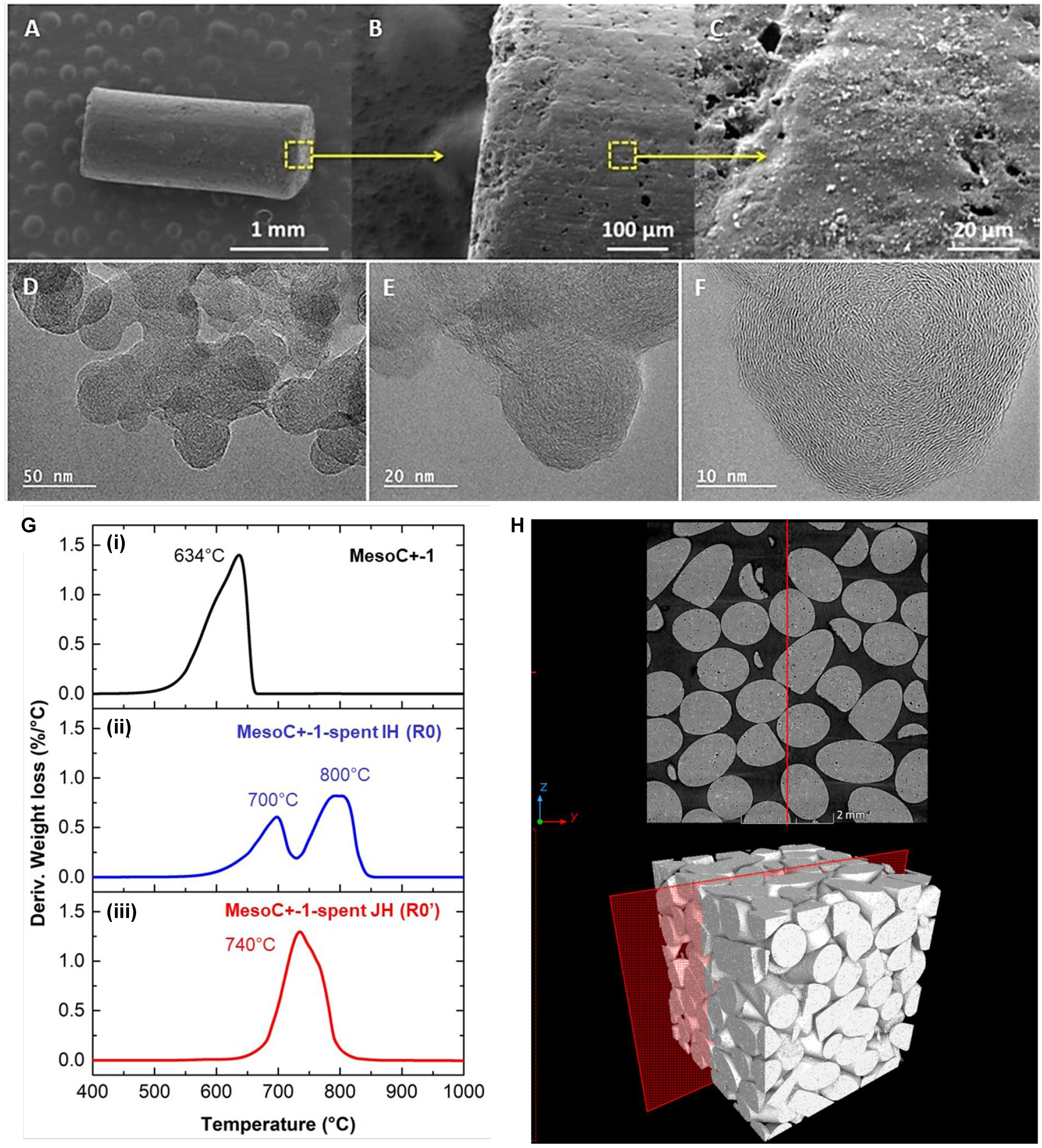
Figure 1. (A-C) SEM and (D-F) TEM micrographs of the pristine MESOC+-1 carbon catalyst with different magnifications; (G) TGA spectra of the fresh (i) and spent catalysts after CMD test under direct IH (ii) and indirect JH (iii) mode; (H) CT micrographs of the fresh catalyst bed showing the loosely packed bed (37 vol.% apparent porosity determined by AutoCAD software) with point contact between the different catalyst extrudates. SEM: Scanning electron microscopy; TEM: transmission electron microscopy; TGA: thermal gravimetric analysis; CMD: catalytic decomposition of methane; IH: induction heating; JH: Joule heating; CT: computed tomography.


