


Robotic resection of the uncinate process of the pancreas
 0
0 Abstract
Watch a video of this article.
BRIEF EXPLANATION
Robotic surgery is becoming increasingly common in pancreatic resection. Due to its retroperitoneal location, rich vascular supply, and limited exposure of the pancreas, pancreatic surgery remains technically challenging. Robotic systems overcome several limitations of conventional laparoscopy and offer a function-preserving option for selected benign or low-grade pancreatic tumors[1,2].
We present the case of a 47-year-old woman with persistent epigastric pain for 1 day and a preoperative diagnosis of intraductal papillary mucinous neoplasm (IPMN). Preoperative magnetic resonance cholangiopancreatography (MRCP) suggested a visible separation between the lesion and the main pancreatic duct [Figure 1A]. Although an exact distance was not available from the imaging report, this finding was considered in the preoperative assessment and further evaluated intraoperatively under high-definition robotic visualization. Following informed consent, robotic resection of the pancreatic uncinate process was performed [Figure 1B]. The gastric wall was suspended using the third robotic arm, the gastrocolic ligament was opened with ultrasonic shears, and the descending and horizontal portions of the duodenum were mobilized. The superior mesenteric vein was dissected from the inferior pancreatic border, and the uncinate process with the tumor was lifted dorsally and cephalad. During the procedure, the inferior pancreatic vascular arcade was preserved, and the inferior pancreaticoduodenal artery was protected until complete tumor separation. Transected branch pancreatic ducts were ligated and closed with 4-0 PDS sutures [Figure 1C]. Figure 1D shows the reinforced resection surface. Our video demonstrates the management in detail.
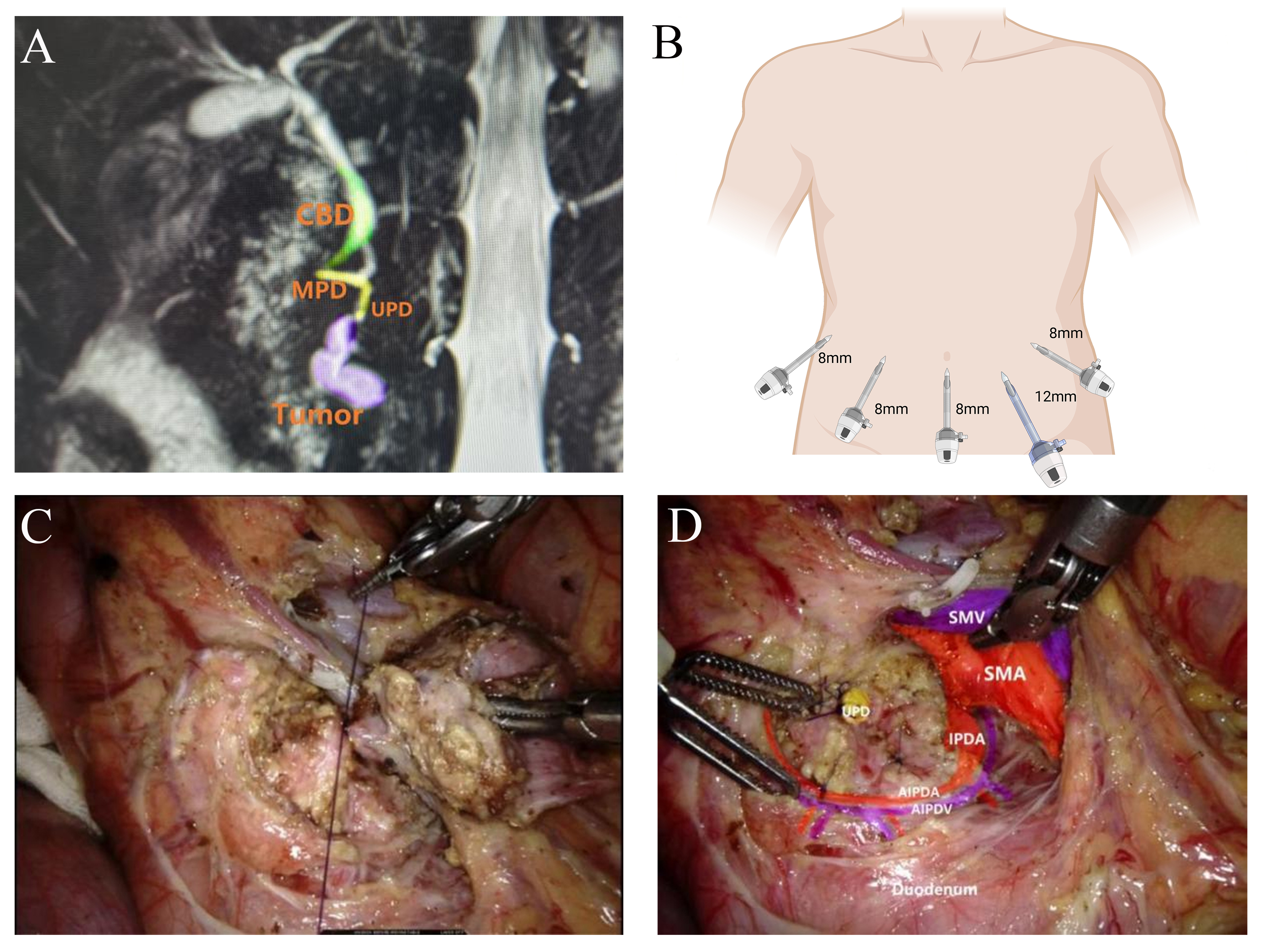
Figure 1. (A) Preoperative MRCP showing the tumor and pancreaticobiliary anatomy; (B) Trocar placement for robotic resection of the pancreatic uncinate process [Created in BioRender. Yan, C. (2026) https://BioRender.com/8c8zom8]; (C) Intraoperative closure of the branch pancreatic duct by ligation; (D) Schematic view of the surgical anatomy after resection. MRCP: Magnetic resonance cholangiopancreatography; CBD: common bile duct; MPD: main pancreatic duct; UPD: uncinate pancreatic ducts; SMV: superior mesenteric vein; SMA: superior mesenteric artery; IPDA: inferior pancreaticoduodenal artery; AIPDA: anterior inferior pancreaticoduodenal artery; AIPDV: anterior inferior pancreaticoduodenal vein.
The key technical challenges are accurate anatomy of the uncinate process, definition of a safe boundary from the main pancreatic duct, and preservation of the pancreaticoduodenal vascular arcade. It is of critical importance to protect the inferior pancreaticoduodenal artery intraoperatively. Additionally, meticulous closure of branch ducts can reduce the risk of postoperative pancreatic fistula. In this case, the postoperative recovery was uneventful, with preserved pancreatic endocrine and exocrine function.
In conclusion, robotic resection of the pancreatic uncinate process is a safe and feasible option for selected patients, enabling minimally invasive pancreatic function preservation.
DECLARATIONS
Authors’ contributions
Made substantial contributions to video editing and production and drafted the initial manuscript: Yan C, Jiang Y
Made substantial contributions to critical review of the video and the final manuscript: Chen G
Availability of data and materials
Not applicable.
AI and AI-assisted tools statement
Not applicable.
Financial support and sponsorship
National Natural Science Foundation of China (82560612); Hunan Provincial Natural Science Foundation Key Project (2026JJ30093); Interdisciplinary Research Program in Medicine and Engineering, The First Affiliated Hospital of University of South China (IRP-M&E-2025-08); University of South China Clinical Research 4310 Program (20224310NHYCG01).
Conflicts of interest
All authors declared that there are no conflicts of interest.
Ethical approval and consent to participate
This single-patient surgical video report was conducted in accordance with the Declaration of Helsinki and did not involve prospective human experimentation or animal research. Formal ethics committee approval was not required according to institutional policy. Informed consent was obtained from the patient.
Consent for publication
Written informed consent was obtained from the patient for the use and publication of anonymized clinical information, intraoperative images, and operative video for academic purposes.
Copyright
© The Author(s) 2026.
Supplementary Materials
Please note: The publisher is not responsible for the content or functionality of any supporting information supplied by the authors. Any queries (other than missing content) should be directed to the corresponding author for the article.
REFERENCES
1. Giuliani T, De Pastena M, Paiella S, et al. Pancreatic enucleation patients share the same quality of life as the general population at long-term follow-up: a propensity score-matched analysis. Ann Surg. 2023;277:e609-16.
Cite This Article
 Open Access
How to Cite
Download Citation
Export Citation File:
Type of Import
Tips on Downloading Citation
Citation Manager File Format
Type of Import
Direct Import: When the Direct Import option is selected (the default state), a dialogue box will give you the option to Save or Open the downloaded citation data. Choosing Open will either launch your citation manager or give you a choice of applications with which to use the metadata. The Save option saves the file locally for later use.
Indirect Import: When the Indirect Import option is selected, the metadata is displayed and may be copied and pasted as needed.
About This Article
Copyright

Data & Comments
Data
0









Comments
Comments must be written in English. Spam, offensive content, impersonation, and private information will not be permitted. If any comment is reported and identified as inappropriate content by OAE staff, the comment will be removed without notice. If you have any queries or need any help, please contact us at support@oaepublish.com.