


fig7
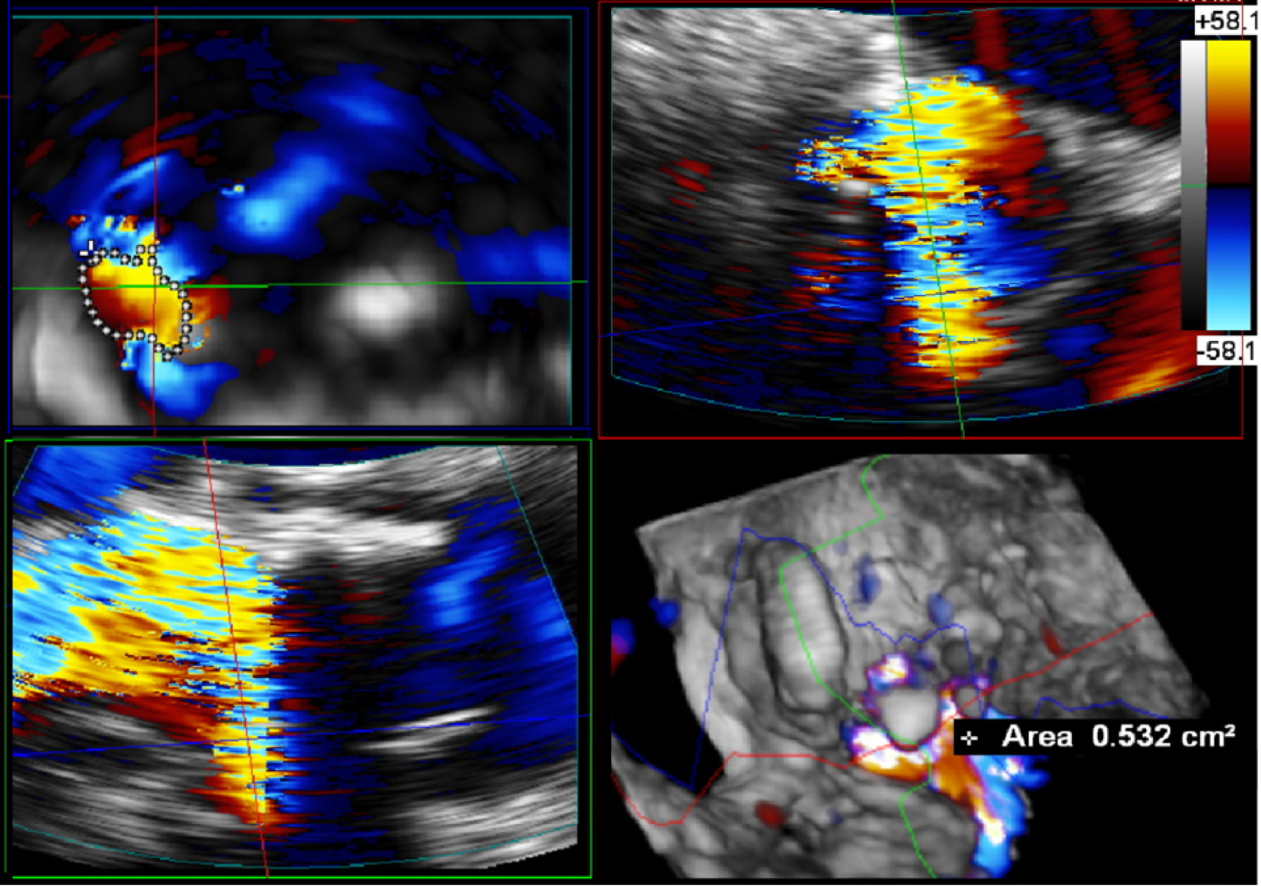
Figure 7. Following AV short-axis imaging, a 3D “zoom modality” with color is applied. The resulting image is presented in four distinct portions: sagittal, coronal, transverse, and whole-volume render. The diastolic frame is carefully selected for optimal visualization. To comprehensively demonstrate the paravalvular leak, the sagittal and coronal planes must be precisely bisected in the transverse plane. By methodically tracing the circular path, a 3D VC of the paravalvular leak can be generated. It is important to note that regurgitation is classified as severe when the VC area exceeds 0.3 cm2. 3D: Three-dimensional; VC: vena contracta.


