


fig6
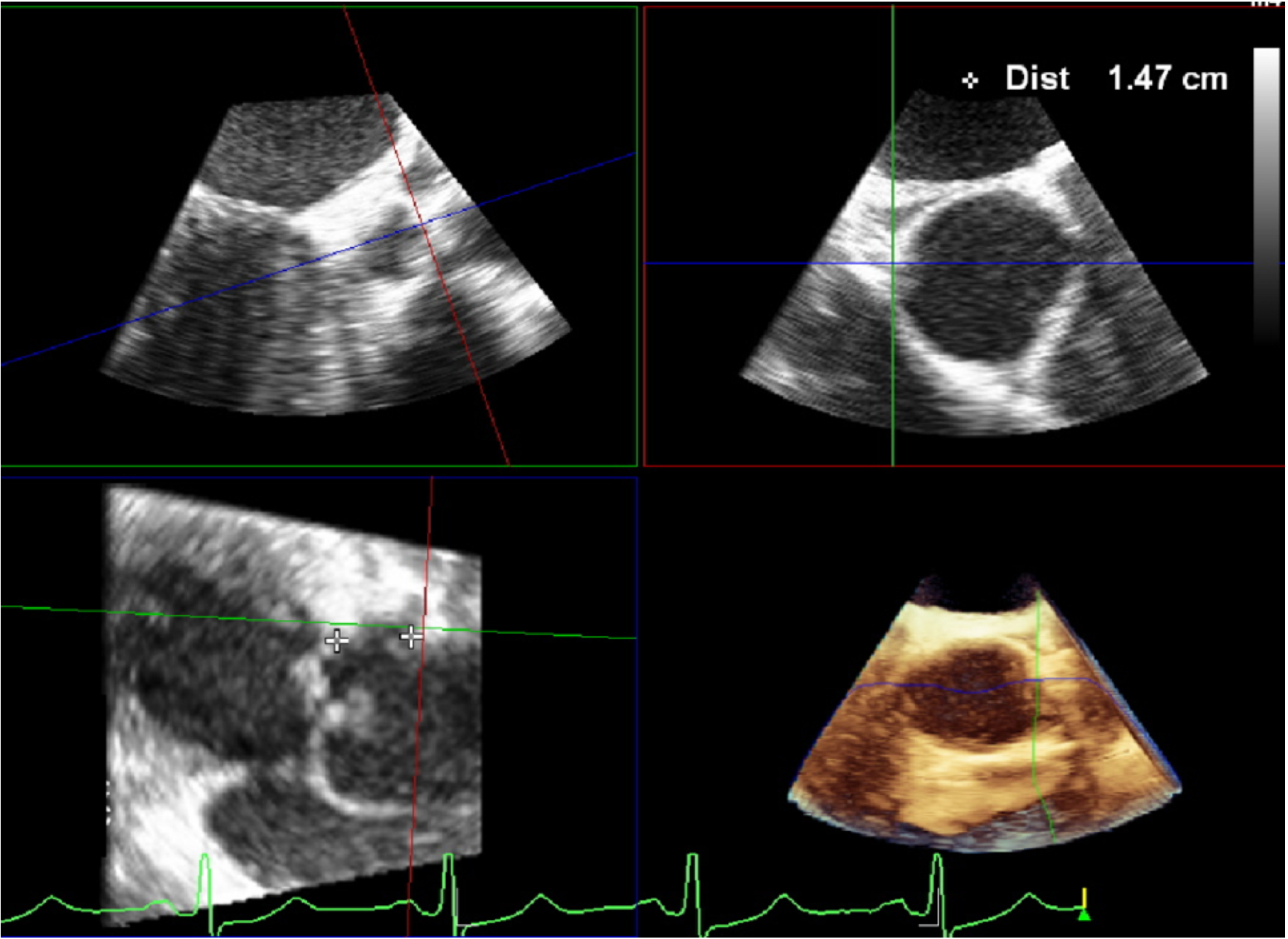
Figure 6. Combine the sagittal and coronal images to cut the AV long axis. In the sagittal plane, search for an indentation at the 10 o’clock position, which represents the left main coronary origin. Then move the marker along the aortic root in coronal view. Rotate the marker in the transverse image anticlockwise until it is perfectly aligned with the left major ostium. Finally, carefully measure the distance between the left main ostium and the left cusp in the sagittal plane. AV: Aortic valve.


