


fig4
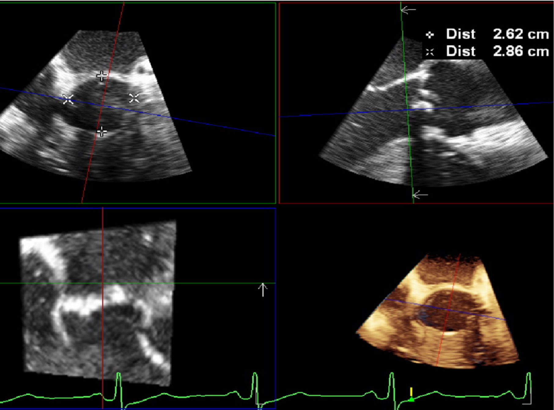
Figure 4. Annular evaluation begins by acquiring a high-quality 2D mid-esophageal short axis (30°-60°) or long axis (110°-120°) image, then selecting the 3D single-beat “zoom modality”. Choose the mid-systolic frame for measurements, as the aortic root is more circular during systole. The sagittal and coronal planes must bisect the AV long axis. The cross-sectional plane should be positioned at the ventricular-aortic junction, with the transverse plane adjusted to display the VBR. 2D: Two-dimensional; 3D: three-dimensional; AV: aortic valve; VBR: virtual basal ring.


