


fig3
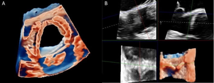
Figure 3. Glass (transparency) allows the imager to adjust the degree of transparency of both cardiac and extra-cardiac structures (Panel A). Multiplanar reconstruction allows live rotation of perpendicular planes on a 3D image to display any desired 2D imaging plane (Panel B). 2D: Two-dimensional; 3D: three-dimensional.


