


fig2
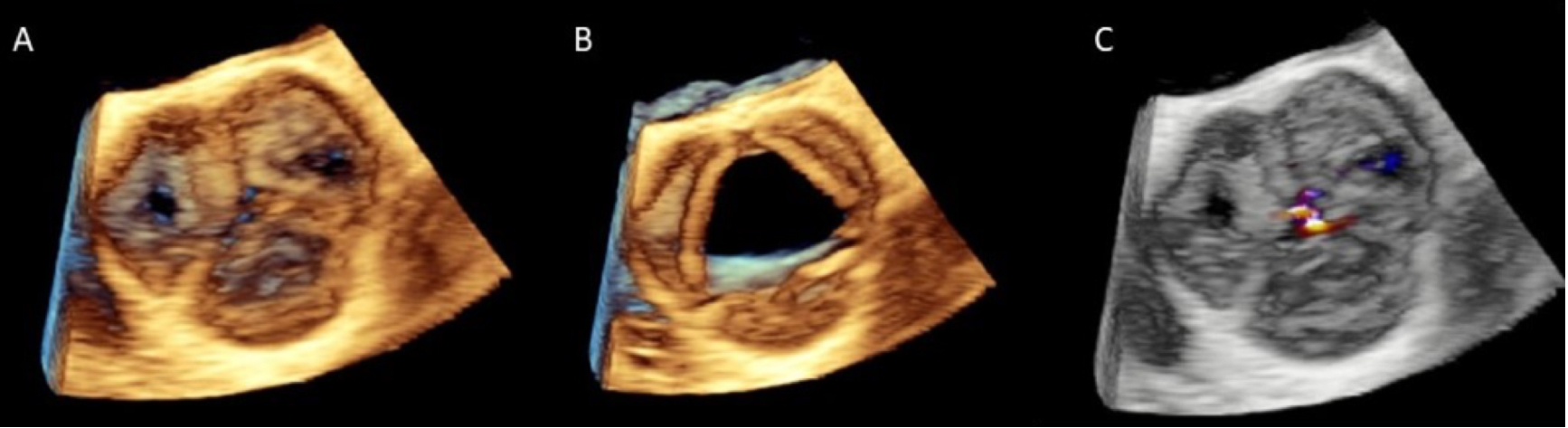
Figure 2. Multi-beat 3D TEE acquisition in zoom modality of the AV in diastole (Panel A) and systole (Panel B). 3D TEE mid-esophageal short-axis view with color-flow Doppler demonstrates moderate AR with a central origin (Panel C). TEE: Transesophageal echocardiography; AR: aortic regurgitation.


