


fig1
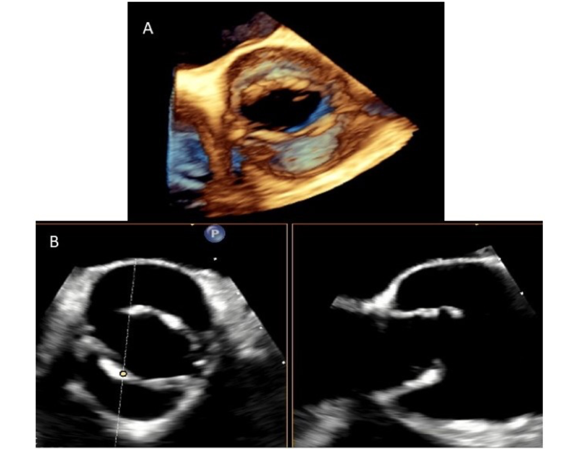
Figure 1. 3D TEE of a BAV in the short-axis (Panel A). X-plane mode at the level of the aortic valve shows BAV without raphe, anteropeposterior phenotype (Panel B). TEE: Transesophageal echocardiography; BAV: bicuspid aortic valve.
Figure 1. 3D TEE of a BAV in the short-axis (Panel A). X-plane mode at the level of the aortic valve shows BAV without raphe, anteropeposterior phenotype (Panel B). TEE: Transesophageal echocardiography; BAV: bicuspid aortic valve.

All published articles are preserved here permanently:
https://www.portico.org/publishers/oae/

